. 2020 May;182(5):1104-1116.
doi: 10.1002/ajmg.a.61531. Epub 2020 Mar 5.

Loss-of-function of Endothelin receptor type A results in Oro-Oto-Cardiac syndrome

Affiliations

Loss-of-function of Endothelin receptor type A results in Oro-Oto-Cardiac syndrome

Amanda Barone Pritchard et al. Am J Med Genet A. 2020 May.

Abstract

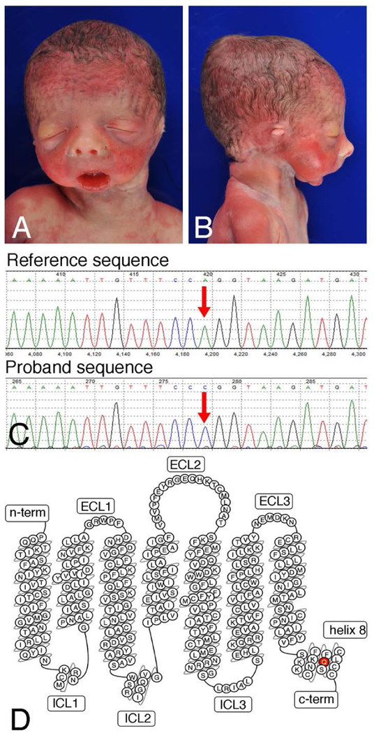
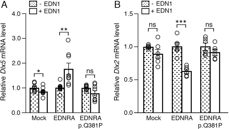
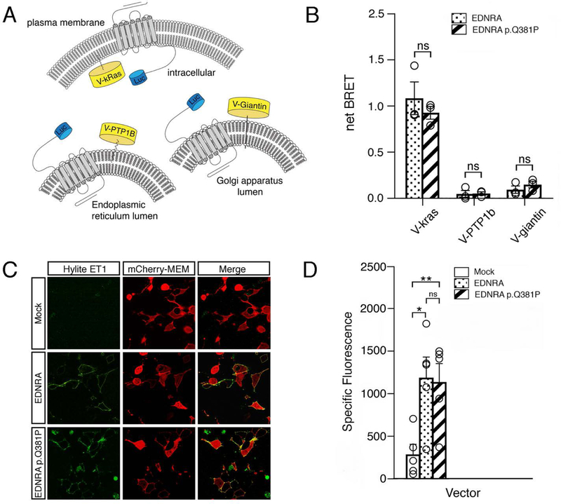
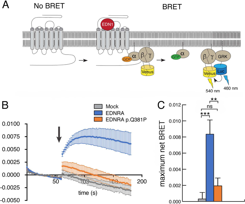
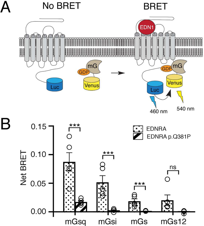
Craniofacial morphogenesis is regulated in part by signaling from the Endothelin receptor type A (EDNRA). Pathogenic variants in EDNRA signaling pathway components EDNRA, GNAI3, PCLB4, and EDN1 cause Mandibulofacial Dysostosis with Alopecia (MFDA), Auriculocondylar syndrome (ARCND) 1, 2, and 3, respectively. However, cardiovascular development is normal in MFDA and ARCND individuals, unlike Ednra knockout mice. One explanation may be that partial EDNRA signaling remains in MFDA and ARCND, as mice with reduced, but not absent, EDNRA signaling also lack a cardiovascular phenotype. Here we report an individual with craniofacial and cardiovascular malformations mimicking the Ednra -/- mouse phenotype, including a distinctive micrognathia with microstomia and a hypoplastic aortic arch. Exome sequencing found a novel homozygous missense variant in EDNRA (c.1142A>C; p.Q381P). Bioluminescence resonance energy transfer assays revealed that this amino acid substitution in helix 8 of EDNRA prevents recruitment of G proteins to the receptor, abrogating subsequent receptor activation by its ligand, Endothelin-1. This homozygous variant is thus the first reported loss-of-function EDNRA allele, resulting in a syndrome we have named Oro-Oto-Cardiac Syndrome. Further, our results illustrate that EDNRA signaling is required for both normal human craniofacial and cardiovascular development, and that limited EDNRA signaling is likely retained in ARCND and MFDA individuals. This work illustrates a straightforward approach to identifying the functional consequence of novel genetic variants in signaling molecules associated with malformation syndromes.

Keywords: Auriculocondylar syndrome; BRET; cardiovascular; micrognathia; neural crest cell.


Figures

Figure 1.
Figure 1.. Phenotypic and genomic analysis.
(a) Anterior and (b) lateral views of the infant’s face demonstrating dysmorphic features including downslanting palpebral fissures, upturned nasal tip, thin upper lip, micrognathia, microstomia, and abnormal external ears. (c) Sanger sequencing confirmation of the c.1142A>C mutation in the proband, resulting in the p.Q381P variant. (d) Bubble diagram of the EDNRA (adaptedfromGPCRdb.org). The p.Q381P mutation in helix 8 is denoted in red. c-term, C-terminus; ECL, extracellular loop; ICL, intracellular loop; n-term, N-terminus.
Figure 2.
Figure 2.. The EDNRA p.Q381P variant cannot induce EDNRA-dependent gene expression.
qRT-PCR analysis of Dlx5 (A) and Dlx2 (B) gene expression in MC3T3-E1 cells following transfection with empty vector (mock), EDNRA or EDNRA p.Q381P and treatment with EDN1. (a) While addition of EDN1 resulted in upregulation of Dlx5 expression in cells with wild type EDNRA, Dlx5 expression was not induced in cells with the EDNRA p.Q381P variant. (b) Addition of EDN1 resulted in downregulation of Dlx2 expression in cells with a wild type EDNRA but not in cells with the EDNRA p.Q381P variant (B). Assays were performed in duplicate or triplicate at least three times. Error bars represent SEM; two-tailed t-test; *p<0.05, **p< 0.005, ***p<0.001; ns., not significant.
Figure 3.
Figure 3.. EDNRA subcellular localization and EDN1 binding is not disrupted by the p.Q381P variant.
(a) Schematic of the BRET assay for subcellular localization, in which EDNRA-Renilla luciferase 8 (RLuc8) is combined with Venus (V)-kras (plasma membrane), Venus-PTP1B (endoplasmic reticulum) or Venus-giantin (Golgi apparatus) to measure BRET in different cellular compartments. (b) Average net BRET of unstimulated HEK293 cells transfected with wildtype EDNRA-RLuc8 or EDNRA p.Q381P-RLuc8 and V-kras, V-PTPB1 or V-giantin. Assays were performed at least three times. Error bars represent SEM.; two-tailed t-test; n.s., not significant. (c) Confocal images of HEK293T cells transfected with mCherry-MEM and pcDNA3.1, wild type EDNRA or EDNRA p.Q381P and incubated with Hilyte fluor-488-ET-1. mCherry-MEM and Hilyte fluor-488-ET-1 co-localized within cells expressing wild type EDNRA or EDNRA p.Q381P but not in empty vector (mock)-transfected cells. Images are representative of ligand binding after a 5 minute incubation. Green and red channels represent Hilyte fluor-488-ET-1 and mCherry-MEM, respectively. (d) HEK293T cells transfected with empty vector (mock), wild type EDNRA or EDNRA p.Q381P were incubated with Hilyte fluor-488-ET-1 and quantitatively analyzed with a fluorescence microplate reader. Specific fluorescence was calculated by subtracting background fluorescence values (from empty wells treated with Hilyte fluor-488-ET-1) from the raw fluorescence values. Assays were performed in triplicate at least three times. Error bars represent SEM; two-tailed t-test *p < 0.05, **p< 0.01; n.s., not significant.
Figure 4.
Figure 4.. The EDNRA p.Q381P variant disrupts G protein activation.
(a) Schematic for the G protein activation BRET reporter assay. EDN1-induced EDNRA activation promotes dissociation of the G protein heterotrimer to Gαq and Gβγ-Venus. Subsequent interaction of Gβγ-Venus with mas-GRKct-nLuc produces BRET, which is an indirect reporter of G protein activation. (b) BRET assay in HEK293T cells transfected with the BRET components shown in (a) and empty vector (mock), wild type EDNRA or EDNRA p.Q381P. Baseline BRET was measured for 30 seconds, at which time cells were treated with EDN1 (indicated by arrow) and measured for another 2 minutes. A robust EDN1-induced BRET response was observed only in cells expressing wildtype EDNRA (blue trace). In the presence of EDNRA p.Q381P (orange trace), BRET was not statistically different from the presence of a mock vector (grey trace). Traces represent the average delta BRET of at least three independent transfections. Error bars at each timepoint represent SEM. (c) Maximum BRET response was quantified as the average of maximum delta BRET values elicited by EDN1. Assays were performed in triplicate at least three times. Error bars indicate SEM; two-tailed t-test; **p < 0.01, ***p< 0.005; n.s., not significant.
Figure 5.
Figure 5.. The EDNRA p.Q381P variant disrupts G protein coupling.
(a) Schematic of the BRET assay for G protein coupling. A Venus-mini G protein can associate with a wild type EDNRA-RLuc8 after addition of EDN1, resulting in BRET. (b) The maximum BRET response was quantified for four classes of mini G proteins with wildtype EDNRA-RLuc8 or EDNRA-RLuc8 p.Q381P. Maximum BRET response for all mini G proteins was lower in cells expressing EDNRA-RLuc8 p.Q381P compared to wildtype EDNRA, though the change for mGs12 was not statistically significant (p=0.0678). Assays were performed in triplicate at least three times. Error bars indicate SEM; two-tailed t-test ***p<0.005; ns, not significant.

Similar articles

Cited by

References

    1. Abe M, Ruest L-B, & Clouthier DE (2007). Fate of cranial neural crest cells during craniofacial development in endothelin-A receptor deficient mice. International. Journal of Develeopmental. Biology, 51, 97–105. doi: 10.1387/ijdb.062237ma - DOI - PMC - PubMed
    1. Charité J, McFadden DG, Merlo GR, Levi G, Clouthier DE, Yanagisawa M, … Olson EN (2001). Role of Dlx6 in regulation of an endothelin-1-dependent, dHAND branchial arch enhancer. Genes and Development, 15, 3039–3049. doi: 10.1101/gad.931701 - DOI - PMC - PubMed
    1. Clouthier DE, Garcia E, & Schilling TF (2010). Regulation of facial morphogenesis by endothelin signaling: insights from mouse and fish. American Journal of Medical Genetics, Part A., 152A, 2962–2973. doi: 10.1002/ajmg.a.33568 - DOI - PMC - PubMed
    1. Clouthier DE, Hosoda K, Richardson JA, Williams SC, Yanagisawa H, Kuwaki T, … Yanagisawa M. (1998). Cranial and cardiac neural crest defects in endothelin-A receptor-deficient mice. Development, 125, 813–824.
    1. Clouthier DE, Passos-Bueno MR, Tavares ALP, Lyonnet S, Amiel J, & Gordon CT (2013). Understanding the basis of Auriculocondylar syndrome: Insights from human, mouse and zebrafish studies. American Journal of Medical Genetics, Part C, 163, 306–317. doi: 10.1002/ajmg.c.31376 - DOI - PMC - PubMed

Publication types

MeSH terms

Substances

Supplementary concepts