Observational Study
. 2020 Mar;7(3):363-374.
doi: 10.1002/acn3.51001. Epub 2020 Mar 4.

Hematoma enlargement characteristics in deep versus lobar intracerebral hemorrhage

Affiliations
Observational Study

Hematoma enlargement characteristics in deep versus lobar intracerebral hemorrhage

Jochen A Sembill et al. Ann Clin Transl Neurol. 2020 Mar.

Abstract

Objective: Hematoma enlargement (HE) is associated with clinical outcomes after supratentorial intracerebral hemorrhage (ICH). This study evaluates whether HE characteristics and association with functional outcome differ in deep versus lobar ICH.

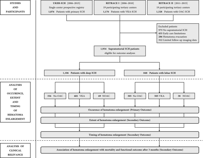
Methods: Pooled analysis of individual patient data between January 2006 and December 2015 from a German-wide cohort study (RETRACE, I + II) investigating ICH related to oral anticoagulants (OAC) at 22 participating centers, and from one single-center registry (UKER-ICH) investigating non-OAC-ICH patients. Altogether, 1954 supratentorial ICH patients were eligible for outcome analyses, which were separately conducted or controlled for OAC, that is, vitamin-K-antagonists (VKA, n = 1186) and non-vitamin-K-antagonist-oral-anticoagulants (NOAC, n = 107). Confounding was addressed using propensity score matching, cox regression modeling and multivariate modeling. Main outcomes were occurrence, extent, and timing of HE (>33%/>6 mL) and its association with 3-month functional outcome.

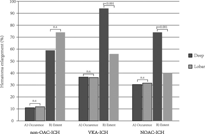
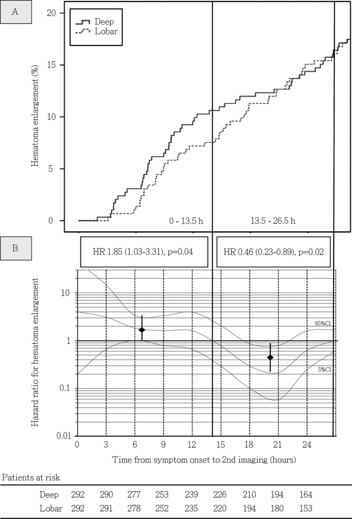
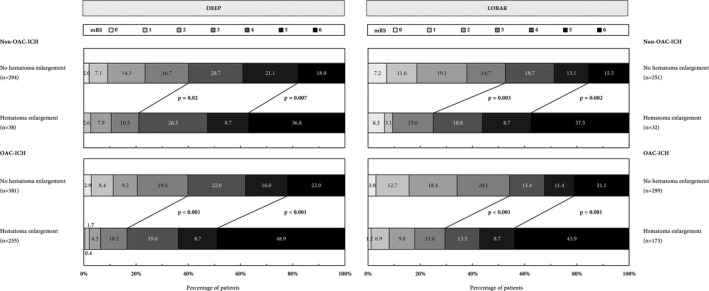
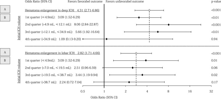
Results: Occurrence of HE was not different after deep versus lobar ICH in patients with non-OAC-ICH (39/356 [11.0%] vs. 36/305 [11.8%], P = 0.73), VKA-ICH (249/681 [36.6%] vs. 183/505 [36.2%], P = 0.91), and NOAC-ICH (21/69 [30.4%] vs. 12/38 [31.6%], P = 0.90). HE extent did not differ after non-OAC-ICH (deep:+59% [40-122] vs. lobar:+74% [37-124], P = 0.65), but both patients with VKA-ICH and NOAC-ICH showed greater HE extent after deep ICH [VKA-ICH, deep: +94% [54-199] vs. lobar: +56% [35-116], P < 0.001; NOAC-ICH, deep: +74% [56-123] vs. lobar: +40% [21-49], P = 0.001). Deep compared to lobar ICH patients had higher HE hazard during first 13.5 h after onset (Hazard ratio [HR]: 1.85 [1.03-3.31], P = 0.04), followed by lower hazard (13.5-26.5 h, HR: 0.46 [0.23-0.89], P = 0.02), and equal hazard thereafter (HR: 0.96 [0.56-1.65], P = 0.89). Odds ratio for unfavorable outcome was higher after HE in deep (4.31 [2.71-6.86], P < 0.001) versus lobar ICH (2.82 [1.71-4.66], P < 0.001), and only significant after small-medium (1st volume-quarter, deep: 3.09 [1.52-6.29], P < 0.01; lobar: 3.86 [1.35-11.04], P = 0.01) as opposed to large-sized ICH (4th volume-quarter, deep: 1.09 [0.13-9.20], P = 0.94; lobar: 2.24 [0.72-7.04], P = 0.17).

Interpretation: HE occurrence does not differ among deep and lobar ICH. However, compared to lobar ICH, HE after deep ICH is of greater extent in OAC-ICH, occurs earlier and may be of greater clinical relevance. Overall, clinical significance is more apparent after small-medium compared to large-sized bleedings.


Conflict of interest statement

The authors have no conflicts of interest related to the contents of the manuscript.

Figures

Figure 1
Figure 1
Study flowchart. Altogether, individual level data from 3,580 spontaneous ICH patients were analyzed to identify 1,954 supratentorial ICH patients eligible for outcome analyses. Data were provided by two parts of a German‐wide observational studies (RETRACE I and II) conducted at 22 participating tertiary centers, and by one single‐center university hospital registry.
Figure 2
Figure 2
Occurrence and extent of intracerebral hematoma enlargement. Occurrence (A) and extent (B) of hematoma enlargement in patients with deep compared to lobar ICH. Hematoma enlargement was defined as an increase in ICH volume of more than 33% or 6 mL from initial to follow‐up imaging. The extent of hematoma enlargement, that is, percentage ICH volume increase, was compared in patients actually suffering hematoma enlargement. Separate analyses were conducted for patients with non‐OAC‐ICH (n = 661), VKA‐ICH (n = 1186), and NOAC‐ICH (n = 107). Abbreviations: ICH, Intracerebral hemorrhage; NOAC, non‐vitamin‐K‐antagonist oral anticoagulant; n.s., not significant; OAC, Oral anticoagulation; VKA, vitamin‐K‐antagonist.
Figure 3
Figure 3
Incidence rates and time‐dependent hazard ratios for hematoma enlargement in patients with deep versus lobar ICH. (A) Incidence rates of hematoma enlargement detected through control imaging during the hyperacute course of deep and lobar ICH management in propensity‐matched cohorts. (B) Time‐dependent hazard ratios for hematoma enlargement in deep versus lobar ICH patients. Adjusted COX proportional hazard models were calculated for propensity‐matched cohorts with additional adjustment for intraventricular hemorrhage and prior oral anticoagulation to visualize the association between time since symptom onset and detection of hematoma enlargement by control imaging in patients dichotomized according to supratentorial ICH location. Hazard ratio estimates (y‐axis) for deep ICH patients were calculated at each hour since symptom onset using time‐patient‐clusters (HR estimate at the median of a 5‐hour interval) of patients with control imaging at a median of the presented hour (x‐axis) and compared with lobar ICH patients with data points within these clusters. To correct for overestimation, we weighted and smoothed hazard ratios by the method of moving averages. The dashed lines indicate time intervals with increased risk for detection of hematoma enlargement identified by the intercept of the adjusted HR median with the HR of 1. Medians and 95% CI displayed as square with whiskers represent hazard ratios for HE in deep compared to lobar ICH during mentioned identified time intervals, that is, 0–13.5 and 13.5–26.5 h. Patients at risk included in both analyses (A + B) are displayed using 3‐hour intervals, showing comparable numbers of patients receiving control imaging at each time point from individual onset of deep or lobar ICH. Abbreviations: CI, Confidence interval; HR, Hazard Ratio; h, hours; ICH, Intracerebral hemorrhage.
Figure 4
Figure 4
Functional outcome of patients with supratentorial Non‐OAC‐ICH and OAC‐ICH comparing patients with and without hematoma enlargement. Distribution of functional outcome and mortality at 3 months using the modified Rankin Scale (mRS, range 0–6, from 0 = no symptoms, to 5 = severe disability, and 6 = dead). Dichotomized comparison of patients with and without hematoma enlargement. The thick lines separate proportion of patients with favorable (mRS 0–3) and unfavorable (mRS 4–6) outcome as well as patients with and without 3‐month mortality. Abbreviations: ICH, Intracerebral hemorrhage; mRS, modified Rankin Scale; OAC, Oral anticoagulation.
Figure 5
Figure 5
Association of hematoma enlargement with 3‐month functional outcome according to ICH location and initial hematoma volume. Forest plots showing association of hematoma enlargement with functional outcome in (A) deep and lobar ICH patients and (B) deep and lobar ICH patients in relation to initial hematoma volume split according to quartiles. Multivariable modeling in both analyses (A + B) included adjustment for relevant outcome predictors, that is, age, Glasgow Coma Scale, oral anticoagulation, initial ICH volume, and intraventricular hemorrhage. Abbreviations: CI, Confidence interval; ICH, Intracerebral hemorrhage.

References

    1. Brouwers HB, Falcone GJ, McNamara KA, et al. CTA spot sign predicts hematoma expansion in patients with delayed presentation after intracerebral hemorrhage. Neurocrit Care 2012;17:421–428. - PMC - PubMed
    1. Flibotte JJ, Hagan N, O'Donnell J, et al. Warfarin, hematoma expansion, and outcome of intracerebral hemorrhage. Neurology 2004;28:1059–1064. - PubMed
    1. Kuramatsu JB, Gerner ST, Schellinger PD, et al. Anticoagulant reversal, blood pressure levels, and anticoagulant resumption in patients with anticoagulation‐related intracerebral hemorrhage. JAMA 2015;24:824–836. - PubMed
    1. Dowlatshahi D, Yogendrakumar V, Aviv RI, et al. Small intracerebral hemorrhages have a low spot sign prevalence and are less likely to expand. Int J Stroke 2016;11:191–197. - PubMed
    1. Al‐Shahi Salman R, Frantzias J, Lee RJ, et al. Absolute risk and predictors of the growth of acute spontaneous intracerebral haemorrhage: a systematic review and meta‐analysis of individual patient data. Lancet Neurol 2018;17:885–894. - PMC - PubMed

Publication types

MeSH terms

LinkOut - more resources