Postnatal development of the entorhinal cortex: A stereological study in macaque monkeys
- PMID: 32134112
- PMCID: PMC7392818
- DOI: 10.1002/cne.24897
Postnatal development of the entorhinal cortex: A stereological study in macaque monkeys
Abstract
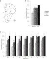
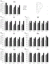
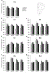
The entorhinal cortex is the main gateway for interactions between the neocortex and the hippocampus. Distinct regions, layers, and cells of the hippocampal formation exhibit different profiles of structural and molecular maturation during postnatal development. Here, we provide estimates of neuron number, neuronal soma size, and volume of the different layers and subdivisions of the monkey entorhinal cortex (Eo, Er, Elr, Ei, Elc, Ec, Ecl) during postnatal development. We found different developmental changes in neuronal soma size and volume of distinct layers in different subdivisions, but no changes in neuron number. Layers I and II developed early in most subdivisions. Layer III exhibited early maturation in Ec and Ecl, a two-step/early maturation in Ei and a late maturation in Er. Layers V and VI exhibited an early maturation in Ec and Ecl, a two-step and early maturation in Ei, and a late maturation in Er. Neuronal soma size increased transiently at 6 months of age and decreased thereafter to reach adult size, except in Layer II of Ei, and Layers II and III of Ec and Ecl. These findings support the theory that different hippocampal circuits exhibit distinct developmental profiles, which may subserve the emergence of different hippocampus-dependent memory processes. We discuss how the early maturation of the caudal entorhinal cortex may contribute to path integration and basic allocentric spatial processing, whereas the late maturation of the rostral entorhinal cortex may contribute to the increased precision of allocentric spatial representations and the temporal integration of individual items into episodic memories.
Keywords: Macaca mulatta; RRID:SCR_000696, California National Primate Research Center Analytical and Resource Core; RRID:SCR_002526, Stereo Investigator; RRID:SCR_002865, SPSS; RRID:SCR_014199, Adobe Photoshop; allocentric spatial memory; episodic memory; hippocampal formation; infantile amnesia; medial temporal lobe; object memory; path integration.
© 2020 Wiley Periodicals, Inc.
Figures


References
-
- Acredolo LP (1978). Development of spatial orientation in infancy. Developmental Psychology, 14(3), 224–234.
-
- Amaral DG, Insausti R, & Cowan WM (1987). The entorhinal cortex of the monkey: I. Cytoarchitectonic organization. J Comp Neurol, 264(3), 326–355. - PubMed
-
- Amaral DG, & Lavenex P (2007). Hippocampal neuroanatomy In Andersen P, Morris RGM, Amaral DG, Bliss TV, & O’Keefe J (Eds.), The hippocampus book (pp. 37–114). Oxford: Oxford University Press.
Publication types
MeSH terms
Grants and funding
LinkOut - more resources
Full Text Sources

