. 2020 Mar 27;40(3):BSR20192620.
doi: 10.1042/BSR20192620.

Down-regulation of protease-activated receptor 2 ameliorated osteoarthritis in rats through regulation of MAPK/NF-κB signaling pathway in vivo and in vitro

Affiliations

Down-regulation of protease-activated receptor 2 ameliorated osteoarthritis in rats through regulation of MAPK/NF-κB signaling pathway in vivo and in vitro

Shichang Yan et al. Biosci Rep. .

Abstract

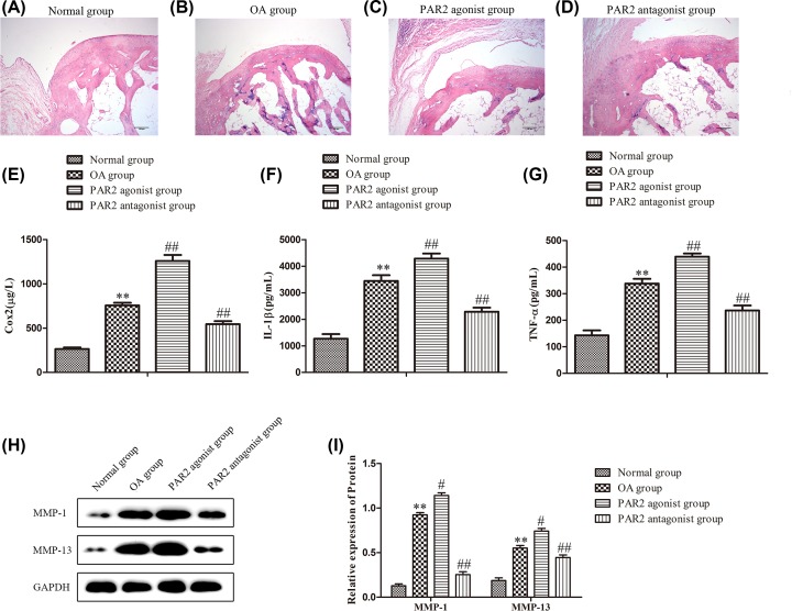
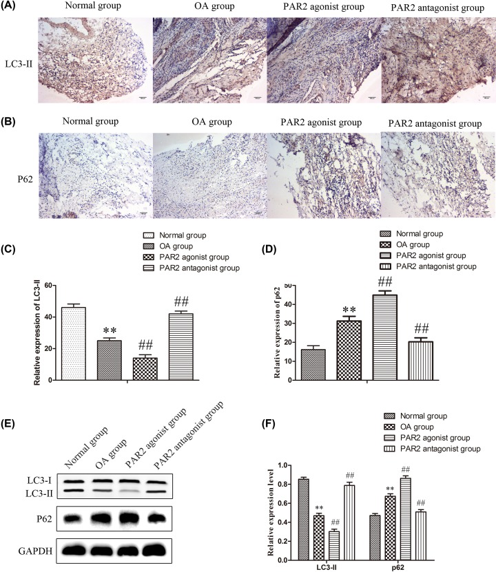
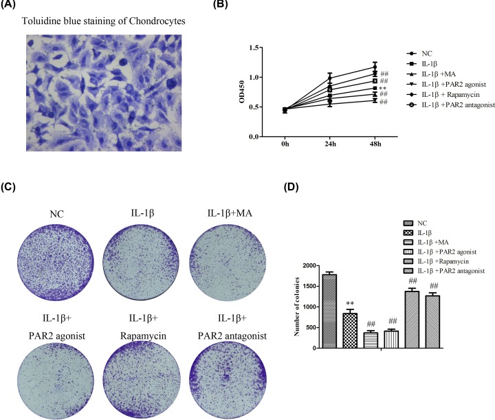
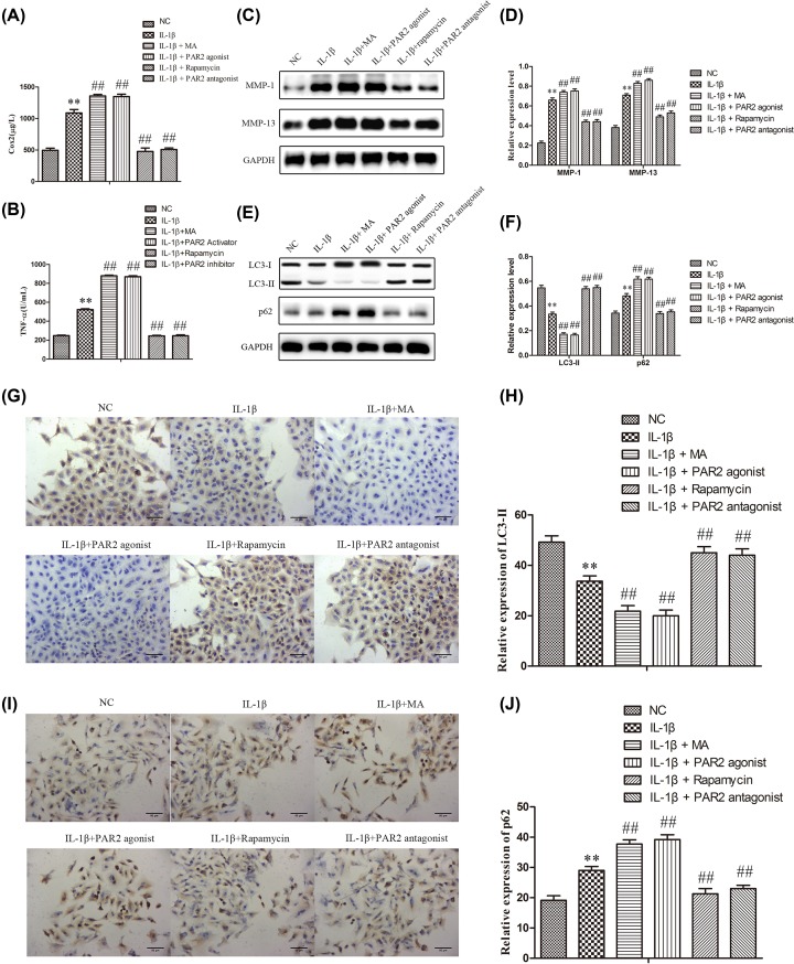
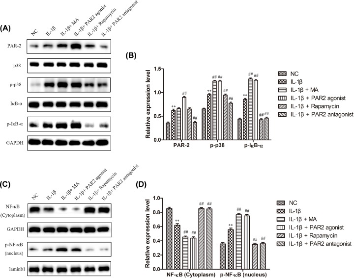
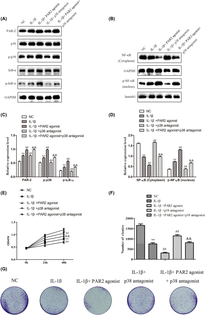
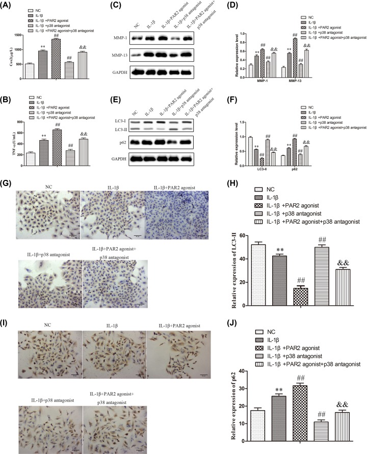
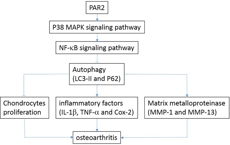
Recently, protease-activated receptor 2 (PAR2) has been proved to be involved in the inflammatory response including osteoarthritis (OA). In the present study, we found that PAR2 antagonist could remarkably improve the pathological condition of OA rats in vivo. In addition, we also found that PAR2 antagonist could suppress the production of inflammatory factors (TNF-α and Cox-2), decrease the levels of MMP-1 and MMP-13, and restrain the levels of P62 proteins and aggravate the expression of LC3-II both in vivo and in vitro. Besides, in vitro, PAR2 antagonist could increase the proliferation and colony formation of chondrocytes induced with IL-1β. Moreover, PAR2 antagonist could decrease the expression of expressions of p-p38, p-IκBα and p-NF-κB in vitro. However, PAR2 agonist exhibited the opposite effects. Furthermore, SB203580, a p38 MAPK inhibitor, could remarkably promote the proliferation of chondrocytes induced with IL-1β, could alleviate the production of TNF-α and Cox-2, could down-regulate the protein expressions of MMP-1 and MMP-13, and could decrease the expression of P62 and increase the expressions of LC3-II of chondrocytes induced with IL-1β. Importantly, SB203580 could reverse the effects of PAR2 agonist on the functions of chondrocytes induced with IL-1β. Taken together, the present data suggest that down-regulation of PAR2 can ameliorate OA through inducing autophagy via regulation of MAPK/NF-κB signaling pathway in vivo and in vitro, and PAR2 can be considered as a potential candidate to treat OA.

Keywords: Autophagy; MAPK/NF-kB signaling pathway; Osteoarthritis; Protease-activated receptor 2.


Conflict of interest statement

The authors declare that there are no competing interests associated with the manuscript.

Figures

Figure 1
Figure 1. Pathology, inflammatory factors, matalloproteinases were regulated by PAR2 in osteoarthritis cartilage tissue in rats
OA rats were injected with PAR2 agonist or PAR2 antagonist through knee joint cavity (AD) The pathological changes were detected by HE staining assay. (EG) The levels of Cox-2, IL-1β and TNF-α were determined using ELISA. (H,I) The levels of MMP-1 and MMP-13 were determined using Western blot and quantification analysis. The values are mean ± SD of three independent experiments. **P<0.01 vs normal group, ##P<0.01 vs OA group.
Figure 2
Figure 2. Relative protein of autophagy was regulated by PAR2 in osteoarthritis cartilage tissue in rats
(A and C) The expression of LC3II was detected by immunohistochemistry and quantification analysis. (B and D) The expression of p62 was detected by immunohistochemistry and quantification analysis. (E and F) The expressions of LC3II and p62 were examined by Western blot and quantification analysis. The values are mean ± SD of three independent experiments. **P<0.01 vs normal group, ##P<0.01 vs OA group.
Figure 3
Figure 3. Cell proliferation is regulated by PAR2 in chondrocytes induced by IL-1β
(A) Primary chondrocytes of rats were extracted, and toluidine blue staining were performed to identify primary chondrocytes. (B) The cell viability was determined by CCK-8 assay. (C and D) The cell proliferation was determined by clony formation assay. The values are mean ± SD of three independent experiments. **P<0.01 vs NC group, ##P<0.01 vs IL-1β treatment group.
Figure 4
Figure 4. Inflammation factor, matrix protease and relative protein of autophagy were regulated by PAR2 in chondrocytes induced by IL-1β
(A and B) Inflammation factor (Cox-2 and TNF-α) were determined using ELISA. (C and D) matrix protease (MMP-1 and MMP-13) were detected by Western blot and quantification analysis. (E and F) Relative protein of autophagy (LC3II and p62) were detected by Western blot and quantification analysis. (GJ) The expression of LC3II and p62 were further confirmed by immunohistochemistry and quantification analysis. The values are mean ± SD of three independent experiments. **P<0.01 vs NC group, ##P<0.01 vs IL-1β treatment group.
Figure 5
Figure 5. MAPK / NF-κB signal pathway participated in the effect of PAR2 on chondrocytes induced by IL-1β
(A and B) The expression levels of PAR2, p38, p-p38, IκB-α, and p- IκB-α were determined by Western blot and quantification analysis. (C and D) The expression levels of p- NF-κB (nucleus) and NF-κB (cytoplasm) were determined by Western blot and quantification analysis. The values are mean ± SD of three independent experiments. **P<0.01 vs NC group, ##P<0.01 vs IL-1β treatment group.
Figure 6
Figure 6. PAR2 regulated chondrocytes proliferation induced by IL-1β by MAPK / NF-κB signal pathway
SB203580, a novel p38 MAPK inhibitor with selective activity against p38 MAPK, was employed to inhibit the p38 MAPK expression in chondrocytes induced by IL-1β. (AD) The expression levels of PAR2, p38, p-p38, IκB-α, p- IκB-α, p- NF-κB (nucleus) and NF-κB (cytoplasm) were determined by Western blot and quantification analysis. (E) The cell viability was determined by CCK-8 assay. (F and G) The cell proliferation was determined by clony formation assay and quantification analysis. The values are mean ± SD of three independent experiments. **P<0.01 vs NC group, ##P<0.01 vs IL-1β treatment group, &&P<0.01 vs IL-1β+ PAR2 agonist.
Figure 7
Figure 7. PAR2 regulates Inflammation factor, matrix protease, and relative protein of autophagy by MAPK / NF-κB in chondrocytes induced by IL-1β
(A and B) Inflammation factor (Cox-2 and TNF-α) were determined using ELISA. (C and D) Matrix protease (MMP-1 and MMP-13) were detected by Western blot and quantification analysis. (E and F) Relative protein of autophagy (LC3II and p62) were detected by Western blot and quantification analysis. (GJ) The expression of LC3II and p62 were further confirmed by immunohistochemistry and quantification analysis. The values are mean ± SD of three independent experiments. **P<0.01 vs NC group, ##P<0.01 vs IL-1β treatment group, &&P<0.01 vs IL-1β+ PAR2 agonist.
Figure 8
Figure 8. The relationship was described among PAR2 and OA
PAR2 could activate MAPK signaling pathway, and then activate NF-κB signaling pathway, which cause the inhibition of chondrocytes proliferation, increase of inflammation factor (IL-1β, Cox-2 and TNF-α), and increase of matrix protease (MMP-1 and MMP-13). Those promote the progression of OA.

Similar articles

Cited by

References

    1. Glyn-Jones S., Palmer A.J., Agricola R. et al. . (2015) Osteoarthritis. Lancet 386, 376–387 10.1016/S0140-6736(14)60802-3 - DOI - PubMed
    1. Flemming D.J. and Gustas-French C.N. (2017) Rapidly Progressive Osteoarthritis: a Review of the Clinical and Radiologic Presentation. Curr. Rheumatol. Rep. 19, 42 10.1007/s11926-017-0665-5 - DOI - PubMed
    1. Martel-Pelletier J., Barr A.J., Cicuttini F.M. et al. . (2016) Osteoarthritis. Nat. Rev. Dis. Primers 2, 16072 10.1038/nrdp.2016.72 - DOI - PubMed
    1. Favero M., El-Hadi H., Belluzzi E. et al. . (2017) Infrapatellar fat pad features in osteoarthritis: a histopathological and molecular study. Rheumatology (Oxford) 56, 1784–1793 10.1093/rheumatology/kex287 - DOI - PubMed
    1. Pereira D., Ramos E. and Branco J. (2015) Osteoarthritis. Acta. Med. Port. 28, 99–106 10.20344/amp.5477 - DOI - PubMed

Publication types

MeSH terms