. 2020 Mar 5;15(3):e0222058.
doi: 10.1371/journal.pone.0222058. eCollection 2020.

Differentiation of human colon tissue in culture: Effects of calcium on trans-epithelial electrical resistance and tissue cohesive properties

Affiliations

Differentiation of human colon tissue in culture: Effects of calcium on trans-epithelial electrical resistance and tissue cohesive properties

Shannon D McClintock et al. PLoS One. .

Abstract

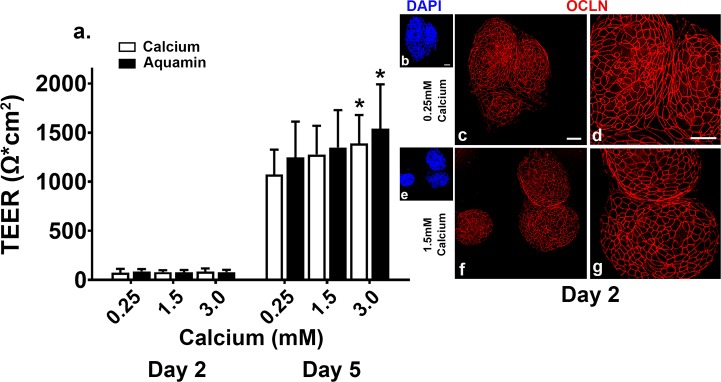
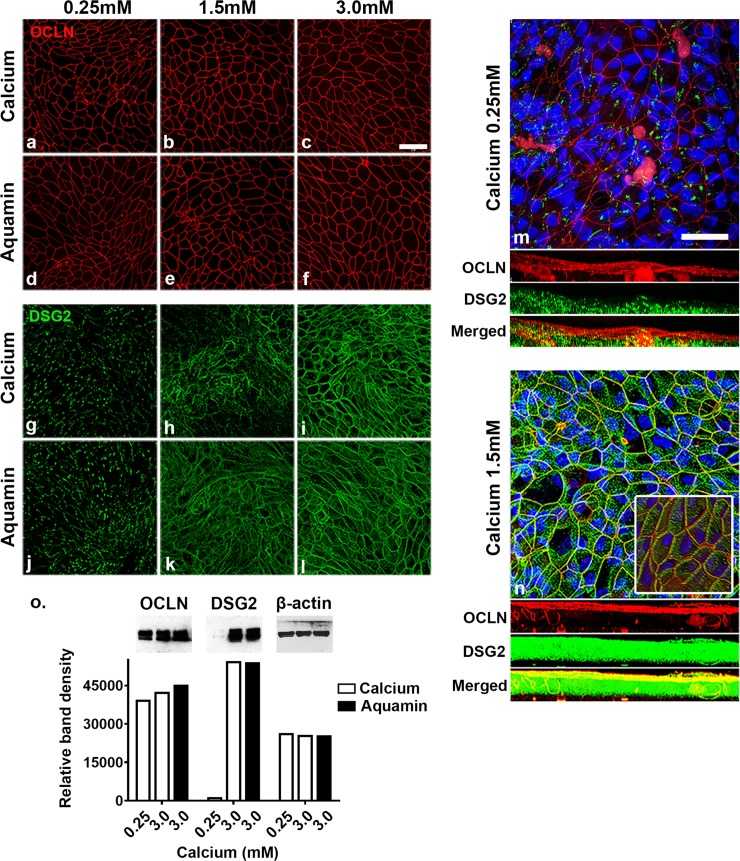
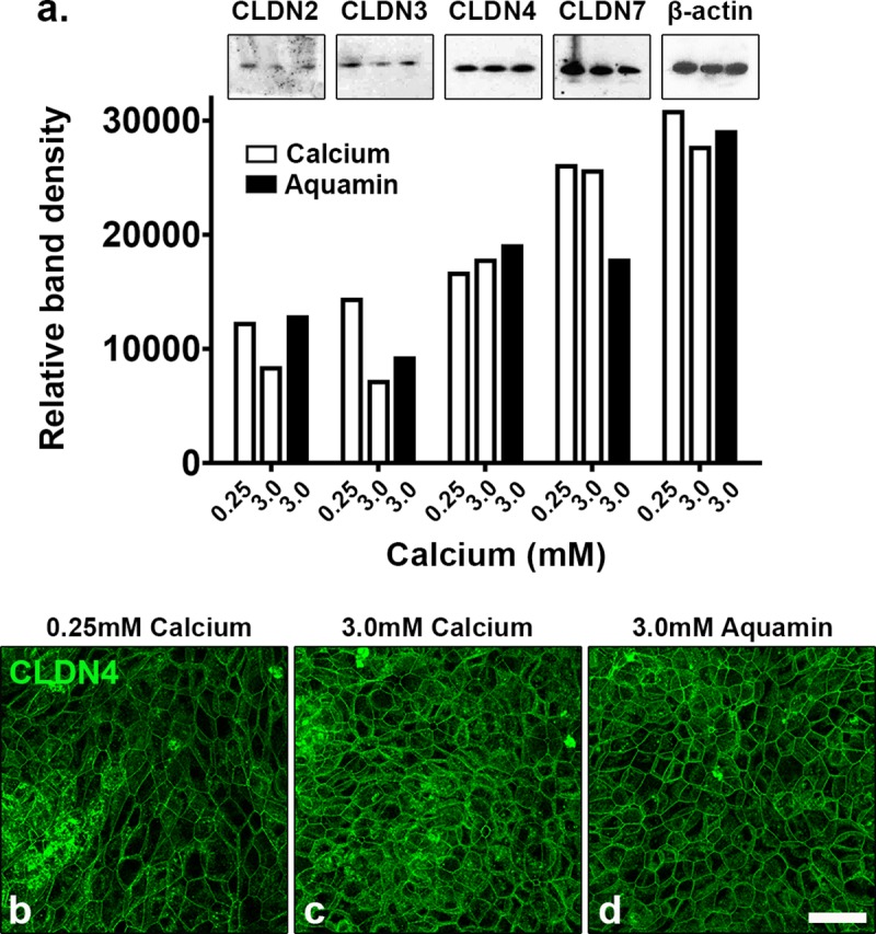
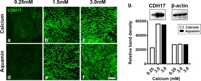
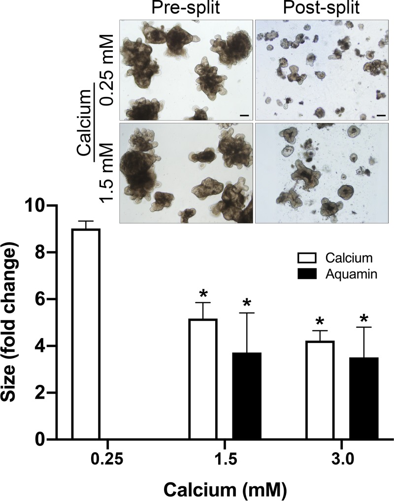
Background and aims: Human colonoid cultures maintained under low-calcium (0.25 mM) conditions undergo differentiation spontaneously and, concomitantly, express a high level of tight junction proteins, but not desmosomal proteins. When calcium is included to a final concentration of 1.5-3.0 mM (provided either as a single agent or as a combination of calcium and additional minerals), there is little change in tight junction protein expression but a strong up-regulation of desmosomal proteins and an increase in desmosome formation. The aim of this study was to assess the functional consequences of calcium-mediated differences in barrier protein expression.

Methods: Human colonoid-derived epithelial cells were interrogated in transwell culture under low- or high-calcium conditions for monolayer integrity and ion permeability by measuring trans-epithelial electrical resistance (TEER) across the confluent monolayer. Colonoid cohesiveness was assessed in parallel.

Results: TEER values were high in the low-calcium environment but increased in response to calcium. In addition, colonoid cohesiveness increased substantially with calcium supplementation. In both assays, the response to multi-mineral intervention was greater than the response to calcium alone. Consistent with these findings, several components of tight junctions were expressed at 0.25 mM calcium but these did not increase substantially with supplementation. Cadherin-17 and desmoglein-2, in contrast, were weakly-expressed under low calcium conditions but increased with intervention.

Conclusions: These findings indicate that low ambient calcium levels are sufficient to support the formation of a permeability barrier in the colonic epithelium. Higher calcium levels promote tissue cohesion and enhance barrier function. These findings may help explain how an adequate calcium intake contributes to colonic health by improving barrier function, even though there is little change in colonic histological features over a wide range of calcium intake levels.


Conflict of interest statement

There is no conflict of interest to declare from any author. Marigot LTD (Cork, Ireland) has provided Aquamin® as a gift for use in the study. This does not, in any way, alter our adherence to PLOS ONE policies on sharing data and materials.

Figures

Fig 1
Fig 1. Trans-epithelial electrical resistance (TEER) values.
Colonoids were plated on transwell membranes and incubated under the indicated conditions. a: At day-2 and -5, electrical resistance across the cell layer was assessed. Values shown are means and standard deviations based on five separate experiments with 3 or 4 samples (individual membranes) per data point in each experiment). Membrane to membrane variability was routinely less than 5%. Data were analyzed for statistical differences using ANOVA followed by unpaired-group comparisons. * indicate statistical significance from calcium at 0.25 mM (p<0.05; calculated by two-tailed unpaired t-test). Inset: Confocal fluorescent microscopic (max projected) images of membranes stained after the day-2 reading with DAPI (panels: b,e) or with antibody to occludin (panels: c,d,f,g). Scale bar = 10 μm. OCLN = occludin.
Fig 2
Fig 2. Confocal fluorescent microscopic images and western blot results at day-5.
Colonoids were plated on transwell membranes and incubated under the indicated conditions. After TEER assessment at day-5, membranes were prepared and stained. a-f: Occludin (max projected); g-l: Desmoglein-2 (max projected). m-n: Occludin and desmoglein-2 (z-stack composites made up of approximately 50 planes per image). Nuclear staining is indicated by blue color (DAPI). The three bands below the main images represent horizontal views of staining through the cell layer at each z-plane. Occludin is red, desmoglein-2 is green and the bright yellow color in the horizontal view of the 1.5 mM calcium image represents a composite of the two proteins. Inset: The inset in the 1.5 mM calcium image is a view of the entire z-stack viewed from a 45o angle to the cell surface in order to provide a 3-dimensional rendering. Scale bar = 10 μm. o: Western blot for occludin and desmoglein-2. 10 μg of protein from each condition was used. β-actin was used as a loading control. Band quantitation was done using ImageJ software. OCLN = occludin; DSG2 = desmoglein-2.
Fig 3
Fig 3. Evaluation of claudin expression by western blotting and confocal fluorescence microscopy.
Colonoids were plated on transwell membranes and incubated under the indicated conditions. After TEER assessment at day-5, lysates from some membranes were prepared for western blotting and additional membranes were prepared for confocal fluorescence microscopy. a: Western blot: 10 μg of protein from each condition was used. β-actin was used as a loading control. Band quantitation was done using ImageJ software. b-d: Claudin-4 (max projected) was assessed by confocal fluorescence microscopy. Scale bar = 10 μm. CLDN = claudin.
Fig 4
Fig 4. Evaluation of cadherin-17 expression by confocal fluorescent microscopy and western blotting.
Colonoids were plated on transwell membranes and treated as indicated. a-f: Confocal fluorescence microscopy. After TEER assessment at day-5, membranes were prepared and stained for cadherin-17 expression (max projected). Scale bar = 10 μm. g: Protein isolated from each condition was assessed for cadherin-17 expression by western blotting. 10 μg of protein from each condition was used. β-actin was used as a loading control. Band quantitation was done using ImageJ software. CDH17 = cadherin-17.
Fig 5
Fig 5. Colonoid cohesion.
Colonoids were maintained for 14 days in culture under the indicated conditions. At the end of the incubation period, the size of multiple individual colonoids was assessed by measuring surface area in phase-contrast images. Following this, the colonoids were harvested and fragmented for subculture. After plating, the fragmented colonoids were again “sized.” Values shown represent the change in surface area means and standard deviations of individual colonoids based on three separate experiments representing colonoids from three different subjects with a minimum of 80 colonoids from each subject assessed per treatment group in both pre- and post-split cultures. Data were compared for statistical differences using ANOVA followed by unpaired-group comparisons. * indicates a difference from 0.25 mM calcium at p<0.05. Inset: Representative examples of pre-split and post-split colonoids. Scale bar = 200 μm.

References

    1. Clayburgh DR, Shen L, Turner JR. A porous defense: the leaky epithelial barrier in intestinal disease. Lab Invest. 2004;84:282–291. 10.1038/labinvest.3700050 - DOI - PubMed
    1. Turner JR. Molecular basis of epithelial barrier regulation: From basic mechanisms to clinical application. Am J Pathol. 2006;169:1901–1909. 10.2353/ajpath.2006.060681 - DOI - PMC - PubMed
    1. Shen L Su L, Turner JR. Mechanisms and functional implication of intestinal barrier defects. Digestive Dis. 2009;27:443–449. - PMC - PubMed
    1. Turner JR. Intestinal mucosal barrier function in health and disease. Nat Rev Immunol. 2009;9:799–809. 10.1038/nri2653 - DOI - PubMed
    1. Camilleri M, Madsen K, Spiller R, Van Meerveld BG, Verne GN. Intestinal barrier function in health and gastrointestinal disease. Neurogastroenterol Motil. 2012;24:503–512. 10.1111/j.1365-2982.2012.01921.x - DOI - PMC - PubMed

Publication types