. 2020 Apr;24(7):3917-3930.
doi: 10.1111/jcmm.14978. Epub 2020 Mar 5.

Monophosphoryl lipid A alleviated radiation-induced testicular injury through TLR4-dependent exosomes

Affiliations

Monophosphoryl lipid A alleviated radiation-induced testicular injury through TLR4-dependent exosomes

Zhe Liu et al. J Cell Mol Med. 2020 Apr.

Abstract

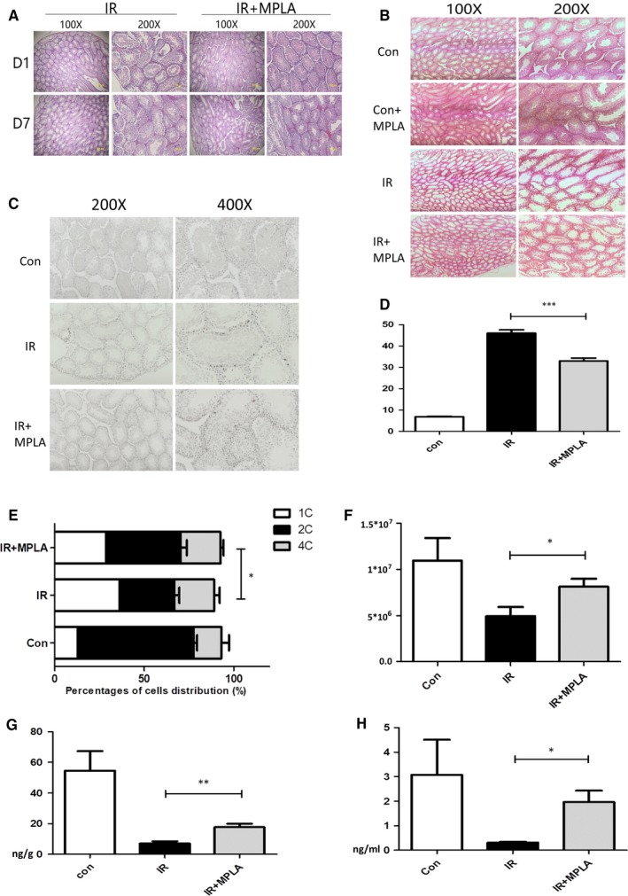
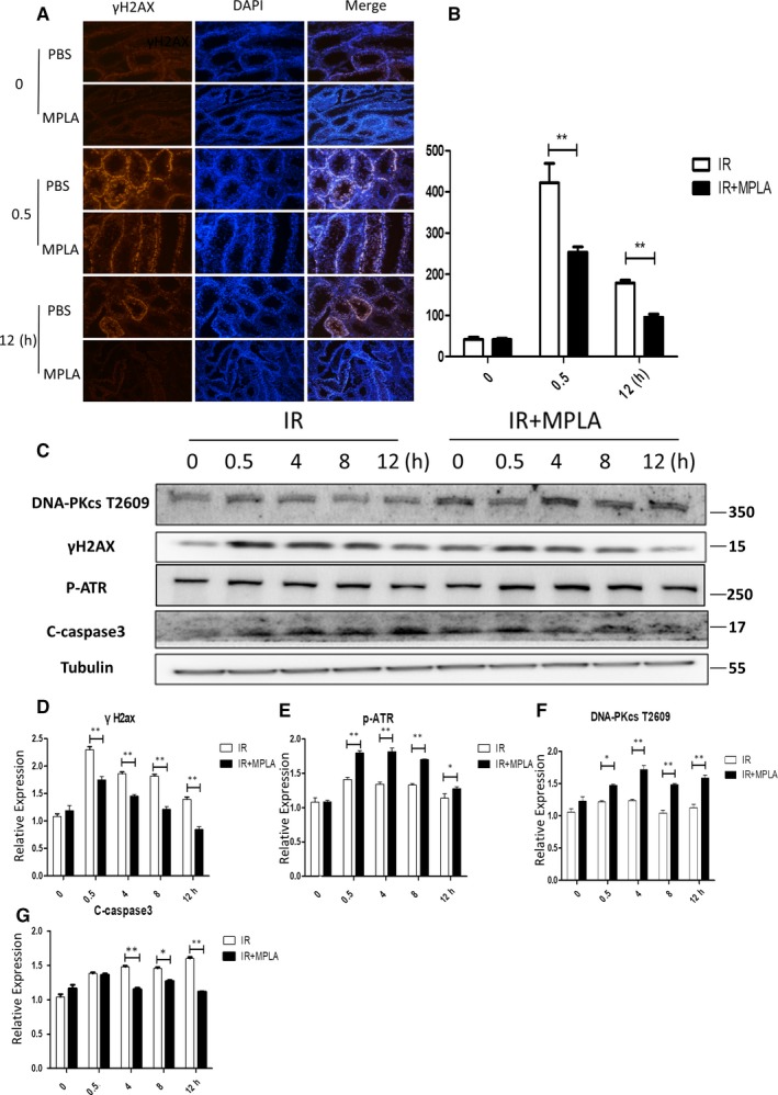
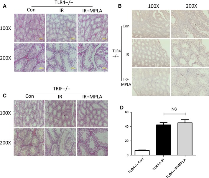
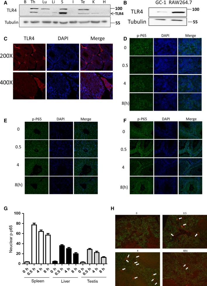
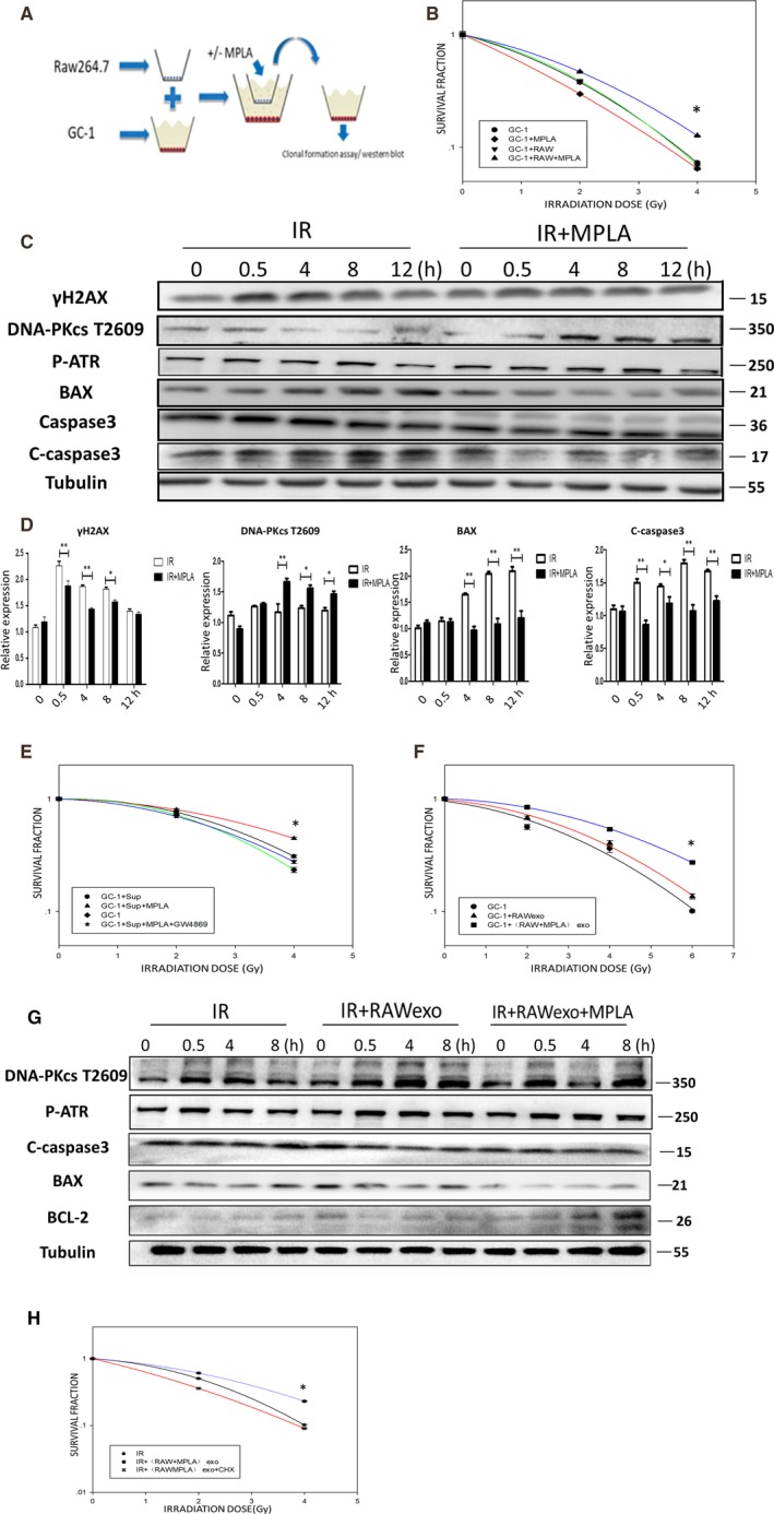
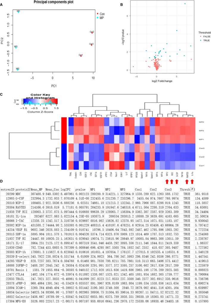
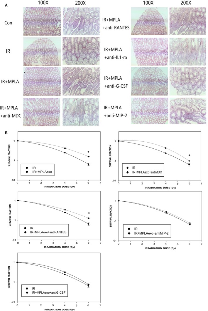
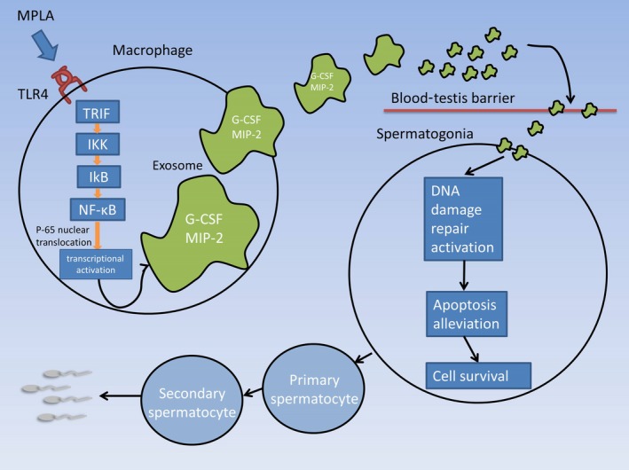
Radiation protection on male testis is an important task for ionizing radiation-related workers or people who receive radiotherapy for tumours near the testicle. In recent years, Toll-like receptors (TLRs), especially TLR4, have been widely studied as a radiation protection target. In this study, we detected that a low-toxicity TLR4 agonist monophosphoryl lipid A (MPLA) produced obvious radiation protection effects on mice testis. We found that MPLA effectively alleviated testis structure damage and cell apoptosis induced by ionizing radiation (IR). However, as the expression abundance differs a lot in distinct cells and tissues, MPLA seemed not to directly activate TLR4 singling pathway in mice testis. Here, we demonstrated a brand new mechanism for MPLA producing radiation protection effects on testis. We observed a significant activation of TLR4 pathway in macrophages after MPLA stimulation and identified significant changes in macrophage-derived exosomes protein expression. We proved that after MPLA treatment, macrophage-derived exosomes played an important role in testis radiation protection, and specially, G-CSF and MIP-2 in exosomes are the core molecules in this protection effect.

Keywords: MPLA; exosome; radioprotection; testis.


Conflict of interest statement

The authors have no conflicts of interest to disclose.

Figures

Figure 1
Figure 1
MPLA treatment before ionizing radiation (IR) alleviate IR injury in mice testis. A, On day 1 and day 7 after 4 Gy IR, testis was isolated and subjected to tissue sectioning and H&E staining. B, Mice were exposed to IR at dose of 2 Gy, at day 21 after irradiation; testis were isolated and subjected to tissue sectioning and H&E staining. C, Testis was isolated at 16 h after IR and fixed with polyformaldehyde; polyformaldehyde‐fixed paraffin‐embedded testis was stained with TUNEL method. D, TUNEL‐positive cells were counted in 200× field of view, ten fields of views were randomly selected in each group, and average numbers of TUNEL‐positive cells were calculated and showed. E, At day 21, sperms in epididymis were calculated by using microscope cell counting method. F, Flow cytometry was used to calculate 1c, 2c and 4c spermatogenic cells. G, H, Mice testosterone in testis and serum was examined by ELISA method 21 days after exposure to 2 Gy IR. Data were presented as mean ± SD (n = 3). *P < .05. **P < .01, ***P < .001
Figure 2
Figure 2
MPLA alleviated apoptosis pathway activation and helps activate DNA damage repair pathway. A, B, Immunofluorescence staining was used to determine γH2AX activation level in testis after IR, γH2AX positive cells were counted and calculated as showed in (B). (C)DNA‐PKcs t2609, γH2AX, p‐ATR, C‐caspase3 expression level was determined by western bolt assay, Tubulin served as a loading control. (D, E, F, G) Protein relative expression level was showed in (D‐G). D, Quantitative data of γH2AX. E, Quantative data of p‐ATR. F, Analysis of DNA‐PKcs T2056. G, Quantative analysis of c‐Caspase 3. Data were presented as mean± SD (n=3). *P < .05. **P < .01, ***P < .001
Figure 3
Figure 3
MPLA protected mice testis from IR injury via TLR4‐Trif–dependent pathway. A, Seven days after TLR4−/− mice exposed to 4Gy IR, mice testis was isolated and subjected to tissue sectioning and H&E staining. B, TUNEL method was used to determine TLR4−/− mice cell apoptosis in testis 16 h after exposure to IR. C, TUNEL‐positive cells were counted in 200X field of views, 10 fields of views were randomly selected in each group, and average numbers of TUNEL‐positive cells were calculated and showed. D, Seven days after TRIF−/− mice exposed to 4 Gy IR, mice testis was isolated and subjected to tissue sectioning and H&E staining. Data were presented as mean ± SD (n = 3). *P < .05. **P < .01, ***P < .001
Figure 4
Figure 4
The distribution of TLR4 and the activation level of TLR4‐NF‐κB pathway in different tissues. A, TLR4 expression level in different organs was examined by Western blot assay, B represents brain, TH represents thymus, Lu represents lung, Li represents liver, S represents spleen, I represents intestine, TE represents testis, K represents kidney, and H represents heart. B, TLR4 expression level in GC‐1 and RAW264.7 was examined by Western blot assay. C, Immunofluorescence method was used to determine TLR4 expression level and distribution in testis. D‐F, Mice were treated with MPLA at dose of 50 µg/kg; spleen (D), liver (E) and testis (F) were isolated at different time‐point and then were subjected to tissue sectioning; and p‐P65 was stained by immunofluorescence method. G, Ten 200× field of views were randomly selected, and cells with p‐P65 nuclear translocation were calculated and showed in G. H, After 50 µg/kg MPLA administration, mice testis was isolated at different time‐point. p‐P65 and F4/80 immunofluorescence co‐staining was used to determine TLR4 activation level in mice macrophages
Figure 5
Figure 5
MPLA produced IR protection effects on spermatogonias depending on RAW264.7 TLR4 activation and RAW264.7‐derived exosomes. A, Schematic of co‐culture system. B, Clonal formation assay was used to determine proliferative capacity of GC‐1 cells in co‐culture system after IR. C, The GC‐1 cells in co‐culture system were treated with or without MPLA. Expression level of γH2AX, DNA‐PKcs t2609, p‐ATR, Bax and C‐caspase3 was examined by Western blot assay. D, Protein relative expression level was calculated as showed. E, Proliferative capacity of GC‐1 was examined by clonal formation assay after RAW264.7 supernatant treatment. F, Proliferative capacity of GC‐1 was examined after RAW264.7‐derived exosomes treatment. G, Expression level of DNA‐PKcs t2609, p‐ATR, Bax, C‐caspase3 and Bcl2 was examined by Western blot assay after RAW264.7‐derived exosomes treatment. H, Proliferative capacity of GC‐1 was tested by clonal formation assay after RAW264.7‐derived exosomes treatment with or without CHX. Data were presented as mean ± SD (n = 3). *P < .05. **P < .01, ***P < .001
Figure 6
Figure 6
The protein expression profile from RAW264.7‐derived exosomes with or without MPLA treatment. A, The result of principle components analysis. B, Cluster heat map result. C, Heat map result. D, The list of proteins with significant expression level changes
Figure 7
Figure 7
G‐CSF and MIP‐2 in exosomes were the main protection effectors in testis radiation protection. A, On day 7 after neutralizing antibody administration and 4 Gy irradiation, mice testis was isolated and subjected to H&E staining. B, Clonal formation assay was used to determine GC‐1 cell viability after exosomes treatment and irradiation exposure with or without neutralizing antibody. Data were presented as mean ± SD (n = 3). *P < .05. **P < .01, ***P < .001
Figure 8
Figure 8
Proposed model for MPLA produced testis radiation protection. In macrophages, TLR4‐TRIF pathway was activated after MPLA administration. Subsequently, NF‐κB subunit p‐65 was phosphorylated and translocated into nuclear, which resulted in expression of multiple cytokines and chemokines. G‐CSF and MIP‐2, as the important mediators in testis radiation protection, were transported by macrophage‐derived exosomes and across the blood‐testis barrier and eventually promoted spermatogonias survival after irradiation

References

    1. Choy H, Gerber DE, Bradley JD, et al. Concurrent pemetrexed and radiation therapy in the treatment of patients with inoperable stage III non‐small cell lung cancer: a systematic review of completed and ongoing studies. Lung Cancer. 2015;87:232‐240. - PubMed
    1. Martin NE, D'Amico AV. Progress and controversies: Radiation therapy for prostate cancer. CA Cancer J Clin. 2014;64:389‐407. - PubMed
    1. Benson AB 3rd, Arnoletti JP, Bekaii‐Saab T, et al. Colon cancer. J Natl Compr Canc Netw. 2011;9:1238‐1290. - PubMed
    1. Bujan L, Walschaerts M, Moinard N, et al. Impact of chemotherapy and radiotherapy for testicular germ cell tumors on spermatogenesis and sperm DNA: a multicenter prospective study from the CECOS network. Fertil Steril. 2013;100:673‐680. - PubMed
    1. Grignard E, Gueguen Y, Grison S, et al. Testicular steroidogenesis is not altered by 137 cesium Chernobyl fallout, following in utero or post‐natal chronic exposure. C R Biol. 2010;333:416‐423. - PubMed

Publication types

MeSH terms