

Maternal Vitamin D Deficiency Causes Sustained Impairment of Lung Structure and Function and Increases Susceptibility to Hyperoxia-induced Lung Injury in Infant Rats
- PMID: 32135073
- PMCID: PMC7328245
- DOI: 10.1165/rcmb.2019-0295OC
Maternal Vitamin D Deficiency Causes Sustained Impairment of Lung Structure and Function and Increases Susceptibility to Hyperoxia-induced Lung Injury in Infant Rats
Abstract
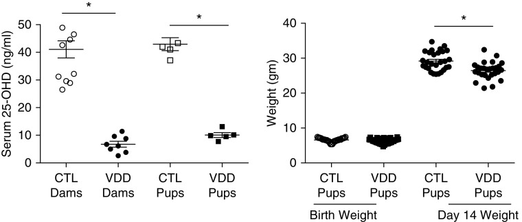
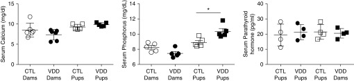
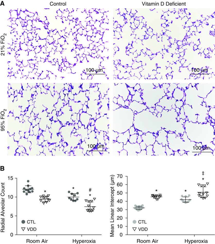
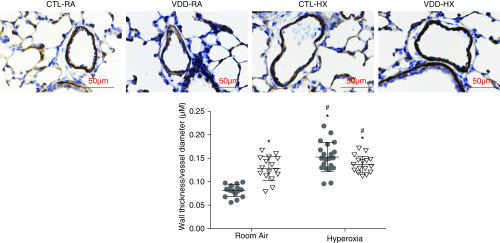
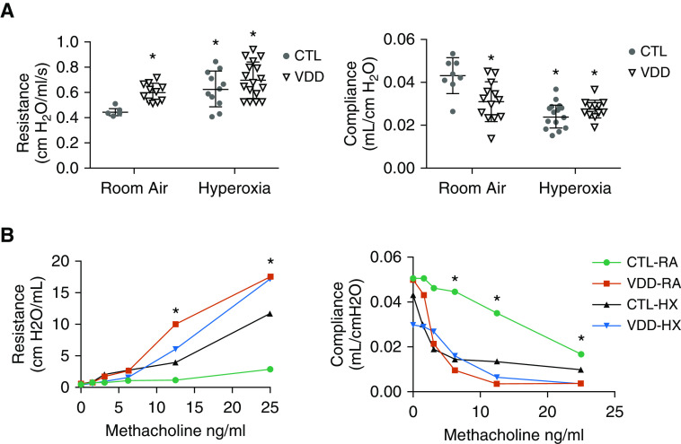
Vitamin D deficiency (VDD) during pregnancy is associated with increased respiratory morbidities and risk for chronic lung disease after preterm birth. However, the direct effects of maternal VDD on perinatal lung structure and function and whether maternal VDD increases the susceptibility of lung injury due to hyperoxia are uncertain. In the present study, we sought to determine whether maternal VDD is sufficient to impair lung structure and function and whether VDD increases the impact of hyperoxia on the developing rat lung. Four-week-old rats were fed VDD chow and housed in a room shielded from ultraviolet A/B light to achieve 25-hydroxyvitamin D concentrations <10 ng/ml at mating and throughout lactation. Lung structure was assessed at 2 weeks for radial alveolar count, mean linear intercept, pulmonary vessel density, and lung function (lung compliance and resistance). The effects of hyperoxia for 2 weeks after birth were assessed after exposure to fraction of inspired oxygen of 0.95. At 2 weeks, VDD offspring had decreased alveolar and vascular growth and abnormal airway reactivity and lung function. Impaired lung structure and function in VDD offspring were similar to those observed in control rats exposed to postnatal hyperoxia alone. Maternal VDD causes sustained abnormalities of distal lung growth, increases in airway hyperreactivity, and abnormal lung mechanics during infancy. These changes in VDD pups were as severe as those measured after exposure to postnatal hyperoxia alone. We speculate that antenatal disruption of vitamin D signaling increases the risk for late-childhood respiratory disease.
Keywords: hyperoxia; lung development; maternal vitamin D deficiency; vitamin D deficiency.
Figures


Comment in
-
Vitamin D: Feel It in More Than Just Your Bones!Am J Respir Cell Mol Biol. 2020 Jul;63(1):11-12. doi: 10.1165/rcmb.2020-0072ED. Am J Respir Cell Mol Biol. 2020. PMID: 32160008 Free PMC article. No abstract available.
Similar articles
-
Maternal vitamin D deficiency induces transcriptomic changes in newborn rat lungs.J Steroid Biochem Mol Biol. 2020 May;199:105613. doi: 10.1016/j.jsbmb.2020.105613. Epub 2020 Jan 30. J Steroid Biochem Mol Biol. 2020. PMID: 32007564
-
Protective effect of vitamin D against hyperoxia-induced lung injury in newborn rats.Pediatr Pulmonol. 2017 Jan;52(1):69-76. doi: 10.1002/ppul.23500. Epub 2016 Jun 13. Pediatr Pulmonol. 2017. PMID: 27291304
-
Maternal Tn Immunization Attenuates Hyperoxia-Induced Lung Injury in Neonatal Rats Through Suppression of Oxidative Stress and Inflammation.Front Immunol. 2019 Apr 4;10:681. doi: 10.3389/fimmu.2019.00681. eCollection 2019. Front Immunol. 2019. PMID: 31019509 Free PMC article.
-
[Vitamin D deficiency in pregnancy and its impact on the fetus, the newborn and in childhood].Rev Paul Pediatr. 2015 Jan-Mar;33(1):104-13. doi: 10.1016/j.rpped.2014.05.004. Epub 2015 Feb 7. Rev Paul Pediatr. 2015. PMID: 25662013 Free PMC article. Review.
-
Vitamin D Deficiency and Oral Health: A Comprehensive Review.Nutrients. 2020 May 19;12(5):1471. doi: 10.3390/nu12051471. Nutrients. 2020. PMID: 32438644 Free PMC article. Review.
Cited by
-
Antenatal Endotoxin Induces Dysanapsis in Experimental Bronchopulmonary Dysplasia.Am J Respir Cell Mol Biol. 2024 Apr;70(4):283-294. doi: 10.1165/rcmb.2023-0157OC. Am J Respir Cell Mol Biol. 2024. PMID: 38207120 Free PMC article.
-
Update in Critical Care 2020.Am J Respir Crit Care Med. 2021 May 1;203(9):1088-1098. doi: 10.1164/rccm.202102-0336UP. Am J Respir Crit Care Med. 2021. PMID: 33734938 Free PMC article. Review. No abstract available.
-
Low gestational vitamin D level and childhood asthma are related to impaired lung function in high-risk children.J Allergy Clin Immunol. 2021 Jul;148(1):110-119.e9. doi: 10.1016/j.jaci.2020.12.647. Epub 2021 Jan 22. J Allergy Clin Immunol. 2021. PMID: 33485958 Free PMC article.
-
Pharmacokinetic modeling of prenatal vitamin D exposure and the impact on offspring asthma and pulmonary function.Biomed Pharmacother. 2025 Feb;183:117859. doi: 10.1016/j.biopha.2025.117859. Epub 2025 Jan 27. Biomed Pharmacother. 2025. PMID: 39874780 Free PMC article.
-
Development of late pulmonary hypertension after antenatal inflammation in experimental bronchopulmonary dysplasia.Pediatr Res. 2025 Jun 28. doi: 10.1038/s41390-025-04223-6. Online ahead of print. Pediatr Res. 2025. PMID: 40581700
References
-
- Haugen M, Brantsaeter AL, Trogstad L, Alexander J, Roth C, Magnus P, et al. Vitamin D supplementation and reduced risk of preeclampsia in nulliparous women. Epidemiology. 2009;20:720–726. - PubMed
-
- Wagner CL, Baggerly C, McDonnell S, Baggerly KA, French CB, Baggerly L, et al. Post-hoc analysis of vitamin D status and reduced risk of preterm birth in two vitamin D pregnancy cohorts compared with South Carolina March of Dimes 2009–2011 rates. J Steroid Biochem Mol Biol. 2016;155:245–251. - PMC - PubMed
Publication types
MeSH terms
Substances
Grants and funding
LinkOut - more resources
Full Text Sources
Medical
