. 2020 May 6;106(3):404-420.e8.
doi: 10.1016/j.neuron.2020.01.042. Epub 2020 Mar 4.

Pathogenic DDX3X Mutations Impair RNA Metabolism and Neurogenesis during Fetal Cortical Development

Affiliations

Pathogenic DDX3X Mutations Impair RNA Metabolism and Neurogenesis during Fetal Cortical Development

Ashley L Lennox et al. Neuron. .

Abstract

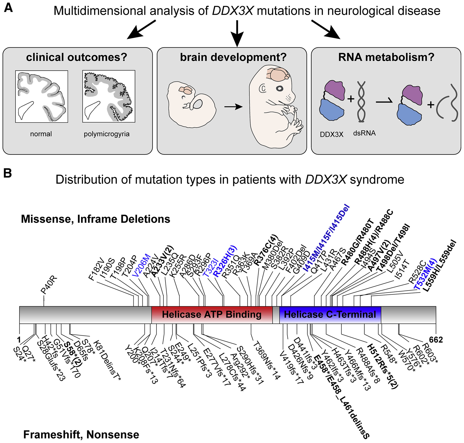
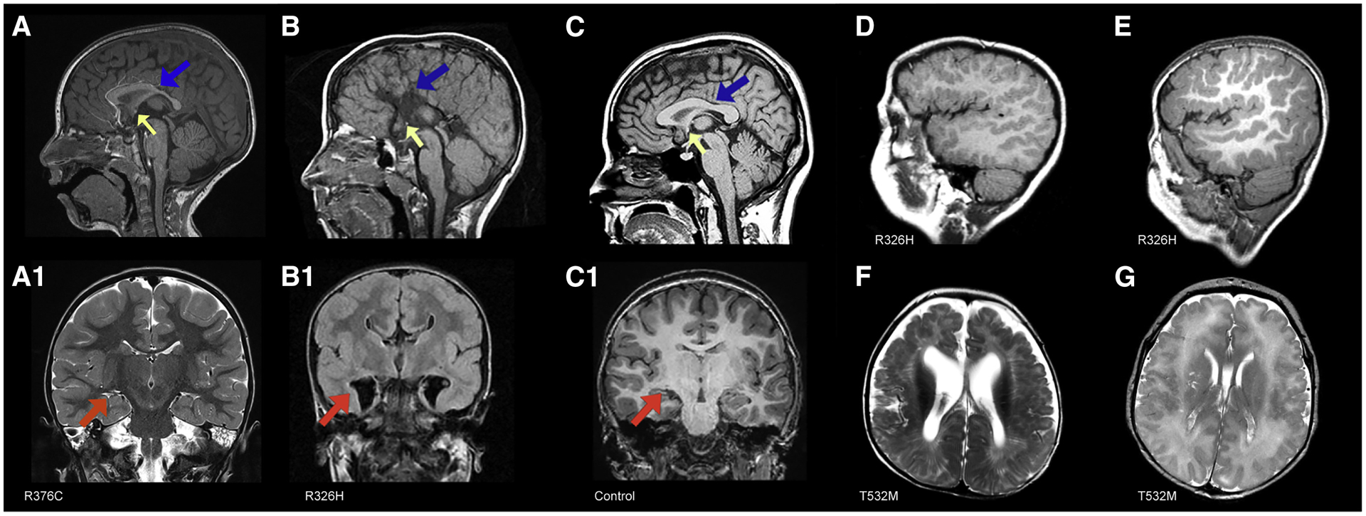
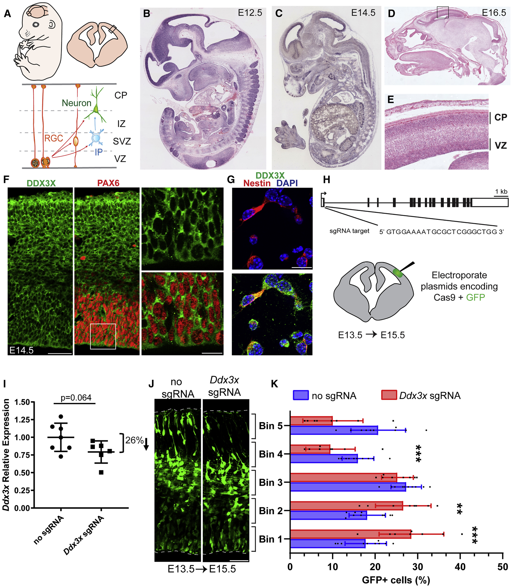
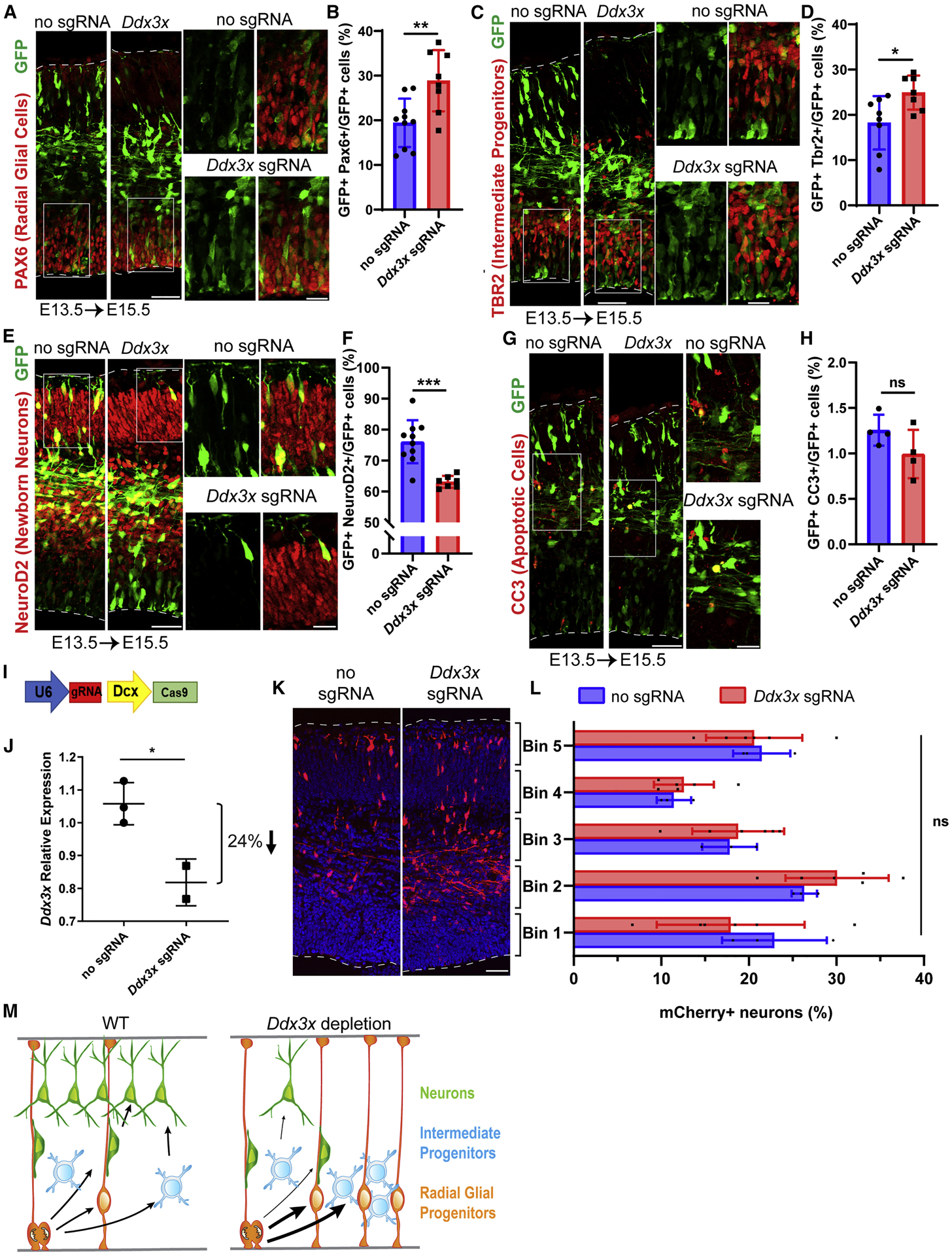
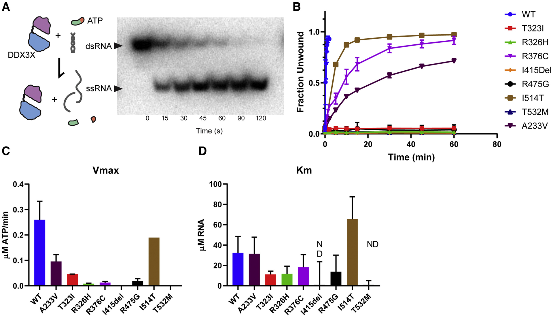
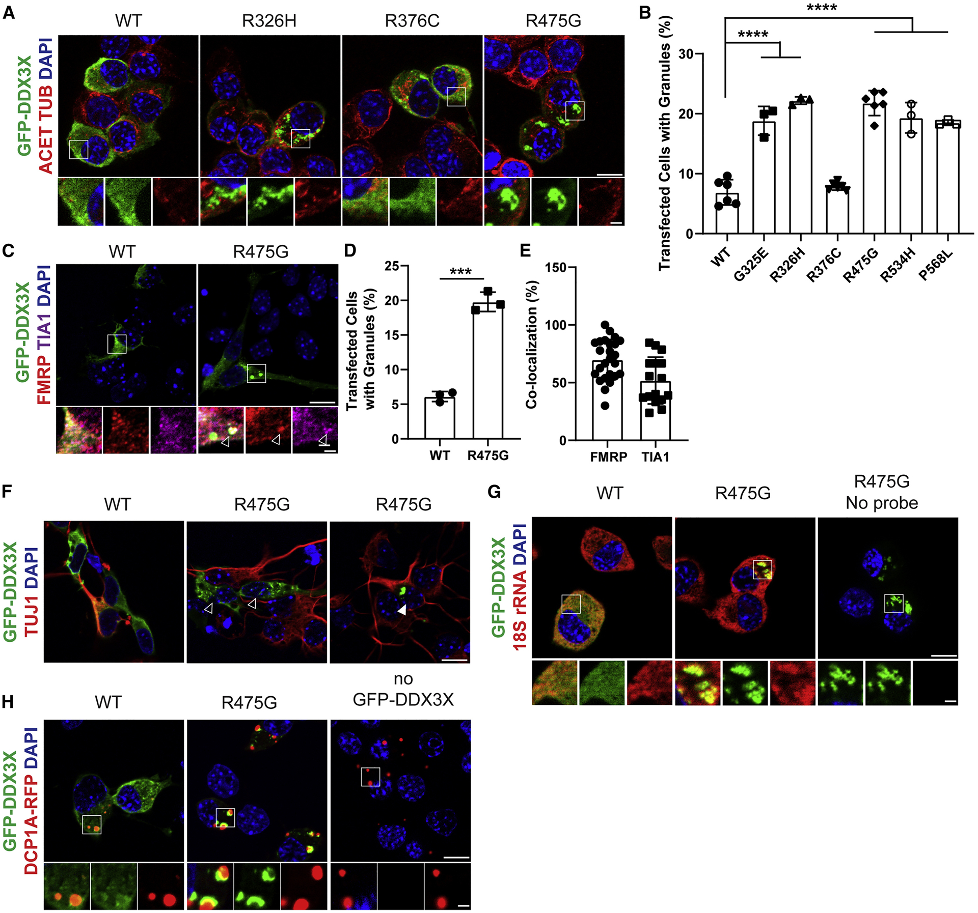
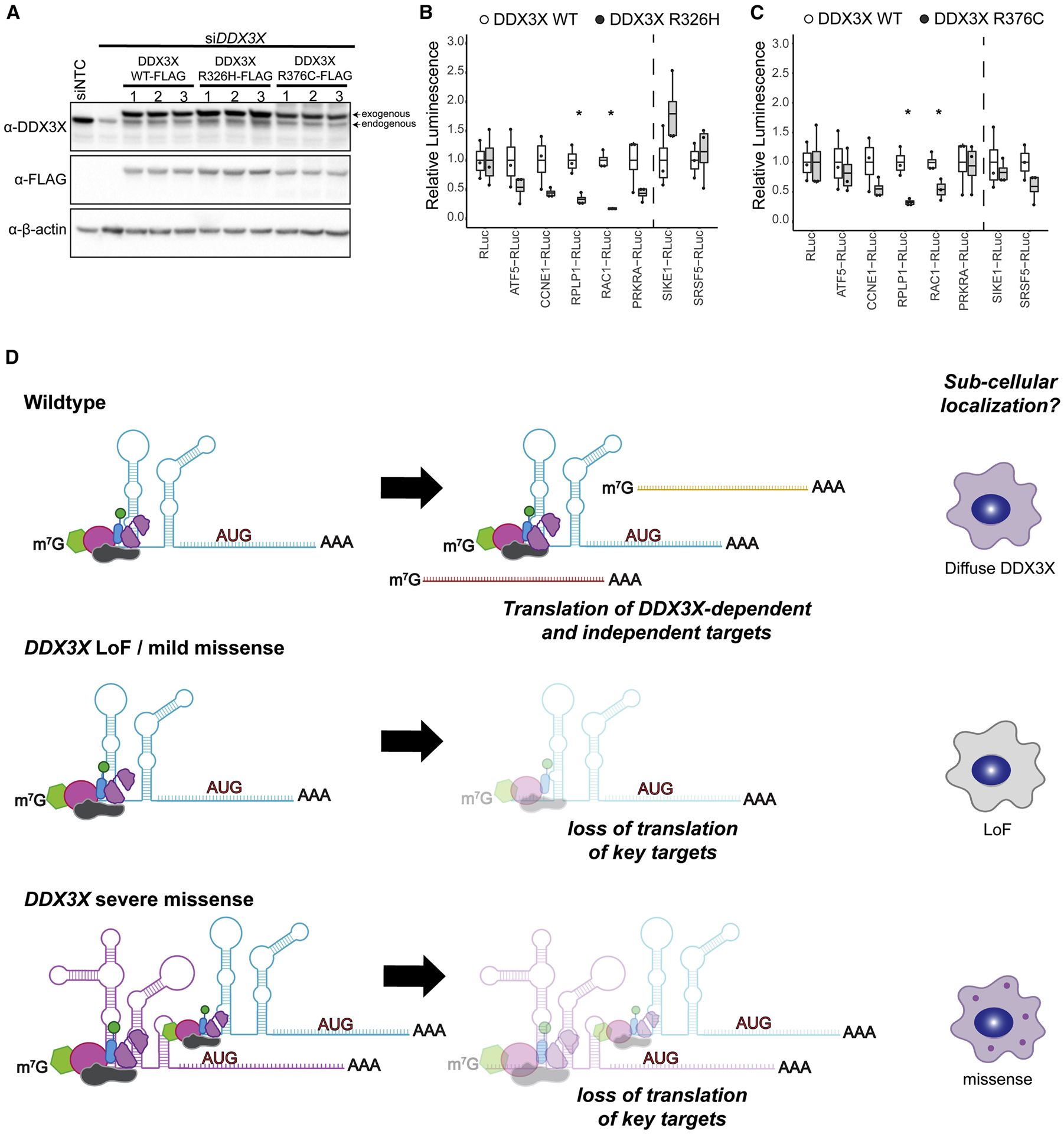
De novo germline mutations in the RNA helicase DDX3X account for 1%-3% of unexplained intellectual disability (ID) cases in females and are associated with autism, brain malformations, and epilepsy. Yet, the developmental and molecular mechanisms by which DDX3X mutations impair brain function are unknown. Here, we use human and mouse genetics and cell biological and biochemical approaches to elucidate mechanisms by which pathogenic DDX3X variants disrupt brain development. We report the largest clinical cohort to date with DDX3X mutations (n = 107), demonstrating a striking correlation between recurrent dominant missense mutations, polymicrogyria, and the most severe clinical outcomes. We show that Ddx3x controls cortical development by regulating neuron generation. Severe DDX3X missense mutations profoundly disrupt RNA helicase activity, induce ectopic RNA-protein granules in neural progenitors and neurons, and impair translation. Together, these results uncover key mechanisms underlying DDX3X syndrome and highlight aberrant RNA metabolism in the pathogenesis of neurodevelopmental disease.

Keywords: DDX3X; autism; corpus callosum; cortical development; helicase; intellectual disability; polymicrogyria; radial glial progenitor; stress granule; translation.


Conflict of interest statement

Declaration of Interests The authors declare no competing interests.

Figures

Figure 1.
Figure 1.. Overview of study and predicted amino acid changes in DDX3X in our cohort
A, Overview of the three questions assessed in this study to understand the role of DDX3X de novo mutations in disease. B, Annotations include the following: Missense and in frame deletions (top); frameshift and nonsense mutations (bottom); Mutations found in patients with polymicrogyria (blue); Recurrent mutations (bolded and indicated by numbers in parentheses). Overall, mutations are enriched in the helicase domains at a rate higher than random chance (p=4.3*10−6 given 87 unique mutation positions including 12 splice sites). Not shown are the 12 splice site mutations (see Table S1).
Figure 2.
Figure 2.. Common brain imaging findings in DDX3X cohort
A, A1 Patient 2612–0 (R376C, mild clinical impairments) at 2 years of age. A, Sagittal image shows hypoplastic corpus callosum with a short, thinned posterior body (blue arrow) and a hypoplastic to absent splenium; small anterior commissure (yellow arrow); inferior genu and rostrum are absent. A1, Coronal image shows mildly diminished white matter volume, normal ventricle size, including temporal horns (red arrow). B, B1, Patient 1090–0 (R326H) at 7 months. B, Sagittal image shows complete agenesis of the corpus callosum and hippocampal commissure (blue arrow) and a small anterior commissure (yellow arrow). B1 Colpocephaly with enlarged keyhole-shaped temporal horns (red arrow). C, C1, Normal MRIs for comparison. C, Mid-sagittal T1-weighted image showing a normal sized corpus callosum (blue arrow) and anterior commissure (yellow arrow). C1, Coronal T1-weighted image shows normal cortical thickness and gyration, normal sized ventricle bodies and temporal horns (red arrow). Saggital images from patients 1090–0 (D) at 7 months and 3437–0 (E) at 4 years (both R326H) with extensive bilateral frontal PMG. Axial images from patients 3072–0 (F) at 6 months and 1954–0 (G) at 3 days of age, (both T532M) and bilateral perisylvian and frontal PMG and enlarged ventricles.
Figure 3.
Figure 3.. Ddx3x is expressed in progenitors and neurons in the embryonic mouse cortex and in vivo disruption alters neurogenesis.
A, Top, cartoon representation of a mouse embryo and coronal cortical section. Bottom, cartoon representation of boxed region above depicting major embryonic cortical cell types examined in this study including radial glial progenitor cells (RGCs), intermediate progenitors (IPs), and neurons. The ventricular zone (VZ), subventricular zone (SVZ), intermediate zone (IZ), and cortical plate (CP) are indicated. B-E, Ddx3x In situ hybridization in sagittal sections of E12.5 (B), E14.5 (C), and E16.5 (D,E) embryos. Box in D is magnified in E. F, G, Immunofluorescence of E14.5 cortical sections (F) co-stained for DDX3X (green), the RGC marker PAX6 (red), and of E14.5 primary cells (G) co-stained for the RGC marker Nestin (red) and DAPI (blue). H, Schematic of mouse Ddx3x gene structure with exons (boxes) and introns (thin lines). Ddx3x sgRNA targets Exon 1 at the indicated sequence. Bottom, schematic of coronal section depicting electroporated region. I, Validation of Ddx3x mRNA knockdown in FACS purified GFP+ cells from E15.5 brains electroporated at E13.5. J, Representative E15.5 coronal sections, electroporated at E13.5, with GFP and either no sgRNA or Ddx3x sgRNA, immunostained for GFP (green). Dotted lines represent ventricular and pial surfaces, and brackets delineate equivalently sized bins. K, Quantification of distribution of GFP-positive cells with Bin1 at the ventricle and Bin5 at the pia. Scale bars: 500 μm and 50 μm (F) and 15 μm (G) and 50 μm (J). Error bars=SD.
Figure 4.
Figure 4.. Ddx3x is required for neuron generation in vivo.
A, C, E, G Sections of E15.5 cortices, electroporated at E13.5, and co-stained with antibodies against GFP (green) and PAX6 (red) (A); TBR2 (red) (C), NeuroD2 (red) (E) and CC3 (red) (G). Boxed regions are shown at higher magnification on the right. B, D, F, H Quantification of percentage of GFP-positive cells expressing PAX6 (B), TBR2 (D), NEUROD2 (F), and CC3 (H). I, Schematic diagram of CRISPR/Cas9 targeting vector under control of neuron-specific Dcx promoter. J, Validation of Ddx3x mRNA knockdown in FACS purified mCherry+ cells from E17.5 brains electroporated at E14.5. K, Representative coronal sections of E17.5 brains coelectroporated at E14.5 with pDcx-mCherry and either pX330-Dcx-Cas9 (no sgRNA) or pX330Dcx-Cas9 plus Ddx3x sgRNA, stained with anti-RFP (red). Dotted lines represent ventricular and pial surfaces, and brackets on the right refer to equivalently sized bins. L, Quantitation of mCherry-positive cells distribution with Bin1 at the ventricle and Bin5 at the pia. M, Schematic model summarizing finding that Ddx3x LoF impairs neuron generation, associated with increased RGCs and IPs. Scale bars: 50 μm, low magnification, 15 μm, high magnification (A,C,E,G, K). Error bars=SD.
Figure 5.
Figure 5.. DDX3X missense mutants exhibit disrupted helicase activity
A, Left, Diagram of DDX3X activity tested in this assay. ATP hydrolysis is necessary for initial binding and release of RNA, but not for RNA unwinding. Right, Non-denaturing gel depicting time course of helicase assay in which amounts of dsRNA (not unwound by DDX3X) and ssRNA (unwinding by DDX3X) are measured. B, Unwinding assay for WT, A233V, T323I, R326H, R376C, I415del, R475G, I514T, and T532 DDX3X. C, D, Graphs depicting Vmax (C) and Km (D). Note, I415del and T532M had unwinding curves which did not vary within the range of tested RNA concentrations (0–40 uM) so Km was not determined (n.d.). The majority of mutants had lower Km (indicating higher affinity for RNA) than WT, with the exception of I514T. Error bars=SD.
Figure 6.
Figure 6.. DDX3X missense mutations induce ectopic RNP granules in neural progenitors.
A, Images of N2A cells transfected for 24 hrs with WT or mutant DDX3X-GFP and immunostained for GFP (green), Acetylated-TUBULIN (red), and DAPI (blue). Below, high magnification images from boxed regions. B, Quantification of percentage of N2A cells containing WT or mutant DDX3X-GFP granules. C, Primary cortical cells transfected for 24 hrs with WT or R475G DDX3X-GFP (green) and co-stained for FMRP (red) and TIA1 (magenta), with granule co-localization granules (arrowhead). D, Quantification of percentage of primary cortical cells containing DDX3X-GFP granules. E, Quantification of DDX3X-granules co-localized with RNA-binding proteins TIA1 or FMRP. F, Primary cortical cells expressing either WT or R475G DDX3X-GFP (green) and stained with TUJ1 (red). Both TUJ1- progenitors (empty arrowhead) and TUJ1+ neurons (filled arrowhead) contain DDX3X granules. G, N2A cells transfected with DDX3X-GFP (green) and probed for 18S rRNA with smFISH probes (red). H, N2A cells transfected with DDX3X-GFP (green) and DCP1A-RFP (red) to mark P-bodies. Scale bars, 10 μm (low magnification) and 2 μm (high magnification) (A, C, F, G, H). Error bars=SD.
Figure 7.
Figure 7.. DDX3X missense mutations alter translation of select targets
A, Western blots depicting DDX3X levels (WT, R326H, or R376C) with simultaneous knockdown of endogenous DDX3X. Endogenous and FLAG-tagged DDX3X is detected. B, C, Translation of in vitro transcribed reporter RNAs in lysates expressing either DDX3X WT, R326H (B) or R376C (C) Signal was normalized to WT. Predicted DDX3X sensitive reporters (ATF5, CCNE1, RPLP1, PRKRA and RAC1) show robust decreases in translation, while control transcripts (SIKE1, SRSF5) right of dotted line demonstrate either no change or modest increases. Box and whisker plot with bars indicating data range, box at upper and lower quartiles, and line at the median. D, Proposed model for mechanism of DDX3X mutants, based upon in vitro biochemical and cell biology studies. Mild missense or DDX3X LoF are impair translation of some targets but do not induce granule formation. Severe DDX3X missense mutations show impaired RNA release and helicase activity, and altered translation of DDX3X-dependent targets. This results in sequestration of RNAs and RNA binding proteins and formation of aberrant RNP granules. Created with BioRender.com.

Comment in

Similar articles

Cited by

References

    1. Achenbach TM (2011). Child Behavior Checklist. In Encyclopedia of Clinical Neuropsychology, Kreutzer JS, DeLuca J, and Caplan B, eds. (New York, NY: Springer New York; ), pp. 546–552.
    1. Amadei G, Zander MA, Yang G, Dumelie JG, Vessey JP, Lipshitz HD, Smibert CA, Kaplan DR, and Miller FD (2015). A Smaug2-Based Translational Repression Complex Determines the Balance between Precursor Maintenance versus Differentiation during Mammalian Neurogenesis. The Journal of neuroscience : the official journal of the Society for Neuroscience 35, 15666–15681. - PMC - PubMed
    1. Ayoub AE, Oh S, Xie Y, Leng J, Cotney J, Dominguez MH, Noonan JP, and Rakic P (2011). Transcriptional programs in transient embryonic zones of the cerebral cortex defined by high-resolution mRNA sequencing. Proc Natl Acad Sci U S A 108, 14950–14955. - PMC - PubMed
    1. Balak C, Benard M, Schaefer E, Iqbal S, Ramsey K, Ernoult-Lange M, Mattioli F, Llaci L, Geoffroy V, Courel M, et al. (2019). Rare De Novo Missense Variants in RNA Helicase DDX6 Cause Intellectual Disability and Dysmorphic Features and Lead to P-Body Defects and RNA Dysregulation. Am J Hum Genet 105, 509–525. - PMC - PubMed
    1. Beal B, Hayes I, McGaughran J, Amor DJ, Miteff C, Jackson V, van Reyk O, Subramanian G, Hildebrand MS, Morgan AT, et al. (2019). Expansion of phenotype of DDX3X syndrome: six new cases. Clinical dysmorphology 28, 169–174. - PubMed

Publication types

LinkOut - more resources