. 2020 Mar 3;12(3):582.
doi: 10.3390/cancers12030582.

Radiosensitization by Hyperthermia: The Effects of Temperature, Sequence, and Time Interval in Cervical Cell Lines

Affiliations

Radiosensitization by Hyperthermia: The Effects of Temperature, Sequence, and Time Interval in Cervical Cell Lines

Xionge Mei et al. Cancers (Basel). .

Abstract

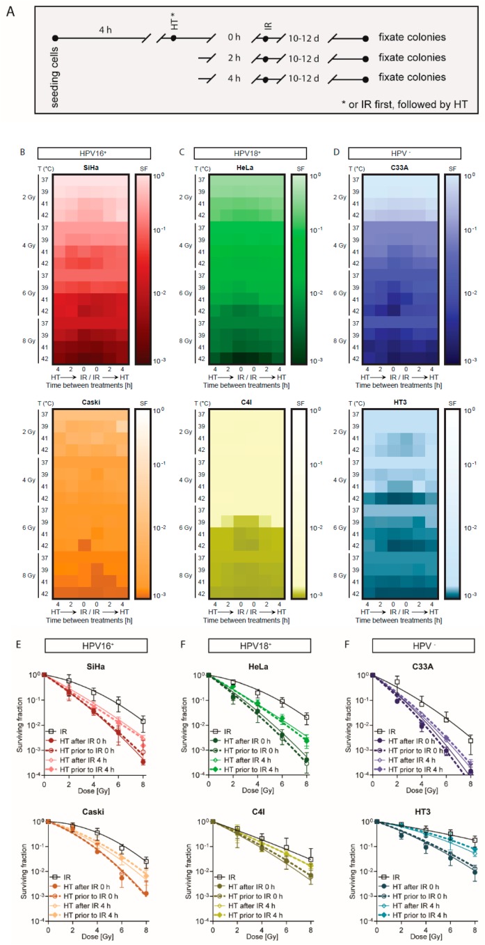
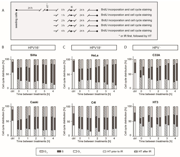
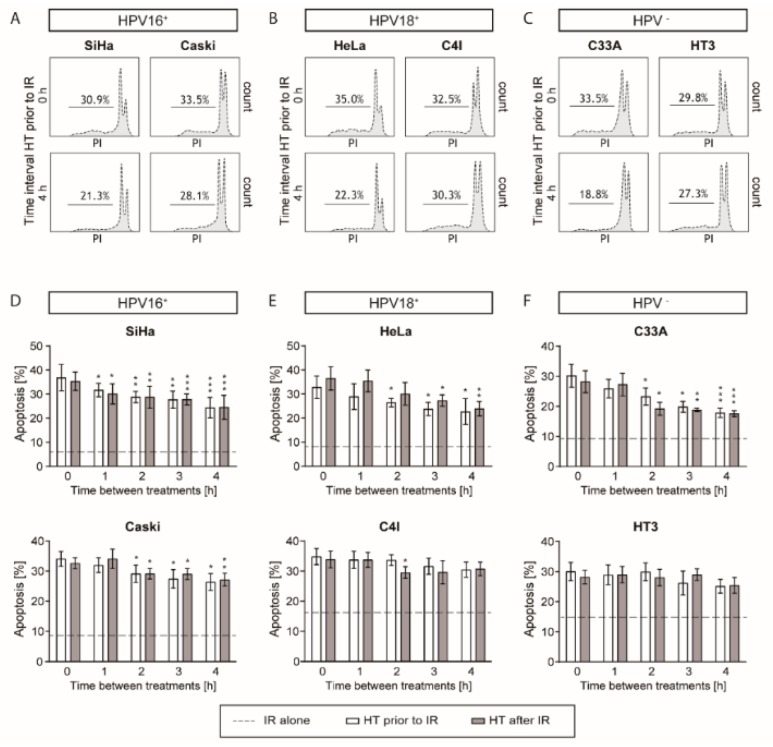
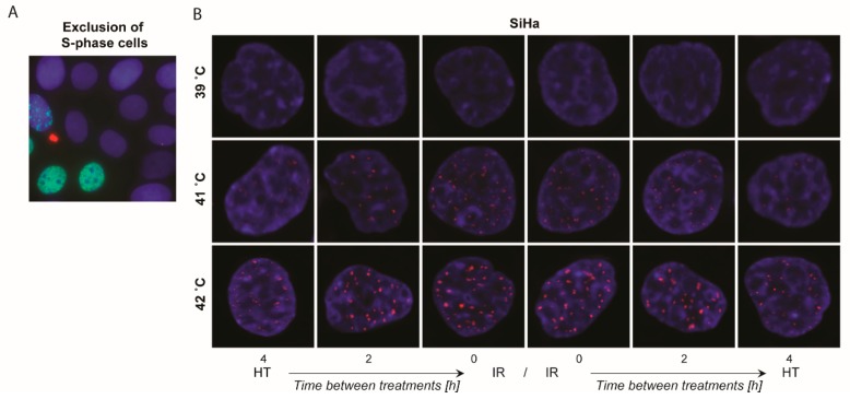
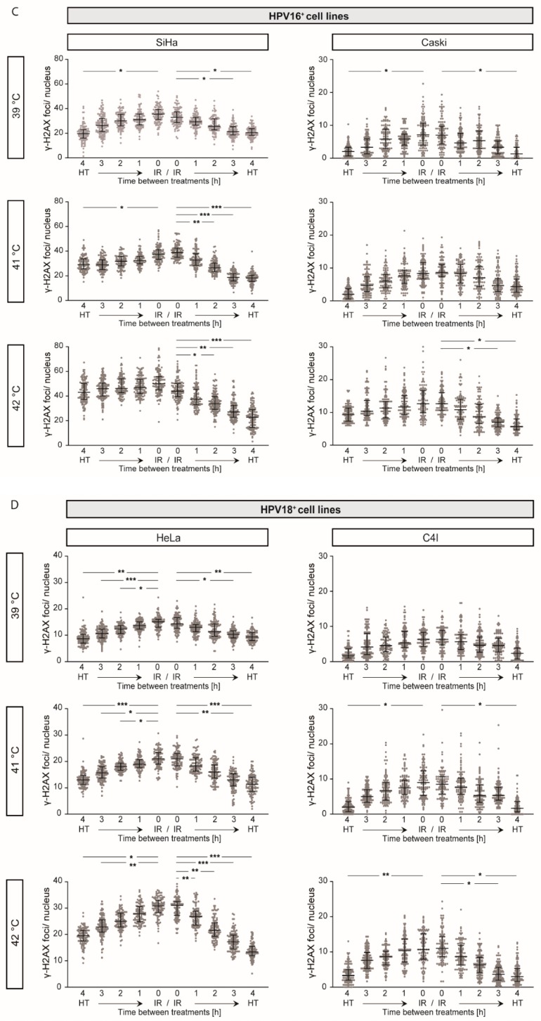
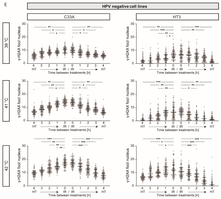
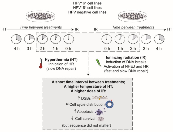
Cervical cancers are almost exclusively caused by an infection with the human papillomavirus (HPV). When patients suffering from cervical cancer have contraindications for chemoradiotherapy, radiotherapy combined with hyperthermia is a good treatment option. Radiation-induced DNA breaks can be repaired by nonhomologous end-joining (NHEJ) or homologous recombination (HR). Hyperthermia can temporarily inactivate homologous recombination. Therefore, combining radiotherapy with hyperthermia can result in the persistence of more fatal radiation-induced DNA breaks. However, there is no consensus on the optimal sequence of radiotherapy and hyperthermia and the optimal time interval between these modalities. Moreover, the temperature of hyperthermia and HPV-type may also be important in radiosensitization by hyperthermia. In this study we thoroughly investigated the impact of different temperatures (37-42 °C), and the sequence of and time interval (0 up to 4 h) between ionizing radiation and hyperthermia on HPV16+: SiHa, Caski; HPV18+: HeLa, C4I; and HPV-: C33A, HT3 cervical cancer cell lines. Our results demonstrate that a short time interval between treatments caused more unrepaired DNA damages and more cell kill, especially at higher temperatures. Although hyperthermia before ionizing radiation may result in slightly more DNA damage, the sequence between hyperthermia and ionizing radiation yielded similar effects on cell survival.

Keywords: human papillomavirus; hyperthermia; ionizing radiation; sequence; time interval.


Conflict of interest statement

The authors declare no conflict of interest.

Figures

Figure 1
Figure 1
A short time interval between ionizing radiation and hyperthermia decreases cell survival. To study the effect of time interval (0, 2, or 4 h) between ionizing radiation (IR) and hyperthermia (HT), clonogenic assays were performed for six cervical carcinoma cell lines. (A) Schematic overview of applied treatments. (BD) Survival fraction is demonstrated in heat maps: (B) HPV16+ cell lines SiHa and Caski, (C) HPV18+ cell lines HeLa and C4I, and (D) HPV-negative cell lines C33A and HT3—a darker color indicates lower cell survival. Per heat map, from top to bottom, different doses (2, 4, 6, and 8 Gy) of ionizing radiation are presented. Within one dose of ionizing radiation, cells treated with different temperatures of hyperthermia (37, 39, 41, and 42 °C). From left to right, within one heat map the left side shows results for hyperthermia applied before ionizing radiation with 0, 2, and 4 hour time interval, while on the right side ionizing radiation was applied before hyperthermia. Means of at least four experiments are presented. (EG) Survival fraction after ionizing radiation (2, 4, 6, and 8 Gy) alone, and hyperthermia (42 °C) combined with ionizing with a 0 hour and 4 hour time interval between the two modalities, in both sequences. (E) HPV16+ cell lines SiHa and Caski, (F) HPV18+ cell lines HeLa and C4I, and (G) HPV-negative cell lines C33A and HT3. Means with standard deviation of at least four independent experiments are presented.
Figure 2
Figure 2
More pronounced G2 arrest after a short time interval between ionizing radiation and hyperthermia. Cell cycle distribution, using BrdU incorporation after different time intervals and sequences of ionizing radiation (IR) and hyperthermia (HT), was performed on six cervical cancer cell lines. (A) Schematic overview of treatment. (B–D) Cell cycle distribution of the cell lines, from left to right: HPV 16+ cell lines SiHa and Caski, HPV18+ cell lines HeLa and C4I, and HPV-negative cell lines C33A and HT3. Untreated samples are marked as control (ctrl). In the samples treated with a short time interval (0 h) between the two therapies, a more pronounced increase in G2 phase was observed and in some cell lines after a time interval (4 h) between the two therapies, the S phase was increased. However, cell cycle distribution was more dependent on the cell line than on the type of HPV. Means of at least four replicates are presented.
Figure 3
Figure 3
Higher levels of apoptosis were detected after shorter time intervals between ionizing radiation (IR) and hyperthermia (HT). Apoptosis levels were measured using the Nicoletti assay. (A–C) Representative flow charts of apoptosis levels of HPV16+, HPV18+, and HPV-negative cell lines. (D–F) Higher levels of apoptosis were observed after a short time interval between ionizing radiation and hyperthermia compared to a longer time interval. Combined hyperthermia with ionizing radiation had a higher level of apoptosis compared to ionizing radiation only, the dotted line shows the levels of cell apoptosis treated with ionizing radiation only. Applying hyperthermia before or after ionizing radiation did not result in any significant differences in SiHa, Caski, HeLa, C4I, C33A, and HT3 cell lines. Means with standard deviation of at least three replicates are presented. * p < 0.05, ** p < 0.01, and *** p < 0.001 indicate the difference between 0 and 1, 2, 3, and 4 h time intervals between ionizing radiation and hyperthermia, or the opposite order of treatments.
Figure 4
Figure 4
Higher levels of DNA damage were observed after treatment with a shorter time interval between ionizing radiation and hyperthermia. (A) Cells in S-phase were excluded to only count the γ-H2AX positive double strand breaks (DSBs) that were radiation-induced. (B) SiHa cells demonstrating the DNA damage using γ-H2AX foci—cells were fixed 24 h after treatments. (C–E) γ-H2AX foci of HPV16+, HPV18+, and HPV-negative cell lines between ionizing radiation (IR; 2 Gy) and hyperthermia (HT) after treatments with different time intervals, different sequences, and various temperatures. Means with standard deviation of at least three replicates are presented, with a minimum of 100 cells per replicate. * p < 0.05, ** p < 0.01, and *** p < 0.001 indicate the difference between 0 and 1, 2, 3, and 4 h time interval between ionizing radiation and hyperthermia, or the opposite order of treatments.
Figure 4
Figure 4
Higher levels of DNA damage were observed after treatment with a shorter time interval between ionizing radiation and hyperthermia. (A) Cells in S-phase were excluded to only count the γ-H2AX positive double strand breaks (DSBs) that were radiation-induced. (B) SiHa cells demonstrating the DNA damage using γ-H2AX foci—cells were fixed 24 h after treatments. (C–E) γ-H2AX foci of HPV16+, HPV18+, and HPV-negative cell lines between ionizing radiation (IR; 2 Gy) and hyperthermia (HT) after treatments with different time intervals, different sequences, and various temperatures. Means with standard deviation of at least three replicates are presented, with a minimum of 100 cells per replicate. * p < 0.05, ** p < 0.01, and *** p < 0.001 indicate the difference between 0 and 1, 2, 3, and 4 h time interval between ionizing radiation and hyperthermia, or the opposite order of treatments.
Figure 5
Figure 5
Summary. Effects of temperature, sequence, and time interval. A shorter time interval between ionizing radiation (IR) and hyperthermia (HT), a higher temperature of hyperthermia, and a higher dose of ionizing radiation were found to be of great importance in cell survival, by causing more residual DNA double strand breaks (DSBs) and higher levels of apoptosis. Either hyperthermia prior to ionizing radiation or hyperthermia after ionizing radiation did not matter on the number of DNA breaks, on the cell cycle distribution, levels of apoptosis, or on cell survival.

References

    1. Šarenac T., Momir M. Cervical cancer, different treatments and importance of bile acids as therapeutic agents in this disease. Front. Pharmacol. 2019;10:484. doi: 10.3389/fphar.2019.00484. - DOI - PMC - PubMed
    1. Datta N.R., Ordonez S.G., Gaipl U.S., Paulides M.M., Crezee H., Gellermann J., Marder D., Puric E., Bodis S. Local hyperthermia combined with radiotherapy and-/or chemotherapy: Recent advances and promises for the future. Cancer Treat. Rev. 2015;41:742–753. doi: 10.1016/j.ctrv.2015.05.009. - DOI - PubMed
    1. Cihoric N., Tsikkinis A., van Rhoon G., Crezee H., Aebersold D.M., Bodis S., Beck M., Nadobny J., Budach V., Wust P., et al. Hyperthermia-related clinical trials on cancer treatment within the ClinicalTrials.gov registry. Int. J. Hyperthermia. 2015;31:609–614. doi: 10.3109/02656736.2015.1040471. - DOI - PubMed
    1. Datta N.R., Rogers S., Klingbiel D., Gomez S., Puric E., Bodis S. Hyperthermia and radiotherapy with or without chemotherapy in locally advanced cervical cancer: A systematic review with conventional and network meta-analyses. Int. J. Hyperthermia. 2016;32:809–821. doi: 10.1080/02656736.2016.1195924. - DOI - PubMed
    1. Lutgens L., van der Zee J., Pijls-Johannesma M., De Haas-Kock D.F., Buijsen J., Mastrigt G.A., Lammering G., De Ruysscher D.K., Lambin P. Combined use of hyperthermia and radiation therapy for treating locally advanced cervix carcinoma. Cochrane Database Syst. Rev. 2010;3:CD006377. doi: 10.1002/14651858.CD006377.pub3. - DOI - PMC - PubMed

LinkOut - more resources