Review
. 2020 Mar 3;9(3):602.
doi: 10.3390/cells9030602.

Designing Novel Therapies to Mend Broken Hearts: ATF6 and Cardiac Proteostasis

Affiliations
Review

Designing Novel Therapies to Mend Broken Hearts: ATF6 and Cardiac Proteostasis

Erik A Blackwood et al. Cells. .

Abstract

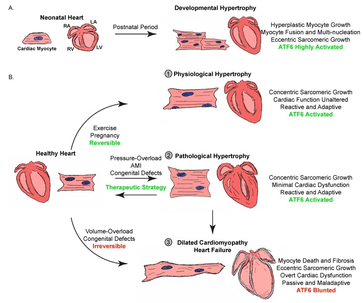
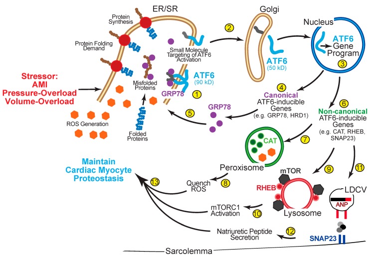
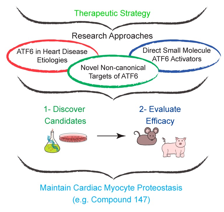
The heart exhibits incredible plasticity in response to both environmental and genetic alterations that affect workload. Over the course of development, or in response to physiological or pathological stimuli, the heart responds to fluctuations in workload by hypertrophic growth primarily by individual cardiac myocytes growing in size. Cardiac hypertrophy is associated with an increase in protein synthesis, which must coordinate with protein folding and degradation to allow for homeostatic growth without affecting the functional integrity of cardiac myocytes (i.e., proteostasis). This increase in the protein folding demand in the growing cardiac myocyte activates the transcription factor, ATF6 (activating transcription factor 6α, an inducer of genes that restore proteostasis. Previously, ATF6 has been shown to induce ER-targeted proteins functioning primarily to enhance ER protein folding and degradation. More recent studies, however, have illuminated adaptive roles for ATF6 functioning outside of the ER by inducing non-canonical targets in a stimulus-specific manner. This unique ability of ATF6 to act as an initial adaptive responder has bolstered an enthusiasm for identifying small molecule activators of ATF6 and similar proteostasis-based therapeutics.

Keywords: ATF6; cardiac myocyte; cardiomyopathy; hypertrophy; proteostasis; small molecule; therapy; transcriptional regulation; unfolded protein response (UPR).


Conflict of interest statement

The authors declare no conflict of interest.

Figures

Figure 1
Figure 1
Types of cardiac hypertrophy. (A) Cross-section drawings are shown to demonstrate the growth of the heart during pre- and postnatal development. This developmental hypertrophy is depicted showing increases in both atrial (RA and LA) and ventricular (RV and LV) chamber blood volume (pink) and wall thickness (red areas are myocardium). Developmental cardiac hypertrophy from the neonatal stage to adulthood is characterized by both hyperplasia and hypertrophy, and since ATF6 is expressed in robust quantities during this period of development, the ER protein folding machinery is sufficient to support the protein-folding load. (B) The adult healthy heart undergoes three main types of cardiac hypertrophy: ① Physiological hypertrophy is an adaptive and reactive process of concentric growth in response to chronic exercise and pregnancy. ATF6 is robustly activated by this process, and the ER protein folding machinery is sufficient to support this form of hypertrophy. ② Pathological hypertrophy is considered an adaptive and reactive process of concentric growth in response to pressure-overload or AMI. In the acute compensatory stages of this concentric growth, ATF6 is robustly activated by this process, and the ER protein folding machinery is sufficient to support this form of hypertrophy. This form of cardiac hypertrophy is reversible and a potential target of ATF6-based therapeutics. ③ Dilated cardiomyopathy and heart failure are either a result of chronic pathological hypertrophy or congenital defects. This is a passive process characterized by chamber dilatation and cardiac myocyte apoptosis and fibrosis. ATF6 and the protein folding machinery are not sufficient at this stage of maladaptive growth.
Figure 2
Figure 2
ATF6 activation and gene program induction. ① In its inactive state, ATF6 is a 90kD ER transmembrane protein that is anchored in the membrane in oligomers via GRP78 and intermolecular disulfide bonding. ② Upon stressors, like cardiac hypertrophy that increases protein synthesis and the protein folding demand or AMI that elevates cellular levels of reactive oxygen species (ROS), GRP78 dissociates from the ER luminal domain of ATF6 and the disulfide bonds are reduced allowing monomerization of ATF6, which allows the 90 kD form of ATF6 to translocate to the Golgi, where is it cleaved by S1P and S2P to liberate the N-terminal approximately 400 amino acids (50 kD) of ATF6 from the ER membrane. It is this unique sequence of activation steps that open a window of small molecule targeting and discovery of ATF6-based therapeutics. ③ The clipped form of ATF6 has a nuclear localization sequence, which facilitates its movement to the nucleus where it binds to specific regulatory elements in ATF6-responsive genes, such as ER stress response elements (ERSEs), and induces the ATF6 gene program. ④ The canonical ATF6 gene program comprises genes that encode proteins that localize to the ER, such as the chaperone, GRP78, or components of ERAD, HRD1 ⑤, where they fortify ER protein folding. ⑥ The non-canonical ATF6 gene program comprises genes that encode proteins not typically categorized as ER stress-response proteins, many of which localize to regions of the cell outside the ER. ⑦ Catalase is a potent antioxidant that localizes to the lumen of peroxisomes where it functions to ⑧ quench ROS. ⑨ Rheb is a small GTPase located on the surface of lysosomes that when bound to mTOR, ⑩ promotes mTORC1 activation and sustains protein synthesis. ⑪ SNAP23 is a t-SNARE protein crucial for ⑫ secretion of natriuretic peptides via large dense-core vesicles (LDCV) in response to hemodynamic stress. ⑬ Both the canonical and non-canonical ATF6 gene programs coordinate to maintain cardiac myocyte proteostasis.
Figure 3
Figure 3
Effect of cardiac myocyte-specific ATF6 deletion in mouse hearts subjected to acute pressure-overload. Mice in which ATF6 has been selectively deleted in cardiac myocytes were subjected to an acute model of pressure overload-induced cardiac hypertrophy. Confocal immunocytofluorescence microscopy analysis of mouse heart sections is shown for a cardiac myocyte marker, Cardiac troponin T (red), protein amyloid oligomers (green), and nuclei (blue). The accumulation of misfolded protein aggregates indicates the necessity of ATF6 to support the protein folding load during concentric cardiac hypertrophy.
Figure 4
Figure 4
Approach to developing novel ATF6-based therapeutics. The therapeutic framework for developing ATF6-based therapeutics is conceptually simple in design. Three main research approaches are prioritized: (1) Expanding the scope of experimental animal models of various etiologies of heart disease in which ATF6 is studied. (2) Discovering other non-canonical targets of ATF6. (3) Using chemical biology to identify potent and specific small molecule activators of ATF6. Coordinately, these research approaches will converge into a streamlined experimental approach of (1) preliminary testing of lead small molecule activators in cell models of disease, in vitro, and (2) evaluating efficacy of these lead small molecule activators in small and large animal models, in vivo.

References

    1. Benjamin E.J., Virani S.S., Callaway C.W., Chamberlain A.M., Chang A.R., Cheng S., Chiuve S.E., Cushman M., Delling F.N., Deo R. American Heart Association Council on Prevention Statistics and Stroke Statistics Heart Disease and Stroke Statistics-2018 Update: A Report From the American Heart Association. Circulation. 2018;137:467–492. doi: 10.1161/CIR.0000000000000558. - DOI - PubMed
    1. Gheorghiade M., Braunwald E. Hospitalizations for heart failure in the United States—A sign of hope. JAMA. 2011;306:1705–1706. doi: 10.1001/jama.2011.1510. - DOI - PubMed
    1. Rosenzweig A. Medicine. Cardiac regeneration. Science. 2012;338:1549–1550. doi: 10.1126/science.1228951. - DOI - PubMed
    1. Kannel W.B. Vital epidemiologic clues in heart failure. J. Clin. Epidemiol. 2000;53:229–235. doi: 10.1016/S0895-4356(99)00135-3. - DOI - PubMed
    1. Hobbs R.E. Guidelines for the Diagnosis and Management of Heart Failure. Am. J. Ther. 2004;11:467–472. doi: 10.1097/01.mjt.0000127150.21184.01. - DOI - PubMed

Publication types

MeSH terms

Substances