. 2020 Mar 3;21(5):1715.
doi: 10.3390/ijms21051715.

GABAergic Input Affects Intracellular Calcium Levels in Developing Granule Cells of Adult Rat Hippocampus

Affiliations

GABAergic Input Affects Intracellular Calcium Levels in Developing Granule Cells of Adult Rat Hippocampus

Davide Lattanzi et al. Int J Mol Sci. .

Abstract

In the dentate gyrus (DG) of the mammalian hippocampus, granule neurons are generated from neural stem cells (NSCs) throughout the life span and are integrated into the hippocampal network. Adult DG neurogenesis is regulated by multiple intrinsic and extrinsic factors that control NSC proliferation, maintenance, and differentiation into mature neurons. γ-Aminobutyric acid (GABA), released by local interneurons, regulates the development of neurons born in adulthood by activating extrasynaptic and synaptic GABAA receptors. In the present work, patch-clamp and calcium imaging techniques were used to record very immature granule cells of adult rat dentate gyrus for investigating the actual role of GABAA receptor activation in intracellular calcium level regulation at an early stage of maturation. Our findings highlight a novel molecular and electrophysiological mechanism, involving calcium-activated potassium channels (BK) and T-type voltage-dependent calcium channels, through which GABA fine-tunes intracellular calcium homeostasis in rat adult-born granule neurons early during their maturation. This mechanism might be instrumental in promoting newborn cell survival.

Keywords: GABA input; T-type voltage-dependent calcium channels; adult rat; calcium-activated potassium channels; hippocampus; immature neurons; membrane potential oscillations.


Conflict of interest statement

The authors declare no conflict of interest.

Figures

Figure 1
Figure 1
Functional features of immature dentate granule cells recorded in whole-cell configuration. (A,B) Voltage-clamp recordings (V-Holding = −70 mV, [Cl]i = 34 mM) of the evoked response to MPP stimulation (top) and spontaneous activity (bottom) in two different immature neurons. (C,D) Current-clamp recordings of intrinsic excitability in response to depolarizing current steps in the same recorded cell. TTX-sensitive rudimentary spike (threshold about −26 mV), and isolated and very slow Ni2+-sensitive spikes (threshold about −58 mV) were elicited by depolarizing current steps starting from membrane potential of −80 mV (C). Depolarizing current steps starting from resting membrane potential (−45 mV) were not able to elicit T-type VDCC spike due to their inactivation (D).
Figure 2
Figure 2
NMDA-induced BK channel activation via NMDARs in immature granule cells. Representative NMDA-induced current at −20 mV holding potential before and after paxilline 10 µM bath application.
Figure 3
Figure 3
Membrane potential oscillations regulate the intracellular calcium levels. (A) Representation of single-cell Ca2+ imaging method and fluorescence image of a recorded immature neuron. The intracellular solution contained 34 mM Cl- and 100 µM Fluo4. (B) Resting membrane potential oscillations recorded in current-clamp mode (top trace) and low-noise voltage-clamp recording at the same potential in the same cell (bottom trace). (C) Representative course of cell body fluorescence in a recorded immature neuron obtained by switching recording mode from current-clamp (C-C) to voltage-clamp (V-C) and vice versa. (D) Significative decrease of [Ca2+]I obtained by switching from current-clamp mode to voltage-clamp mode; the reduction reversed by switching back to current-clamp mode (RM one-way ANOVA F (2, 5) = 15.28 p < 0.01, Tukey’s posthoc test (*) V-C vs. both C-C, p < 0.05).
Figure 4
Figure 4
Membrane potential oscillations in immature granule cells. (A) Representative graph of membrane potential noise distribution recorded in immature neurons before and after 200 µM nickel application to the bath perfusion. (B) Robust spontaneous hyperpolarizing events recorded in current-clamp mode in immature neuron, showing a mean resting membrane potential of −49 mV. A T-type VDCC spike-like event is enlarged in the box.
Figure 5
Figure 5
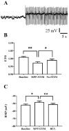
GABAAR activation by MPP stimulation regulates intracellular calcium concentration. (A) Typical response evoked by MPP stimulation (2.5 Hz) recorded in current-clamp mode. MPP stimulation induced a constant slight depolarization shifting membrane potential close to the Cl- equilibrium. Even if the trace is disturbed by artifacts, it is possible to note the reduction of potential oscillations during stimulation. (B) Prolonged MPP stimulation (3 min) induced a significant and reversible decrease of [Ca2+]I and membrane depolarization, as well (C). F/F0, RM one-way ANOVA F (2, 12) = 11.38 p < 0.01, Tukey’s post hoc test (**) baseline vs. MPP-STIM p < 0.01, (*) MPP-STIM vs. No-STIM p < 0.05. RMP, RM one-way ANOVA F (2, 12) = 9.374 p < 0.01, Tukey’s post hoc test (*) baseline vs. MPP-STIM p < 0.05, (**) MPP-STIM vs. No-STIM p < 0.01.
Figure 6
Figure 6
BK channels and T-type VDCC interaction. (A) Hyperpolarizing spontaneous burst recorded in current-clamp mode in immature neuron with resting membrane potential mean value of −52 mV. (B) Ni2+-sensitive T-type VDCC spikes induced by hyperpolarizing current steps starting from resting membrane potential. (C) Paxilline-sensitive after-hyperpolarization induced by T-type VDCC spike. (D) BK-mediated after-hyperpolarization, quantified as the slope of calcium spike repolarization (inset in the graph), recorded using different intracellular calcium chelators. Two-way RM ANOVA with Paxilline treatment and Intracellular buffer as independent variables; Paxilline treatment F (1, 18) = 26.82 p < 0.01, Intracellular buffer F (2, 18) = 9.256 p < 0.01. Tukey’s multiple comparisons test (**) baseline vs. 10 µM paxilline without buffer p < 0.01, (**) baseline vs. 10 µM paxilline 5 mM EGTA p < 0.01, (**) baseline without buffer vs. baseline 3 mM BAPTA p < 0.01, (**) baseline 5 mM EGTA vs. baseline 3 mM BAPTA p < 0.01.
Figure 7
Figure 7
Resting membrane potential oscillations and intracellular calcium concentration. Membrane potential oscillations decreased by increasing calcium buffer capacity. One-way ANOVA F (2, 17) = 16.28 p < 0.01, Tukey’s multiple comparisons test (**) without buffer vs. 5 mM EGTA p < 0.01, (**) without buffer vs. 3 mM BAPTA p < 0.01.
Figure 8
Figure 8
Schematic illustration showing the implication of BK channels, T-type VDCC, and GABAAR in intracellular calcium level regulation. (A) When the extracellular GABA concentration is low, GABAARs are closed, immature neurons have a very high IR, and generate membrane potential oscillations through BK and T-type calcium channel activity. Specifically, robust BK-induced hyperpolarizations elicit T-type VDCC voltage sensor activation and gate opening, which increases calcium entry. In turn, calcium influx quickly activates BK channels, due to their physical interaction (according to the literature [17]), inducing hyperpolarizations. This mechanism is facilitated by low calcium buffer levels present in immature neurons. (B) When the extracellular GABA concentration increases, GABAARs are activated, inducing membrane current shunt. BK-induced hyperpolarization is damped, preventing T-type VDCC opening. Therefore, an increased hippocampal activity through GABAAR action can promote a lowering of intracellular calcium level. This figure was prepared using the Neuroscience-PPT-Toolkit-Suite of Motifolio Inc., USA, and Servier Medical Art (https://smart.servier.com/).

References

    1. Gonçalves J.T., Schafer S.T., Gage F.H. Adult Neurogenesis in the Hippocampus: From Stem Cells to Behavior. Cell. 2016;167:897–914. doi: 10.1016/j.cell.2016.10.021. - DOI - PubMed
    1. Ambrogini P., Lattanzi D., Ciuffoli S., Agostini D., Bertini L., Stocchi V., Santi S., Cuppini R. Morpho-functional characterization of neuronal cells at different stages of maturation in granule cell layer of adult rat dentate gyrus. Brain Res. 2004;1017:21–31. doi: 10.1016/j.brainres.2004.05.039. - DOI - PubMed
    1. Dieni C.V., Chancey J.H., Overstreet-Wadiche L.S. Dynamic functions of GABA signaling during granule cell maturation. Front. Neural Circuits. 2013;6:11. doi: 10.3389/fncir.2012.00113. - DOI - PMC - PubMed
    1. Bao H., Asrican B., Li W., Gu B., Wen Z., Lim S.A., Haniff I., Ramakrishnan C., Deisseroth K., Philpot B., et al. Long-Range GABAergic Inputs Regulate Neural Stem Cell Quiescence and Control Adult Hippocampal Neurogenesis. Cell Stem Cell. 2017;21:604–617. doi: 10.1016/j.stem.2017.10.003. - DOI - PMC - PubMed
    1. Tozuka Y., Fukuda S., Namba T., Seki T., Hisatsune T. GABAergic excitation promotes neuronal differentiation in adult hippocampal progenitor cells. Neuron. 2005;47:803–815. doi: 10.1016/j.neuron.2005.08.023. - DOI - PubMed

LinkOut - more resources