. 2020 Mar 3;21(5):1733.
doi: 10.3390/ijms21051733.

MicroRNA-24-3p Targets Notch and Other Vascular Morphogens to Regulate Post-ischemic Microvascular Responses in Limb Muscles

Affiliations

MicroRNA-24-3p Targets Notch and Other Vascular Morphogens to Regulate Post-ischemic Microvascular Responses in Limb Muscles

Micol Marchetti et al. Int J Mol Sci. .

Abstract

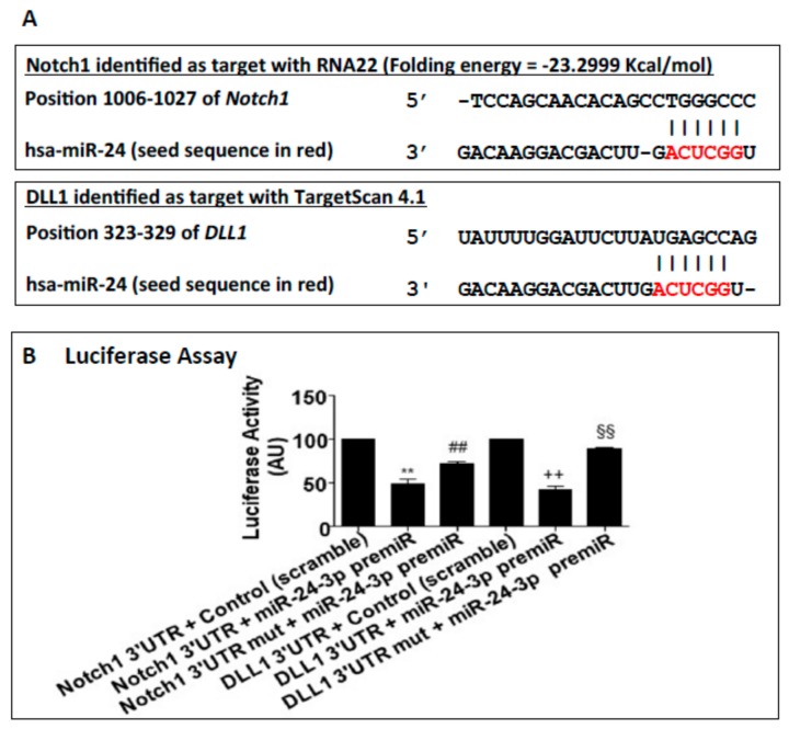
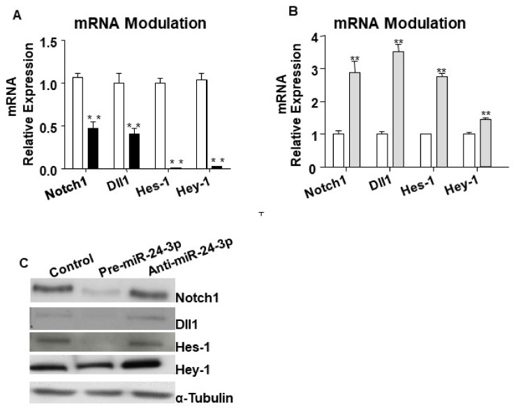
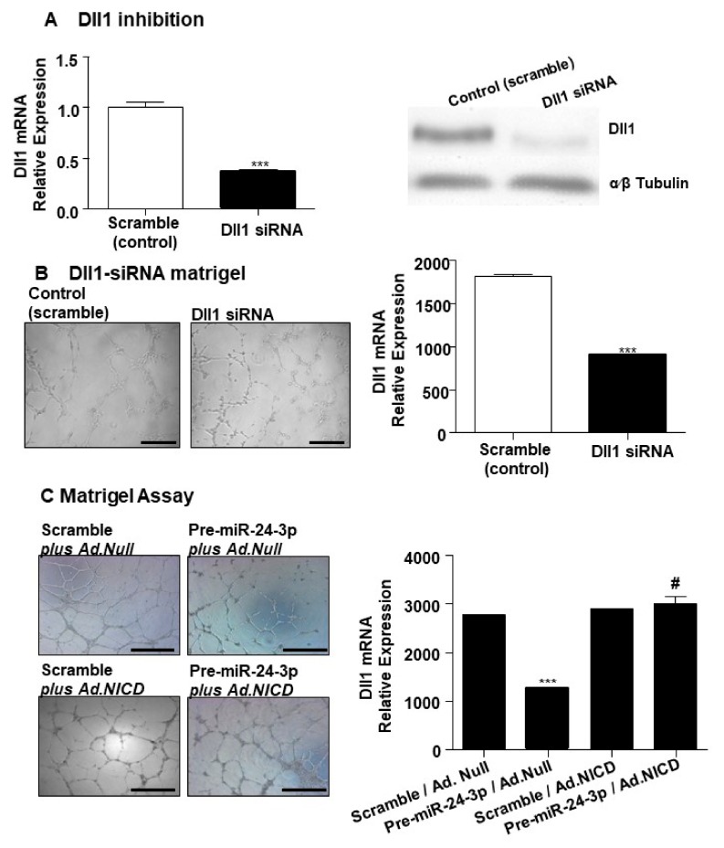
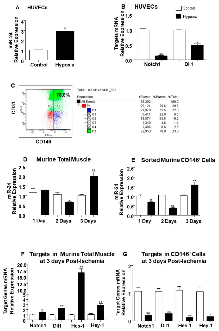
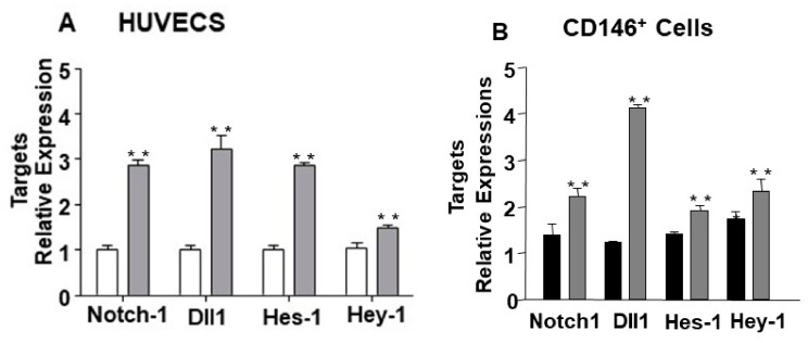
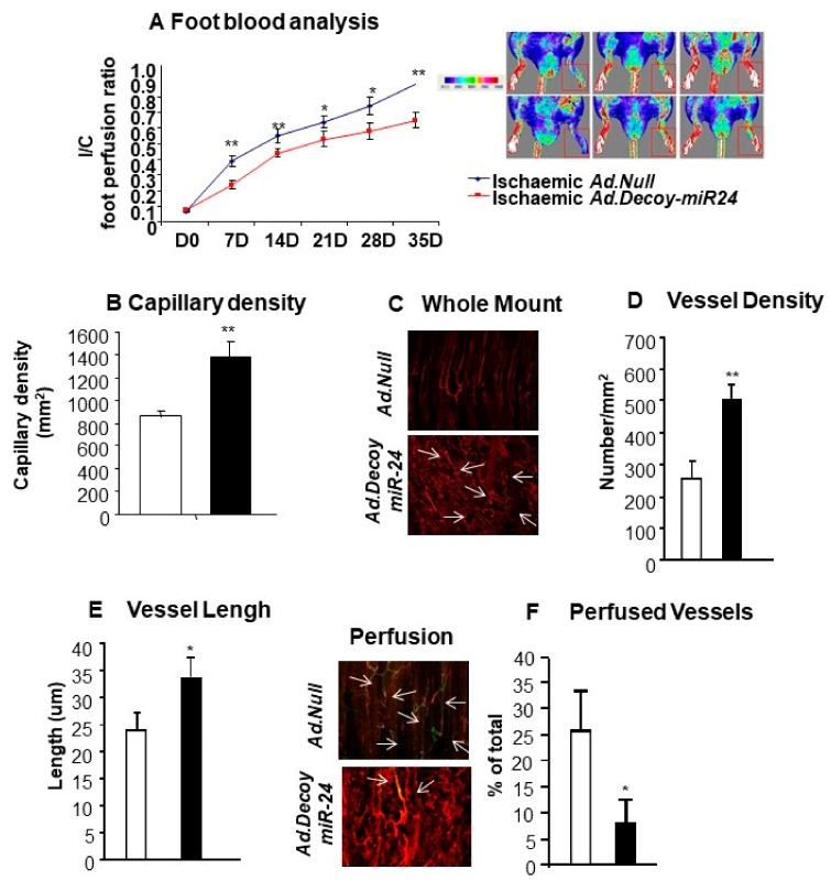
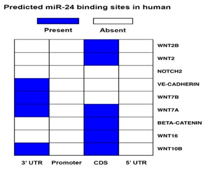
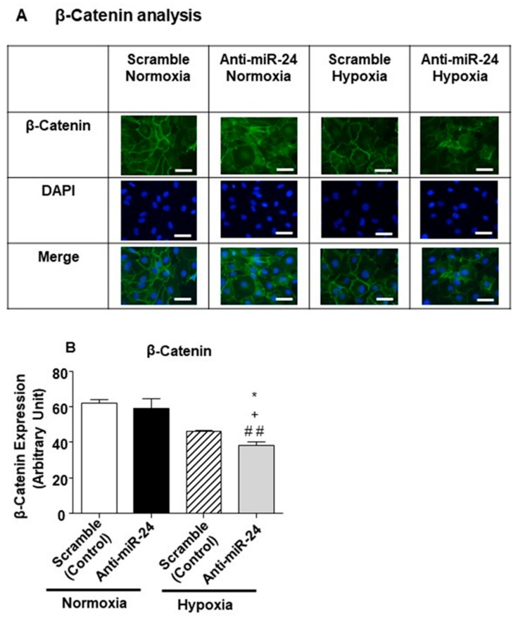
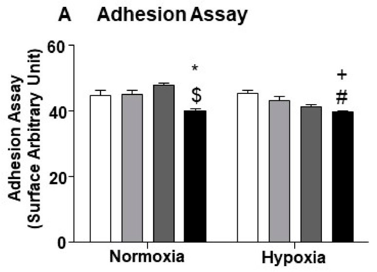
MicroRNAs (miRs) regulate complex processes, including angiogenesis, by targeting multiple mRNAs. miR-24-3p-3p directly represses eNOS, GATA2, and PAK4 in endothelial cells (ECs), thus inhibiting angiogenesis during development and in the infarcted heart. miR-24-3p is widely expressed in cardiovascular cells, suggesting that it could additionally regulate angiogenesis by acting on vascular mural cells. Here, we have investigated: 1) new miR-24-3p targets; 2) the expression and the function of miR-24-3p in human vascular ECs; 3) the impact of miR-24-3p inhibition in the angiogenesis reparative response to limb ischemia in mice. Using bioinformatics target prediction platforms and 3'-UTR luciferase assays, we newly identified Notch1 and its Delta-like ligand 1 (Dll1) to be directly targeted by miR-24-3p. miR-24-3p was expressed in human ECs and pericytes cultured under normal conditions. Exposure to hypoxia increased miR-24-3p in ECs but not in pericytes. Transfection with a miR-24-3p precursor (pre-miR-24-3p) increased miR-24-3p expression in ECs, reducing the cell survival, proliferation, and angiogenic capacity. Opposite effects were caused by miR-24-3p inhibition. The anti-angiogenic action of miR-24-3p overexpression could be prevented by simultaneous adenovirus (Ad)-mediated delivery of constitutively active Notch intracellular domain (NICD) into cultured ECs. We next demonstrated that reduced Notch signalling contributes to the anti-angiogenic effect of miR-24-3p in vitro. In a mouse unilateral limb ischemia model, local miR-24-3p inhibition (by adenovirus-mediated miR-24-3p decoy delivery) restored endothelial Notch signalling and increased capillary density. However, the new vessels appeared disorganised and twisted, worsening post-ischemic blood perfusion recovery. To better understand the underpinning mechanisms, we widened the search for miR-24-3p target genes, identifying several contributors to vascular morphogenesis, such as several members of the Wingless (Wnt) signalling pathway, β-catenin signalling components, and VE-cadherin, which synergise to regulate angiogenesis, pericytes recruitment to neoformed capillaries, maturation, and stabilization of newly formed vessels. Among those, we next focussed on β-catenin to demonstrate that miR-24-3p inhibition reduces β-catenin expression in hypoxic ECs, which is accompanied by reduced adhesion of pericytes to ECs. In summary, miR-24-3p differentially targets several angiogenesis modulators and contributes to autonomous and non-autonomous EC crosstalk. In ischemic limbs, miR-24-3p inhibition increases the production of dysfunctional microvessels, impairing perfusion. Caution should be observed in therapeutic targeting of miR-24-3p.

Keywords: Notch; angiogenesis; endothelial cells; limb ischemia; miR-24-3p; β-catenin.


Conflict of interest statement

The authors have no conflicting financial interest.

Figures

Figure 1
Figure 1
Target-gene prediction and primers sequences: (A) Analysis of the seed sequence of miR-24-3p (red letters) in the 3′-UTR of Notch1 and Dll1 with RNA22 software and Targetscan 4.1. Gray part: binding strength. (B) To validate the relation between miR-24-3p and the predicted targets, the luciferase assay was performed. HEK293 cells were transfected with pre-miR-24-3p or control and the reporter gene for Notch1 3′-UTR or Dll1 3′-UTR or their respective mutants. Experiments were performed in triplicate and repeated three times. Values are means ± SEM. ** p < 0.001 vs. Notch1 3′-UTR + control; # # p < 0.01 vs. Notch1 3′-UTR + pre-miR-24; ++ p < 0.05 vs. Dll1 3′-UTR+ control; $$ p < 0.01 vs. Dll1 3′-UTR + pre-miR-24.
Figure 2
Figure 2
Notch1 and Dll1 direct targets of miR-24-3p and Notch pathway modulation in human umbilical vein endothelial cells (HUVECs). To validate the relation between miR-24-3p and the predicted targets Notch1 and Dll1, mRNA and proteins analyses were performed in HUVECs transfected with premiR-24-3p, anti-miR-24-3p, or control. Hes-1 and Hey-1 were also evaluated, as reporters of the Notch signalling activity. Bar graph (A) shows HUVEC transfected with pre-miR-24-3p (black columns) compared to control (scramble, white columns). Bar graph (B) shows HUVEC transfected with miR-24-3p inhibitor (light grey columns) compared to control (white columns). mRNA levels were normalised to 18S and quantified employing the 2ΔΔCt method and is presented as changes from the values in the respective control group. Experiments were performed in triplicate and repeated three times. Values are means ± SEM. Τ ** p < 0.01 vs. Notch1 3′-UTR + control. Panel (C) shows protein expressions of Notch1, Dll1, Hes-1, and Hey-1 forcing and inhibiting the expression of miR-24-3p. For Western blot analyses, α tubulin was used as house-keeping protein.
Figure 3
Figure 3
Dll1 inhibition in HUVECs affects the network-formation capability, while Notch intracellular domain (NICD) over-expression rescues this angiogenic defect. (A, left) Bar graph shows Dll1 mRNA expression in HUVECs transfected with Dll1-siRNA (black column) compared to HUVECs transfected with scramble sequence (control). (A, right) Western Blot shows the Dll1 protein expression in HUVECs transfected with Dll1-siRNA compared to HUVECs transfected with scramble sequence (control). α-β Tubulin is presented as house-keeping protein. (B, left) Photomicrographs of representative fields show the endothelial networks formed by HUVECs transfected with Dll1-siRNA compared to control group. (B, right) Bar graph represents the quantification of the total length of cord-like structures formed by HUVECs transfected with Dll1-siRNA (black column) compared to control group (scramble, white column). (C, left) Photomicrographs of representative fields show the endothelial network formed by HUVECs differently treated as indicated: HUVECs transfected with scramble and infected with Ad.Null; transfected with pre-miR-24-3p miR-24-3p and infected with Ad.Null; transfected with scramble and infected with Ad.NICD; and HUVECs transfected with pre-miR-24-3p and infected with Ad.NICD. Bar graph (C) represents the photomicrographs’ conditions expressing the total length of tube-like structures. Dll1 mRNA expression was normalised to 18S. Experiments were performed in triplicate and repeated three times. Values are means ± SEM. *** p < 0.001 vs. Scramble in A nd B) and vs. Scramble/Ad.Null (in C); # p < 0.05 vs. Pre-miR-24-3p/Ad.Null (in C).
Figure 4
Figure 4
Modulation of miR-24-3p, Notch-1, and Dll1 mRNA expression by in vitro hypoxia and limb ischemia. (A) Bar graph shows miR-24-3p expression changes induced in endothelial cells (ECs) by culture for 48 h in hypoxia (1% O2, black column) versus standard cell culture conditions (21% O2, white column). (B) Bar graph shows Notch1 mRNA and Dll1 mRNA modulation by hypoxia in the same cells preparations used for the analyses shown in Panel A. (C) FACS analysis plot shows murine sorted CD146+ cells stained with FITC-conjugated CD146, APC-conjugated CD31 antibodies. The cells were extracted from pools of two adductor muscles. (D) Bar graph shows miR-24-3p expressional changes in murine total muscle samples at 1, 2, and 3 days post-ischemia (black columns) compared to murine non-ischemic control samples at matched time-point (white columns). (E) Bar graph shows miR-24-3p modulation in murine ECs sorted from total adductor muscle samples at 1, 2, and 3 days post-ischemia (black columns) compared to contra-lateral non-ischemic samples (white columns). (F) Bar graph shows miR expressional changes in Notch1, Dll1, Hes-1, and Hey-1 in mouse muscle harvested at 3 days post-ischemia (black column) compared to contra-lateral non-ischemic samples (white column). (G) Bar graph shows mRNA expression changes in Notch1, Dll1, Hes-1, and Hey-1 in murine ECs sorted from adductor muscle at 3 days post-ischemia (black column) compared to murine ECs sorted from non-ischemic muscles (white column). miR-24-3p expression was normalised to Snu6, and data are compared to the matched time-point group by the 2ΔΔCt method. Notch1, Dll1, Hes-1, and Hey-1 mRNA expression values were corrected to 18S. Experiments were performed in triplicate and repeated three times. Values are means ± SEM. *p < 0.05, **p < 0.01 vs. control matched time-point samples (n = 10 each group).
Figure 5
Figure 5
Impact of miR-24-3p inhibition on Notch pathway components’ miR-24-3p expression in cultured HUVECs and microvascular ECs exposed to limb ischemia. (A) Bar graph shows miR-24-3pNotch-1, Dll1, and down-stream targets Hes-1 and Hey-1 mRNAs in HUVEC transfected with miR-24-3p inhibitor (light grey columns) compared to control (white columns). (B) Bar graph shows the modulations of Notch-1, Dll1, Hes-1, and Hey-1 mRNA in CD146 positive cells isolated by ischemic adductor muscle at 3 days post-ischemia treated with in vivo miR-24-3p inhibitor (Ad.Decoy.miR-24-3p, grey bar) compared to scramble control (Ad. Null, black bar). Gene expression was normalised to 18S. Cell experiments were performed in triplicate and repeated three times. Analyses on muscular cells were developed using n = 10 mice per group. Values are means ± SEM. ** p < 0.005 vs. the respective control.
Figure 6
Figure 6
Effects of miR-24-3p inhibition in the ischemic limb. (A) Line graph shows the blood flow analysis in ischemic mice injected with Ad.Null (blue line) or Ad.Decoy-miR-24-3p (red line) (n = 12 animals per each group). (B) Bar graph shows increased capillary density after Ad.Decoy-mediated inhibition of miR-24-3p in the ischemic adductor muscle. Photomicrographs (C) and bar graphs (D and E) show the effect of miR-24-3p inhibition on the vascular organization (C), density (D), and length (E) in the ischemic adductor muscle. Photomicrographs and bar graph (F) show the percentage of perfused vessels in muscles treated with either AdDecoy.miR-24-3p or Ad.Null at 21 days of ischemia. Ad.Decoy.miR-24-3p treatment is shown in black, Ad.Null in white. Values are means ± SEM. ** p < 0.01 and * p < 0.01 vs. Ad.Null.
Figure 7
Figure 7
miR-24-3p and Wnt/β-catenin binding prediction. The table shows the target genes prediction related to the Wnt/β-catenin pathway in human (left) and mouse. The prediction was performed using mirWalk software.
Figure 8
Figure 8
β-catenin modulation in in vitro model of hypoxia. (A) The panel shows representative pictures of β-catenin staining in HUVECs treated with miR-24-3p inhibitor and exposed to hypoxia (1% O2) or kept under standard culture conditions for 48 h. Scale bars corresponds to 50 um (B) Bar graph shows the quantification of the staining, where the black bar represents HUVECs treated with miR-24-3p inhibitor, and the white bar represents HUVECs treated with scramble (transfection control). Adhesion assay was performed in quintuplicate and repeated two times; β-catenin staining was performed in triplicate and repeated three times. Values are means ± SEM. * p < 0,05 vs. scramble hypoxia, + p < 0.05 vs. anti-miR-24-3p in standard culture conditions; ## p < 0.01 vs. scramble in standard culture conditions.
Figure 9
Figure 9
Reduced miR24 in ECs affects the adhesion between ECs and pericyte. miR-24-3 under standard (5% O2) or hypoxic culture conditions. Groups are pre-miR-24-3p transfected ECs miR-24-3p (black column); scramble control (dark grey column); vehicle control of transfection (light grey); and untreated cells (white column). Adhesion assay was performed using five wells per treatment and repeated two times. Values are means ± SEM. * p < 0.05 vs. untreated cells in normoxia; + p < 0.05 vs. untreated Cells in hypoxia; $ p < 0.05 vs. scramble in normoxia; # p < 0,05 vs. scramble control in hypoxia.

Similar articles

Cited by

References

    1. Bartel D.P. MicroRNAs: Genomics, biogenesis, mechanism, and function. Cell. 2004;116:281–297. doi: 10.1016/S0092-8674(04)00045-5. - DOI - PubMed
    1. Ørom U.A., Lund A.H. Experimental identification of microRNA targets. Gene. 2010;451:1–5. doi: 10.1016/j.gene.2009.11.008. - DOI - PubMed
    1. El Azzouzi H., Leptidis S., Doevendans P.A., Leon J., De Windt L.J. HypoxamiRs: Regulators of cardiac hypoxia and energy metabolism. Trends Endocrinol. Metab. 2008;12:1426–1431. doi: 10.1016/j.tem.2015.06.008. - DOI - PubMed
    1. Ivan M., Adrian L., Harris A.L., Martelli F., Kulshreshtha R. Hypoxia response and microRNAs: No longer two separate worlds. J. Cell. Mol. Med. 2008;12:1426–1431. doi: 10.1111/j.1582-4934.2008.00398.x. - DOI - PMC - PubMed
    1. Lorenzen J.M., Kaucsar T., Schauerte C., Schmitt R., Rong S., Hübner A., Scherf K., Fiedler J., Martino F., Kumarswamy R., et al. MicroRNA-24 antagonism prevents renal ischemia reperfusion injury. J. Am. Soc. Nephrol. 2014;25:2717–2729. doi: 10.1681/ASN.2013121329. - DOI - PMC - PubMed

MeSH terms