

Positive staining of the immunoligand B7-H6 in abnormal/transformed keratinocytes consistently accompanies the progression of cervical cancer
- PMID: 32138659
- PMCID: PMC7059382
- DOI: 10.1186/s12865-020-0341-9
Positive staining of the immunoligand B7-H6 in abnormal/transformed keratinocytes consistently accompanies the progression of cervical cancer
Abstract
Background: B7-H6 has been revealed as an endogenous immunoligand expressed in a variety of tumors, but not expressed in healthy tissues. Heretofore, no studies have been reported describing B7-H6 in women with cervical cancer. To investigate this question, our present study was conducted.
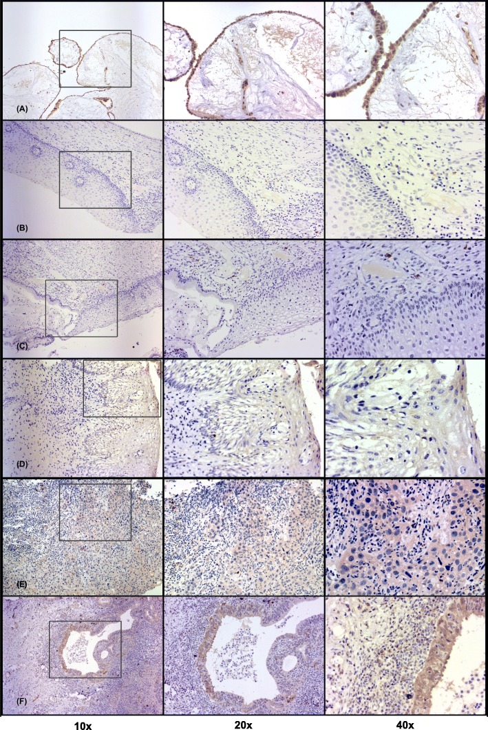
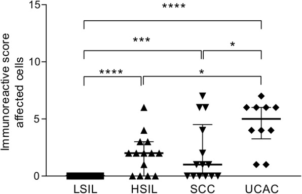
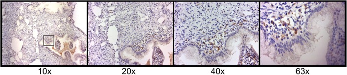
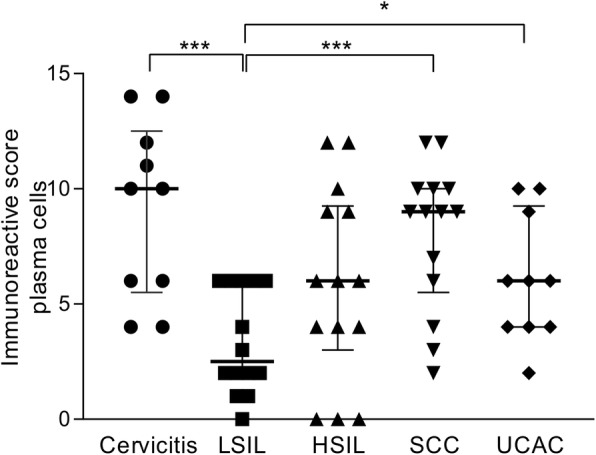
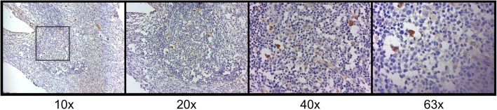
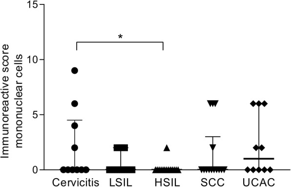
Results: This retrospective study comprised a total of 62 paraffinized cervical biopsies, which were distributed in five groups: low-grade squamous intraepithelial lesions (LSIL), high-grade squamous intraepithelial lesions (HSIL), squamous cervical carcinoma (SCC), uterine cervical adenocarcinoma (UCAC), and a group of cervicitis (as a control for non-abnormal/non-transformed cells). Cervical sections were stained by immunohistochemistry to explore the expression of B7-H6, which was reported according to the immunoreactive score (IRS) system. We observed a complete lack of B7-H6 in LSIL abnormal epithelial cells. Interestingly, B7-H6 began to be seen in HSIL abnormal epithelial cells; more than half of this group had B7-H6 positive cells, with staining characterized by a cytoplasmic and membranous pattern. B7-H6 in the SCC group was also seen in the majority of the sections, showing the same cytoplasmic and membranous pattern. Strong evidence of B7-H6 was notably found in UCAC tumor columnar cells (in 100% of the specimens, also with cytoplasmic and membranous pattern). Moreover, consistent B7-H6 staining was observed in infiltrating plasma cells in all groups.
Conclusions: B7-H6 IRS positively correlated with disease stage in the development of cervical cancer; additionally, B7-H6 scores were found to be even higher in the more aggressive uterine cervical adenocarcinoma, suggesting a possible future therapeutic target for this cancer type.
Keywords: B7-H6; B7H6; Cervical cancer; Cervical intraepithelial lesions; NKp30; Therapeutic target.
Conflict of interest statement
The authors declare that they have no competing interests.
Figures
Similar articles
-
B7-H6, an immunoligand for the natural killer cell activating receptor NKp30, reveals inhibitory effects on cell proliferation and migration, but not apoptosis, in cervical cancer derived-cell lines.BMC Cancer. 2020 Nov 10;20(1):1083. doi: 10.1186/s12885-020-07608-4. BMC Cancer. 2020. PMID: 33172426 Free PMC article.
-
B7-H6 as a Diagnostic Biomarker for Cervical Squamous Cell Carcinoma.Genet Test Mol Biomarkers. 2021 Jul;25(7):463-470. doi: 10.1089/gtmb.2020.0313. Genet Test Mol Biomarkers. 2021. PMID: 34280008
-
Epithelial and tumor-associated endothelial expression of B7-H3 in cervical carcinoma: relation with CD8+ intraepithelial lymphocytes, FIGO stage, and phosphohistone H3 (PHH3) reactivity.Int J Gynecol Pathol. 2015 Mar;34(2):187-95. doi: 10.1097/PGP.0000000000000116. Int J Gynecol Pathol. 2015. PMID: 25675190
-
[Research Advances of A New Co-stimulatory Molecule-B7 Homolog 6--Review].Zhongguo Shi Yan Xue Ye Xue Za Zhi. 2016 Feb;24(1):295-8. doi: 10.7534/j.issn.1009-2137.2016.01.057. Zhongguo Shi Yan Xue Ye Xue Za Zhi. 2016. PMID: 26913440 Review. Chinese.
-
The B7 Family Member B7-H6: a New Bane of Tumor.Pathol Oncol Res. 2018 Oct;24(4):717-721. doi: 10.1007/s12253-017-0357-5. Epub 2017 Oct 31. Pathol Oncol Res. 2018. PMID: 29086181 Review.
Cited by
-
Innate lymphoid cells in early tumor development.Front Immunol. 2022 Aug 12;13:948358. doi: 10.3389/fimmu.2022.948358. eCollection 2022. Front Immunol. 2022. PMID: 36032129 Free PMC article. Review.
-
Immune checkpoints and cancer immunotherapies: insights into newly potential receptors and ligands.Ther Adv Vaccines Immunother. 2023 Aug 30;11:25151355231192043. doi: 10.1177/25151355231192043. eCollection 2023. Ther Adv Vaccines Immunother. 2023. PMID: 37662491 Free PMC article. Review.
-
B7-H6 is a new potential biomarker and therapeutic target of T-lymphoblastic lymphoma.Ann Transl Med. 2021 Feb;9(4):328. doi: 10.21037/atm-20-5308. Ann Transl Med. 2021. PMID: 33708955 Free PMC article.
-
NKp30 Receptor Upregulation in Salivary Glands of Sjögren's Syndrome Characterizes Ectopic Lymphoid Structures and Is Restricted by Rituximab Treatment.Front Immunol. 2021 Sep 14;12:706737. doi: 10.3389/fimmu.2021.706737. eCollection 2021. Front Immunol. 2021. PMID: 34594326 Free PMC article.
-
BCL-2 mutant B7H6-CAR-T cells synergized with venetoclax for treating small cell lung cancer.J Immunother Cancer. 2025 May 7;13(5):e010073. doi: 10.1136/jitc-2024-010073. J Immunother Cancer. 2025. PMID: 40341023 Free PMC article.
References
-
- Winer RL, Hughes JP, Feng Q, Xi LF, Cherne S, O'Reilly S, Kiviat NB, Koutsky LA. Early natural history of incident, type-specific human papillomavirus infections in newly sexually active young women. Cancer Epidemiol Biomarkers Prev. 2011;20(4):699–707. doi: 10.1158/1055-9965.EPI-10-1108. - DOI - PMC - PubMed
-
- Kumar N. Cervical cancer; a nightmare for womanhood: review o f recent advances. Women’s Health Gynaecol. 2016;9:30–34.
