

Ménétrier's disease in childhood: a case report from China
- PMID: 32138711
- PMCID: PMC7059724
- DOI: 10.1186/s12887-020-2005-6
Ménétrier's disease in childhood: a case report from China
Abstract
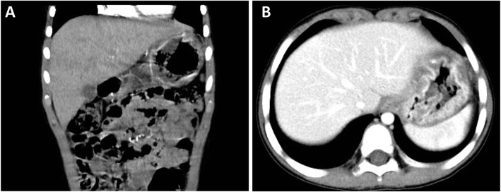
Background: Ménétrier's disease (MD) is a protein-losing gastropathy characterized by gastric hypertrophy, foveolar hyperplasia and hypoalbuminemia. MD is uncommon in childhood with nonspecific clinical symptoms, and the exact cause of pediatric MD is still unclear.
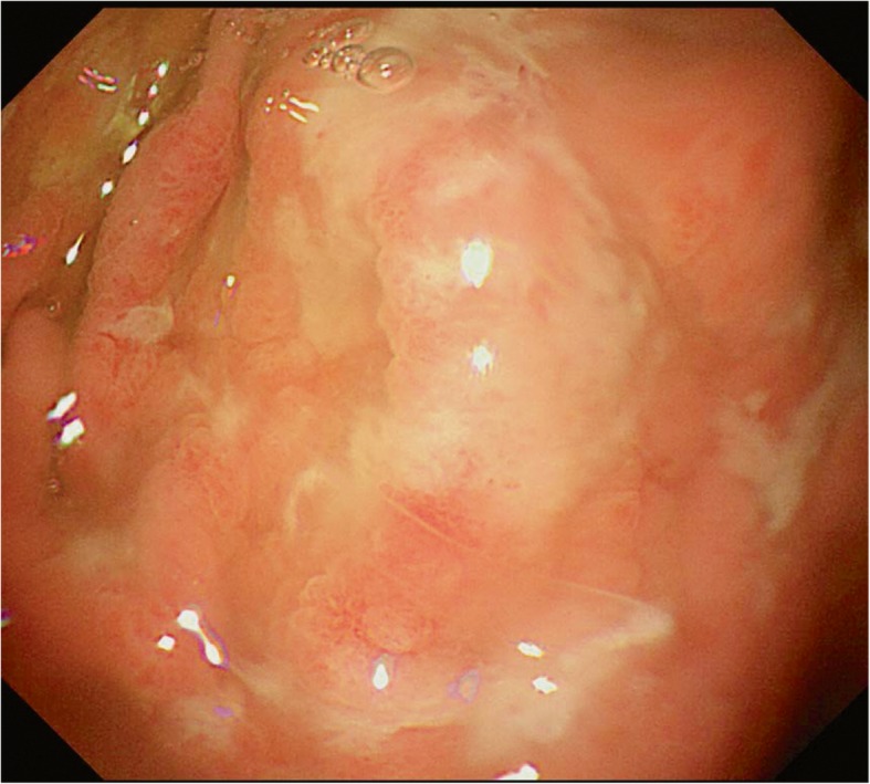
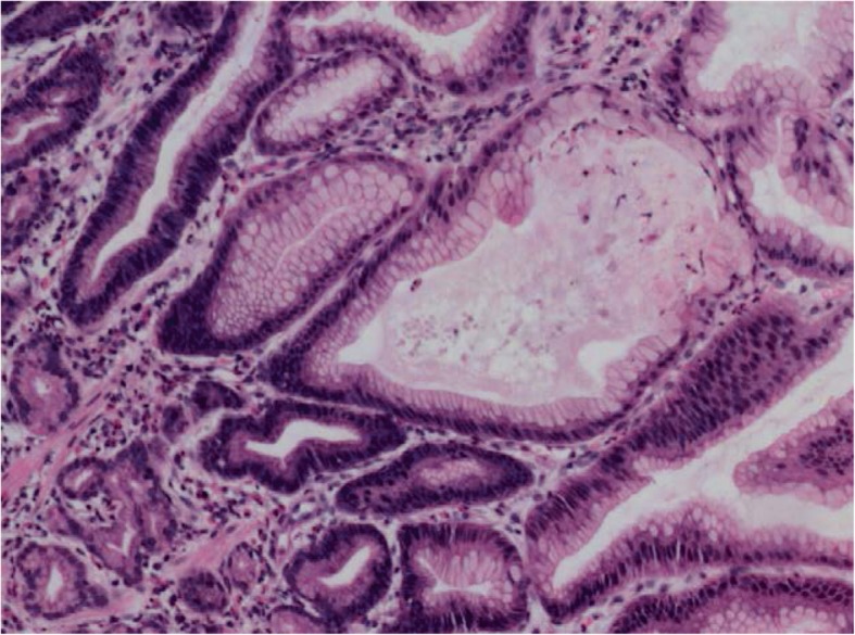
Case presentation: Here, we reported a 4 year and 10-month boy presenting with MD from China. The patient was suffered with vomiting, abdominal pain, hypoproteinemia and edema. Laboratory tests showed that the boy was infected with Clostridium difficile (CD). Gastrointestinal endoscopy revealed giant gastric folds, and histological gastric biopsies showed foveolar hyperplasia with glandular atrophy, infiltration of eosinophils in the lamina propria of the patient. Finally, the boy was recovered after supportive therapy with intravenous albumin and CD eradication.
Conclusion: For the nonspecific clinical symptoms of MD, gastrointestinal endoscopic evaluations with gastric tissue biopsies are required to establish the diagnosis of MD in children with unexplained hypoalbuminemia.
Keywords: Child; Hypertrophy; Hypoalbuminemia; Ménétrier’s disease.
Conflict of interest statement
The authors declare that they have no competing interests.
Figures
References
-
- Ménétrier P. Des polyadenomes gastriques et deleurs rapports avec le cancer del’estomac. Arch Physiol Normal Pathol. 1888;1:232–262.
Publication types
MeSH terms
Grants and funding
- 81870373/National Natural Science Foundation of China/International
- 81500449/National Natural Science Foundation of China/International
- SHDC12017115/Shanghai Hospital Development Center New Frontier Technology Joint Research Project/International
- 2017ZZ02019/Shanghai Municipal Commission of Health and Family Planning/International
LinkOut - more resources
Full Text Sources
