. 2020 May;69(5):954-964.
doi: 10.2337/db19-1106. Epub 2020 Mar 5.

Role of Proinsulin Self-Association in Mutant INS Gene-Induced Diabetes of Youth

Affiliations

Role of Proinsulin Self-Association in Mutant INS Gene-Induced Diabetes of Youth

Jinhong Sun et al. Diabetes. 2020 May.

Abstract

Abnormal interactions between misfolded mutant and wild-type (WT) proinsulin (PI) in the endoplasmic reticulum (ER) drive the molecular pathogenesis of mutant INS gene-induced diabetes of youth (MIDY). How these abnormal interactions are initiated remains unknown. Normally, PI-WT dimerizes in the ER. Here, we suggest that the normal PI-PI contact surface, involving the B-chain, contributes to dominant-negative effects of misfolded MIDY mutants. Specifically, we find that PI B-chain tyrosine-16 (Tyr-B16), which is a key residue in normal PI dimerization, helps confer dominant-negative behavior of MIDY mutant PI-C(A7)Y. Substitutions of Tyr-B16 with either Ala, Asp, or Pro in PI-C(A7)Y decrease the abnormal interactions between the MIDY mutant and PI-WT, rescuing PI-WT export, limiting ER stress, and increasing insulin production in β-cells and human islets. This study reveals the first evidence indicating that noncovalent PI-PI contact initiates dominant-negative behavior of misfolded PI, pointing to a novel therapeutic target to enhance PI-WT export and increase insulin production.


Figures

Figure 1
Figure 1
PI Tyr-B16 plays an important role in PI dimerization. A: Ribbon-and-stick diagram showing the insulin dimer with the aromatic side chain of B-chain Tyr-16 near the center of the homodimerization surface. B: Residues contributing to the accessible surface area (ASA) at the site of contact between two insulin molecules (B-chain residues B9-B29) were identified from the insulin crystal structure using the Protein-Protein Interaction Server. C: Two-chain insulin analogs bearing either Tyr-B16 (black line) or AspB16 (red line) were synthesized (see research design and methods), and retention times from 0.6 mmol/L solutions were measured in a zinc-free buffer by size-exclusion chromatography. The data indicate that the AspB16 analog forms at least three times more monomer than the Tyr-B16 analog. D: Single-chain TyrB16-DesDi insulin analog or AspB16-DesDi insulin analog were synthesized (49 residues, see research design and methods). The aromatic region from one-dimensional 1H–NMR spectra at 700 MHz and 25°C reveals resonance broadening due to self-association of TyrB16-DesDi single-chain insulin analog (bottom), which is notably mitigated in the AspB16-DesDi single-chain insulin analog (top).
Figure 2
Figure 2
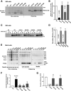
Asp (D) or Ala (A), but not Pro (P), substitution at PI Tyr-B16 does not affect folding and secretion of PI. A and B: 293 cells transfected with empty vector (EV) or plasmids encoding PI-WT, C(A7)Y, Y(B16)A, Y(B16)D, or Y(B16)P were metabolic labeled for 30 min without chase. The newly synthesized PI was IP by anti-insulin followed by analysis of tris-tricine-urea-SDS-PAGE under both nonreducing and reducing conditions. Native PI with correct disulfide pairing recovered under nonreducing conditions as well as total synthesized PI recovered under reducing conditions were quantified and analyzed in B (n = 3). The relative recovery of native PI in the cells expressing PI-WT was set to 100%. C: 293 cells transfected with plasmids encoding PI-WT, C(A7)Y, Y(B16)A, Y(B16)D, or Y(B16)P were metabolic labeled for 30 min followed by 0 or 2 h chase as indicated. The chase media (M) were collected and cells (C) were lysed. Biosynthesis and secretion of mutant PIs were analyzed by IP with anti-insulin followed by 4–12% NuPage. D: The results from C (n = 3) were quantified; the secretion efficiency of PI-WT and mutants was calculated, and that of PI-WT was set to 100%. E: Min6 cells were transiently transfected to express Myc-tagged PI-WT or mutants as indicated. Misfolded DLPCs in the cell lysates analyzed by 4–12% NuPage under both reducing and nonreducing conditions (left and middle panels). The media were incubated overnight and were used to analyzed secretion efficiency of PI-WT and mutants (right panel). *Refers to nonspecific bands. F: In three independent experiments shown in E, the disulfide-linked dimer and trimer under nonreducing condition and total PI-WT or mutants under reducing condition were quantified. The ratios (dimer + trimer/total PI) were calculated and that of PI-WT was set to 1. G: The secretion efficiency (media/cell lysates) of PI-WT and mutants from three independent experiments shown in E was calculated, and that of the PI-WT was set to 100%. ns, not significant.
Figure 3
Figure 3
Substitution of Tyr with Asp at PI B16 limits formation of heterodimerization and DLPCs. A: 293 cells were transfected with plasmids encoding untagged PI-WT, Myc-PI-WT, or PI mutants as indicated. Oxidative (Ox) folding, disulfide-linked PI dimers, and high-molecular-weight DLPCs were analyzed under nonreducing condition (left panel). Total amount of PI-WT and Myc-PI-WT or mutants were analyzed under reducing (Re) condition (right panel). B: Min6 cells were transfected with plasmids encoding Myc-tagged PI-WT or mutants as indicated. Oxidative folding of PI-WT and mutants were analyzed under nonreducing conditions (left panel). The heterodimer formed by Myc-tagged PI-mutant with endogenous PI is indicated by the arrow. The total amount of Myc-tagged PI-WT and mutants were analyzed under reducing condition (right panel). D, homo-dimers formed by untagged PI; D′ refers to homo-dimers formed by Myc-PI; H, hetero-dimers formed by untagged PI with Myc-PI.
Figure 4
Figure 4
Substitution of Tyr with Asp at PI B16 limits abnormal interactions between WT PI and diabetes causing C(A7)Y mutant PI. A: 293 cells were cotransfected with plasmids encoding untagged PI-WT, or GFP-tagged PI bearing the C(A7)Y mutation [GFP-PI-C(A7)Y], or GFP-tagged PI bearing both the C(A7)Y mutation as well as an intragenic suppressor mutation, Y(B16)D [GFP-PI-C(A7)Y/Y(B16)D] at a DNA ratio of 1:1. At 48 h after transfection, the cells were lysed in a co-IP buffer; 90% of the total lysates were IP with anti-GFP. IP GFP-PI and co-IP PI-WT were resolved in 4–12% NuPage under reducing condition along with 10% of the total lysates, transferred to the nitrocellulose membrane, and blotted with anti-human PI, which can recognize both untagged human PI and GFP-PI. B: The co-IP PI-WT and that in the total lysates were quantified using Image J. The percent of co-IP PI-WT in the total lysates was calculated and that of co-IP by GFP-PI-C(A7)Y set as 100%. C: Min6 cells transfected with plasmids encoding GFP-PI-C(A7)Y or GFP-PI-C(A7)Y/Y(B16)D were lysed in a co-IP buffer and IP with anti-GFP. The endogenous PI in the total lysates and co-IP with anti-GFP were resolved in 4–12% NuPage under reducing condition, transferred to the nitrocellulose membrane, and blotted with anti-mouse PI, which can recognize both endogenous mouse PI and GFP-PI. D: The co-IP endogenous PI and that in the total lysates were quantified using Image J. The percent of co-IP endogenous PI in the total lysates was calculated and that of co-IP by GFP-PI-C(A7)Y set as 100%.
Figure 5
Figure 5
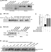
Mutations at PI B16 do not improve folding of diabetes causing PI-C(A7)Y but alleviate its transdominant-negative effect on coexpressed PI-WT. A: 293 cells cotransfected with indicated plasmids were labeled with 35Met/Cys for 30 min without chase. Biosynthesis and folding of newly synthesized PI-WT and mutants were analyzed by tris-tricine-urea-SDS-PAGE under both nonreducing and reducing conditions. B: 293 cells transfected and pulse-labeled as in A were chased 0 or 2 h. The chase media (M) were collected and cells (C) were lysed and analyzed by IP with anti-insulin followed by 4–12% NuPage under reducing conditions. C: Min6 cells were transfected with plasmids encoding Myc-tagged PI-WT or mutants as indicated. At 48 h after transfection, the media, which were cultured overnight, were collected, and the cells were lysed. The secretion efficiency of PI-WT and mutants was analyzed by Western blotting using anti-Myc. D: 293 cells were cotransfected with a plasmid encoding human PI-WT (human PI-WT) and either mouse PI-WT (mouse PI-WT) or mutants as indicated. The secretion of human PI-WT in the presence of mouse PI-WT or mutants was measured using human PI-specific RIA (mean ± SD, n = 3). E: 293 cells were cotransfected with a plasmid encoding untagged PI-WT and Myc-tagged PI-WT or mutants as indicated. At 48 h after transfection, the cells were labeled with 35Met/Cys for 30 min with 0 or 2 h chase. The cell lysates and chase media were collected and IP with anti-insulin. The secretion efficiency of untagged PI-WT in the presence or absence of Myc-PI-WT or mutants were analyzed under reducing conditions. proins, proinsulin.
Figure 6
Figure 6
PI B16 mutations function as an intragenic suppressor of the dominant-negative effects of MIDY PI-C(A7)Y and alleviate β-cell ER stress. A: INS1 cells were transfected with plasmids encoding GFP-tagged PI-WT and mutants as indicated. At 48 h after transfection, the cells were fixed and permeabilized. Confocal immunofluorescence microscopy was performed after double-labeling with anti-GFP (green) and anti-insulin (red). Whereas the cells expressing GFP-C(A7)Y exhibited a diminution of endogenous insulin staining (middle panels, white arrows), in the cells expressing a GFP-C(A7)Y/Y(B16)D endogenous insulin was restored (bottom panels, white arrowheads). B: The 30 residues of the B-chain of human PI, mouse PI 2 (Ins2), and mouse PI 1 (Ins1) were aligned. The highly conserved domain (B10-B28) that are involved PI dimerization is highlighted in yellow. C: 293 cells were cotransfected with plasmids encoding Myc-tagged human PI-WT with untagged mouse mutants. The media were cultured overnight and were collected at 48 h after transfection. Secretion of Myc-tagged human PI-WT in the presence of untagged mouse PI mutants was examined by Western blotting using anti-PI. D: Min6 cells were cotransfected with plasmids encoding human PI-WT with either mouse PI-WT or mutants at a DNA ratio of 1:3. At 48 h after transfection, human insulin content in the transfected cells was measured using human insulin-specific RIA (mean ± SD, n = 3). ***P < 0.0001 comparing the human insulin content in the cells expressing mouse PI-C(A7)Y with that of PI-WT and PI-C(A7)Y/Y(B16)D. E: Human islets were transduced with adenoviruses expressing Myc-tagged mouse PI-WT, C(A7)Y, or C(A7)Y/Y(B16)D, as indicated. At 48 h after infection, human insulin content in the infected human islets was measured by human insulin-specific ELISA. Human PI content was measured by densitometry of Western blotting. The relative contents of human PI and insulin in human islets infected with mouse PI-WT were set to 100%. ***P values comparing the human PI content (P = 0.0023) and human insulin content (P = 0.0004), respectively, in the islets infected with mouse PI-C(A7)Y with that of infected with PI-WT. F: Min6 cells were triple-transfected with plasmids encoding firefly luciferase driven by a BiP promoter, cytomegalovirus-renilla luciferase, and either PI-WT or mutants at a DNA ratio of 1:2:4. At 48 h after transfection, luciferase activities in transfected cells were measured as described in research design and methods (mean ± SD, n = 4). ***P < 0.0001 comparing BiP luciferase in the cells expressing PI-C(A7)Y with that of expressing PI-WT.
Figure 7
Figure 7
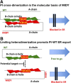
A working model of misfolded mutant PI attacking the dimerization surface of bystander PI-WT as a control point for PI ER export and insulin production. A: PI forms dimers in the ER. In the β-cells that coexpress PI-WT (blue) and misfolded mutant PI (red), the PI dimerization surface of the mutant can abnormally interact with that of the coexpressed bystander PI-WT, which impairs the folding and the ER export of PI-WT. B: Protecting the PI dimerization surface may serve as a potential theropeutic target to limit transdominant effects of misfolded mutant PI, alleviating ER stress and β-cell failure.

Similar articles

Cited by

References

    1. Liu M, Sun J, Cui J, et al. . INS-gene mutations: from genetics and beta cell biology to clinical disease. Mol Aspects Med 2015;42:3–18 - PMC - PubMed
    1. Liu M, Hodish I, Haataja L, et al. . Proinsulin misfolding and diabetes: mutant INS gene-induced diabetes of youth. Trends Endocrinol Metab 2010;21:652–659 - PMC - PubMed
    1. Colombo C, Porzio O, Liu M, et al. .; Early Onset Diabetes Study Group of the Italian Society of Pediatric Endocrinology and Diabetes (SIEDP) . Seven mutations in the human insulin gene linked to permanent neonatal/infancy-onset diabetes mellitus. J Clin Invest 2008;118:2148–2156 - PMC - PubMed
    1. Edghill EL, Flanagan SE, Patch A-M, et al. .; Neonatal Diabetes International Collaborative Group . Insulin mutation screening in 1,044 patients with diabetes: mutations in the INS gene are a common cause of neonatal diabetes but a rare cause of diabetes diagnosed in childhood or adulthood. Diabetes 2008;57:1034–1042 - PMC - PubMed
    1. Støy J, Edghill EL, Flanagan SE, et al. .; Neonatal Diabetes International Collaborative Group . Insulin gene mutations as a cause of permanent neonatal diabetes. Proc Natl Acad Sci U S A 2007;104:15040–15044 - PMC - PubMed

Publication types