. 2020 Mar 6:26:e919714.
doi: 10.12659/MSM.919714.

Long Non-Coding RNA Taurine Upregulated Gene 1 (TUG1) Downregulation Constrains Cell Proliferation and Invasion through Regulating Cell Division Cycle 42 (CDC42) Expression Via MiR-498 in Esophageal Squamous Cell Carcinoma Cells

Affiliations

Long Non-Coding RNA Taurine Upregulated Gene 1 (TUG1) Downregulation Constrains Cell Proliferation and Invasion through Regulating Cell Division Cycle 42 (CDC42) Expression Via MiR-498 in Esophageal Squamous Cell Carcinoma Cells

Zhifeng Wang et al. Med Sci Monit. .

Abstract

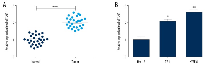
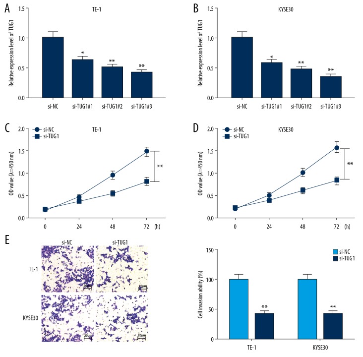
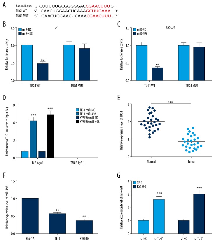
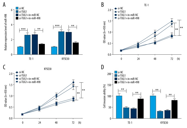
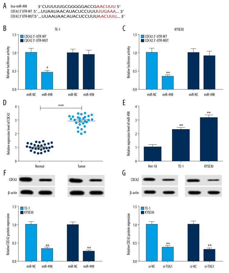
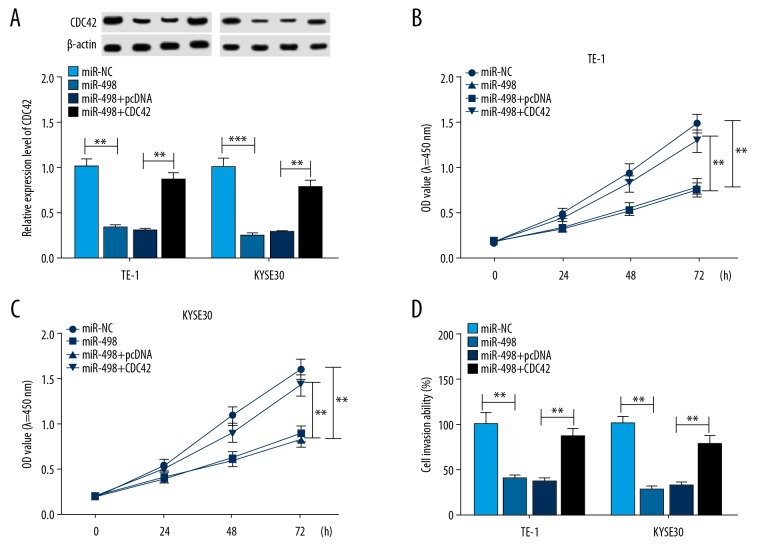
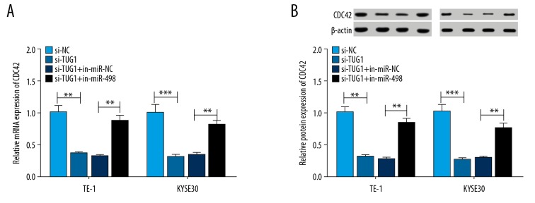
BACKGROUND Esophageal squamous cell carcinoma (ESCC) is a malignant tumor of the gastrointestinal tract. Taurine upregulated gene 1 (TUG1), a long non-coding (lnc) RNA, also known as LIN00080 or TI-227H, was connected with the tumorigenesis of various diseases. Hence, we plumed the role and molecular mechanism of TUG1 in the progression of ESCC. MATERIAL AND METHODS Expression patterns of TUG1, microRNA-498 (miR-498), and cell division cycle 42 (CDC42) mRNA were assessed using quantitative real time polymerase chain reaction (qRT-PCR). The expression level of CDC42 protein was evaluated via western blot analysis. Cell proliferation and invasion were determined with Cell Counting Kit-8 (CCK-8) assay or Transwell assay. The relationship between miR-498 and TUG1 or CDC42 was predicted by online bioinformatics database LncBase Predicted v.2 or microT-CDS and confirmed through dual-luciferase reporter system or RNA immunoprecipitation assay (RIP). RESULTS TUG1 and CDC42 were upregulated while miR-498 was strikingly decreased in ESCC tissues and cells (P<0.0001). Besides, TUG1 suppression blocked the proliferation and invasion of ESCC cells (P<0.001). Importantly, TUG1 decrease restrained CDC42 expression via binding to miR-498 in ESCC cells. Also, the suppressive impacts of TUG1 silencing on the proliferation and invasion of ESCC cells were mitigated by miR-498 reduction. Meanwhile, the repression of proliferation and invasion induced by miR-498 elevation was weakened by CDC42 overexpression. CONCLUSIONS Inhibition of TUG1 hampered cell proliferation and invasion by downregulating CDC42 via upregulating miR-498 in ESCC cells. Thus, TUG1 might be an underlying therapeutic target for ESCC.


Conflict of interest statement

Conflicts of interest

None.

Figures

Figure 1
Figure 1
Expression levels of TUG1 in ESCC tissues and cells. (A) The expression of TUG1 in ESCC tissues and adjoining normal esophageal tissues were analyzed using qRT-PCR. *** P<0.001 (B) qRT-PCR was applied to analyze TUG1 expression in ESCC and Het-1A cells. * P<0.05 and ** P<0.01. GAPDH was deemed as the endogenous for TUG1. Data are shown as a mean±SD from 3 independent experiments. Student’s t test assessed the significance of the differences. qRT-PCR – quantitative real time polymerase chain reaction; TUG1 – taurine upregulated gene 1; ESCC – esophageal squamous cell carcinoma; qRT-PCR – quantitative real-time polymerase chain reaction; GAPDH – glyceraldehyde 3-phosphate dehydrogenase; SD – standard deviation.
Figure 2
Figure 2
TUG1 silencing suppressed the proliferation and invasion of ESCC cells. (A, B) Knockdown efficiency of TUG1 was proved by qRT-PCR in TE-1 and KYSE30 cells. TE-1 and KYSE30 cells were transfected with 3 si-RNAs (si#TUG1-1, si#TUG2, si#TUG3) or si-NC. GAPDH was used as the endogenous for TUG1. * P<0.05 and ** P<0.01. (C, D) CCK-8 assay was employed to analyze the role of TUG1 knockdown on the proliferation of TE-1 and KYSE30 cells. ** P<0.01. (E) Transwell assay was employed to analyze the role of TUG1 downregulation on the invasion of TE-1 and KYSE30 cells. ** P<0.01. Data are shown as a mean±SD from 3 independent experiments. Student’s t-test was employed to analyze the significance of the differences. TUG1 – taurine upregulated gene 1; ESCC – esophageal squamous cell carcinoma; qRT-PCR – quantitative real-time polymerase chain reaction; GAPDH – glyceraldehyde 3-phosphate dehydrogenase; CCK-8 – Cell Counting Kit-8; SD – standard deviation.
Figure 3
Figure 3
TUG1 interacted with miR-498 in ESCC cells. (A) The binding sites between miR-498 and TUG1 were predicted by LncBase Predicted v.2. (B, C) The luciferase activity of TUG1-MUT or TUG1-WT in TE-1 and KYSE30 cells transfected with miR-498 or TUG1-WT was assessed with dual-luciferase reporter assay. ** P<0.01. (D) The interaction between miR-498 and TUG1 in TE-1 and KYSE30 cells was assessed through RIP assay. *** P<0.001. (E, F) qRT-PCR analysis of miR-498 expression levels in ESCC tissues and cells. *** P<0.001 and ** P<0.01. (G) The effect of TUG1 knockdown on the expression of miR-498 in TE-1 and KYSE30 cells was analyzed via qRT-PCR. *** P<0.001. U6 snRNA was used as the endogenous for miR-498. Data are shown as a mean±SD from 3 independent experiments. Student’s t-test assessed the significance of the differences. TUG1 – taurine upregulated gene 1; ESCC – esophageal squamous cell carcinoma; MUT – mutant; WT – wild type; RIP – RNA immunoprecipitation; qRT-PCR – quantitative real time polymerase chain reaction; SD – standard deviation.
Figure 4
Figure 4
Inhibition of miR-498 attenuated the effects of TUG1 knockdown on ESCC cells. (A) qRT-PCR was executed for the detection of the expression level of miR-498 in both TE-1 and KYSE30 cells. *** P<0.001 and ** P<0.01. (A–D) TE-1 and KYSE30 cells were transfected with si-NC, si-TUG1, si-TUG1+in-miR-498, or si-TUG1+in-NC, respectively. (B–D) CCK-8 or Transwell invasion assays analysis of the proliferation and invasion capacities of TE-1 and KYSE30 cells. ** P<0.01. U6 snRNA was used as the endogenous for miR-498. Data are shown as a mean±SD from 3 independent experiments. Student’s t-test assessed the significance of the differences. TUG1 – taurine upregulated gene 1; ESCC – esophageal squamous cell carcinoma; qRT-PCR – quantitative real time polymerase chain reaction; CCK-8 – Cell Counting Kit-8; SD – standard deviation.
Figure 5
Figure 5
MiR-498 targeted CDC42 in ESCC cells. (A) The predicted binding sites of CDC42 in miR-498 by microT-CDS. (B, C) The luciferase activity of CDC42 3′URT-MUT or CDC42 3′URT-WT was estimated in TE-1 and KYSE30 cells transfected with miR-498 or miR-NC using dual-luciferase reporter assay. * P<0.05 and ** P<0.01. (D, E) qRT-PCR was utilized for the detection of the level of CDC42 mRNA in ESCC tissues and cells. *** P<0.001 and ** P<0.01. GAPDH was used as the endogenous for CDC42. (E) Western blot analysis of the role of miR-498 on CDC42 protein expression. ** P<0.01. (F) Western blot analysis of the effect of knockdown of TUG1 on CDC42 protein expression. ** P<0.01. Data are shown as a mean±SD from 3 independent experiments. Student’s t-test assessed the significance of the differences. CDC42 – cell division cycle 42; ESCC – esophageal squamous cell carcinoma; MUT – mutant; WT – wild type; qRT-PCR – quantitative real time polymerase chain reaction; GAPDH – glyceraldehyde 3-phosphate dehydrogenase; SD – standard deviation; TUG1 – taurine upregulated gene 1.
Figure 6
Figure 6
CDC42 overexpression recovered the effect of miR-498 on ESCC cells. (A) Western blot analysis of CDC42 protein expression level in TE-1 and KYSE30 cells. ** P<0.01. (A–D) The miR-NC, miR-498, miR-498+pcDNA, or miR-498+CDC42TE-1 was transfected into KYSE30 cells, respectively. (B–D) CCK-8 or Transwell assays were carried out for the assessment of proliferation and invasion of TE-1 and KYSE30 cells. ** P<0.01. Data are shown as a mean±SD from 3 independent experiments. Student’s t-test assessed the significance of the differences. CDC42 – cell division cycle 42; ESCC – esophageal squamous cell carcinoma; CCK-8 – Cell Counting Kit-8; SD – standard deviation.
Figure 7
Figure 7
Knockdown of TUG1 downregulated CDC42 via miR-498 in ESCC cells. (A) qRT-PCR was executed for evaluation of the expression of CDC42 in TE-1 and KYSE30 cells. ** P<0.01. GAPDH was used as the endogenous for CDC42. (A, B) The si-NC, si-TUG, si-TUG1+in-miR-NC, or si-TUG1+in-miR-498TE-1 was transfected into TE-1 and KYSE30 cells, respectively. (B) Western blot analysis was conducted for the assessment of CDC42 protein expression in TE-1 and KYSE30 cells. ** P<0.01. Data are shown as a mean±SD from 3 independent experiments. Student’s t-test assessed the significance of the differences. TUG1 – taurine upregulated gene 1; CDC42 – cell division cycle 42; ESCC – esophageal squamous cell carcinoma; GAPDH – glyceraldehyde 3-phosphate dehydrogenase; qRT-PCR – quantitative real time polymerase chain reaction; SD – standard deviation.

Similar articles

Cited by

References

    1. Kollarova H, Machova L, Horakova D, et al. Epidemiology of esophageal cancer – an overview article. Biomed Pap Med Fac Univ Palacky Olomouc Czech Repub. 2007;151:17–20. - PubMed
    1. Siegel RL, Miller KD, Jemal A. Cancer statistics, 2015. Cancer J Clin. 2015;65:5–29. - PubMed
    1. Pennathur A, Gibson MK, Jobe BA, et al. Oesophageal carcinoma. Lancet. 2013;381:400–12. - PubMed
    1. Rustgi AK, El-Serag HB. Esophageal carcinoma. N Engl J Med. 2014;371:2499–509. - PubMed
    1. Zhu ZJ, Hu Y, Zhao YF, et al. Early recurrence and death after esophagectomy in patients with esophageal squamous cell carcinoma. Ann Thorac Surg. 2011;91:1502–8. - PubMed