. 2020 Mar 5;10(1):4099.
doi: 10.1038/s41598-020-60966-8.

Effects of chloromethylisothiazolinone/methylisothiazolinone (CMIT/MIT) on Th2/Th17-related immune modulation in an atopic dermatitis mouse model

Affiliations

Effects of chloromethylisothiazolinone/methylisothiazolinone (CMIT/MIT) on Th2/Th17-related immune modulation in an atopic dermatitis mouse model

Han-Na Go et al. Sci Rep. .

Abstract

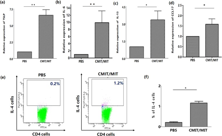
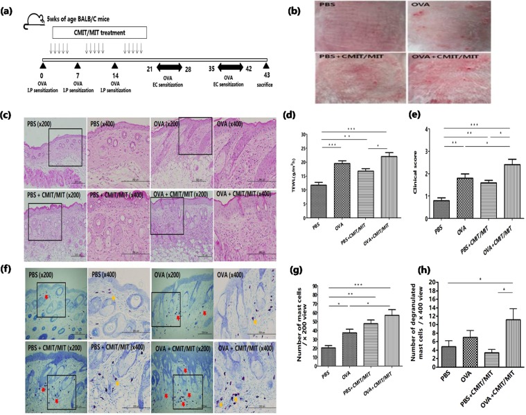
Exposure to chloromethylisothiazolinone/methylisothiazolinone (CMIT/MIT) has been associated with allergic contact dermatitis and occupational asthma. Despite this association however, no study has investigated the effects of CMIT/MIT exposure on the development of atopic dermatitis (AD). This study was conducted to investigate the influence of epicutaneous exposure to CMIT/MIT on AD in a mouse model and the underlying biological mechanisms. BALB/C mice were exposed to CMIT/MIT for 3 weeks and AD was developed using ovalbumin (OVA) epidermal sensitization. CMIT/MIT epicutaneous exposure in normal mice significantly enhanced AD-like phenotypes (e.g., transepidermal water loss, clinical score, total serum immunoglobulin E level and infiltration of inflammatory cells). In addition, CMIT/MIT exposure significantly augmented the mRNA expression level of T helper (Th) 2-related cytokines (thymic stromal lymphopoietin, interleukin (IL)-6 and IL-13), Th2 chemokine (chemokine (C-C motif) ligand 17) and the population of CD4+IL-4+ cells in the skin. Moreover, mice exposed to CMIT/MIT in the OVA challenge had greater AD-like phenotypes, higher IL-4 and IL-17A skin mRNA expression levels, and a larger population of CD4+IL-4+- and IL-17A+-producing cells in the skin-draining lymph nodes. Our current findings in a mouse model thus suggest that CMIT/MIT exposure may cause AD symptoms through the dysregulation of Th2/Th17-related immune responses.


Conflict of interest statement

The authors declare no competing interests.

Figures

Figure 1
Figure 1
Effects of CMIT/MIT on skin lesions and histology, the epidermal permeability barrier, total IgE serum levels and the number of mast cells in the skin of normal mice. (a) Experimental protocol for the effects of CMIT/MIT exposure in normal mice. (b) Representative images of typical skin lesions. (c) H&E staining. (d) TEWL measurements. (e) Serum levels of total IgE determined by ELISA. (f) Toluidine Blue staining of skin samples. The numbers of mast cells (g) and degranulated mast cells (h) per high-power field were counted. Statistical significance was determined using a t-test. Red and yellow arrows denote the mast cells within the dermis. *P < 0.05, **P < 0.01.
Figure 2
Figure 2
Effects of CMIT/MIT on Th2-related cytokines (TSLP, IL-6 and IL-13), a Th2-related chemokine (CCL17) and CD4+IL-4+ cell populations in normal mice. Skin mRNA levels of (a) TSLP, (b) IL-6, (c) IL-13 and (d) CCL17 were assessed by real-time PCR. (e,f) Frequency of IL-4 with CD4+ cells was assessed by flow cytometry in the skin-draining lymph nodes. Statistical significance was determined using a t-test. *P < 0.05, **P < 0.01.
Figure 3
Figure 3
Effects of CMIT/MIT exposure in an AD mouse model. (a) Experimental protocol for assessing the effects of CMIT/MIT exposure in AD mice. (b) Representative images of typical skin lesions. (c) Histological analysis of H&E-stained skin tissue. (d) TEWL measurements. (e) Clinical score measurements. (f) Toluidine blue staining of skin samples. Numbers of mast cells (g) and degranulated mast cells (h) per high-power field were counted. Red and yellow arrows denote the mast cells within the dermis. Statistical significance was determined using ANOVA and a Newman-Keuls multiple comparison test. *P < 0.05, **P < 0.01, ***P < 0.001. I.P, intraperitoneal; EC, epicutaneous.
Figure 4
Figure 4
Effects of CMIT/MIT on serum IgE levels and Th2-/Th17-related responses in an AD mouse model. (a,b) ELISA determinations of (a) the total serum IgE levels and (b) the OVA-specific serum IgE levels. (cf) Skin mRNA expression, assessed by real-time PCR, of (c) TSLP, (d,e) Th2-related cytokines (d, IL-4; e, IL-13) and (f) the Th17-related cytokine, IL-17A. Statistical significance was determined using ANOVA and the Newman-Keuls multiple comparison test. *P < 0.05, **P < 0.01, ***P < 0.001. ND, non-detection.
Figure 5
Figure 5
Effects of CMIT/MIT exposure on (a,b) CD4+IL-17A+ and (a,c) CD4+ IL-4+ cell populations in the skin-draining lymph nodes in the AD mouse model. Statistical significance was determined using ANOVA and the Newman-Keuls multiple comparison test. *P < 0.05, **P < 0.01, ***P < 0.001.

Similar articles

Cited by

References

    1. Novak N, Bieber T, Leung DY. Immune mechanisms leading to atopic dermatitis. J. Allergy Clin. Immunol. 2003;112:S128–139. doi: 10.1016/j.jaci.2003.09.032. - DOI - PubMed
    1. Li C, et al. Development of atopic dermatitis-like skin disease from the chronic loss of epidermal caspase-8. Proc. Natl Acad. Sci. USA. 2010;107:22249–22254. doi: 10.1073/pnas.1009751108. - DOI - PMC - PubMed
    1. Cork Michael J., Robinson Darren A., Vasilopoulos Yiannis, Ferguson Adam, Moustafa Manar, MacGowan Alice, Duff Gordon W., Ward Simon J., Tazi-Ahnini Rachid. New perspectives on epidermal barrier dysfunction in atopic dermatitis: Gene–environment interactions. Journal of Allergy and Clinical Immunology. 2006;118(1):3–21. doi: 10.1016/j.jaci.2006.04.042. - DOI - PubMed
    1. Kim K. Influences of Environmental Chemicals on Atopic Dermatitis. Toxicol. Res. 2015;31:89–96. doi: 10.5487/TR.2015.31.2.089. - DOI - PMC - PubMed
    1. Kathuria P, Silverberg JI. Association of pollution and climate with atopic eczema in US children. Pediatr. Allergy Immunol. 2016;27:478–485. doi: 10.1111/pai.12543. - DOI - PubMed

Publication types