Review
. 2020 Mar;37(1):97-102.
doi: 10.1055/s-0039-3401845. Epub 2020 Mar 4.

Update: Splenic Artery Embolization in Blunt Abdominal Trauma

Affiliations
Review

Update: Splenic Artery Embolization in Blunt Abdominal Trauma

Mangaladevi S Patil et al. Semin Intervent Radiol. 2020 Mar.

Abstract

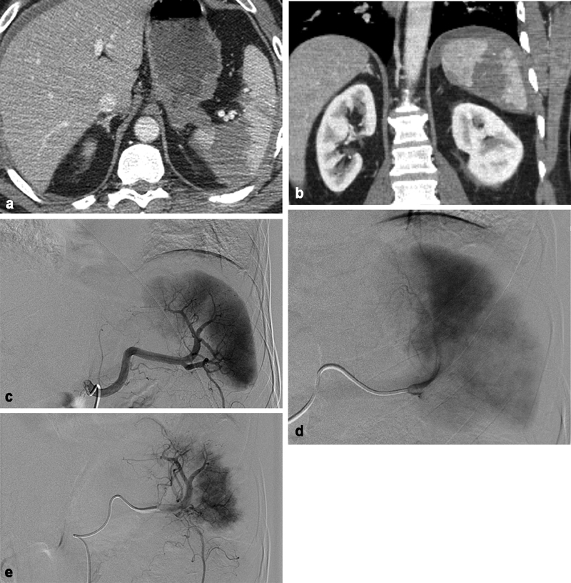
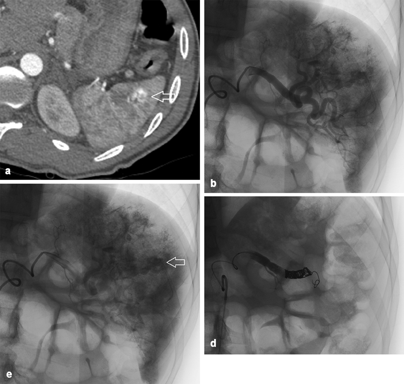
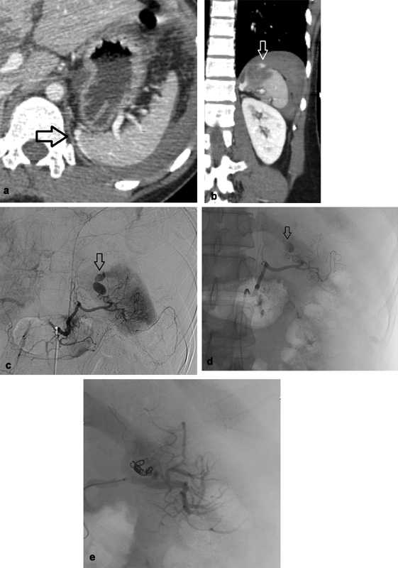
The spleen is the most commonly injured organ after blunt abdominal trauma. Nonoperative management with splenic arterial embolization (SAE) is the current standard of care for hemodynamically stable patients. Current data favor the use of proximal and coil embolization techniques in adults, while observation is suggested in the pediatric population. In this review, the authors describe the most recent evidence informing the clinical indications, techniques, and complications for SAE.

Keywords: blunt splenic injury; interventional radiology; splenic arterial embolization; trauma.


Conflict of interest statement

Conflict of Interest None of the authors have conflicts.

Figures

Fig. 1
Fig. 1
A 50-year-old man who fell from 20 feet. ( a ) Axial CT of the abdomen and ( b ) coronal CT of the abdomen showing extensive splenic contusion/laceration with perisplenic hematoma consistent with a grade IV injury. ( c ) Early-phase splenic angiography. ( d ) Late-phase splenic angiography demonstrating multiple segmental perfusion defects, correlating to known splenic lacerations. Faint petechial contrast pooling in the region of the parenchymal defect. ( e ) Postembolization angiography following Gelfoam embolization from the distal main splenic artery with resultant sluggish flow and peripheral vascular pruning.
Fig. 2
Fig. 2
A 24-year-old individual involved in pedestrian–automobile collision. ( a ) Axial CT showing an arterial blush in the lateral spleen ( arrow ) adjacent to multiple vessels suggestive of a pseudoaneurysm. ( b ) Splenic angiography showing extravascular contrast at multiple sites. ( c ) Delayed imaging on angiography shows contrast pooling ( arrow on one such collection), representative of multifocal pseudoaneurysms. ( d ) Angiography posttreatment. Given the multifocal findings, it was determined to embolize the spleen proximally using multiple coils beyond the dorsal pancreatic artery. Complete vascular occlusion demonstrated.
Fig. 3
Fig. 3
A 29-year-old man involved in motor vehicle crash. ( a ) Axial CT abdomen and ( b ) coronal CT abdomen showing small focal laceration along the superior medial aspect of the spleen with extravasation of contrast ( arrow ). ( c ) Celiac angiography. ( d ) Splenic angiography demonstrating a pseudoaneurysm along the medial aspect of the spleen with active extravasation into the peritoneum ( arrow ). ( e ) Embolization of a distal splenic artery branch supplying the pseudoaneurysm was performed with multiple coils and small aliquots of Gelfoam slurry resulting in no flow seen past the area of embolization, no evidence of remaining pseudoaneurysm or extravasation.

References

    1. Ahuja C, Farsad K, Chadha M. An overview of splenic embolization. AJR Am J Roentgenol. 2015;205(04):720–725. - PubMed
    1. Sclafani S J. The role of angiographic hemostasis in salvage of the injured spleen. Radiology. 1981;141(03):645–650. - PubMed
    1. Bisharat N, Omari H, Lavi I, Raz R. Risk of infection and death among post-splenectomy patients. J Infect. 2001;43(03):182–186. - PubMed
    1. Imbrogno B F, Ray C E. Splenic artery embolization in blunt trauma. Semin Intervent Radiol. 2012;29(02):147–149. - PMC - PubMed
    1. Organ injury scaling 2018 update: spleen, liver, and kidney: erratum. J Trauma Acute Care Surg. 2019;87(02):512. - PubMed