

Case Reports
doi: 10.1016/j.radcr.2019.12.025.
eCollection 2020 May.
Pulmonary adenomyoma presenting as a right cardiophrenic angle mass
Affiliations
- PMID: 32140196
- PMCID: PMC7044680
- DOI: 10.1016/j.radcr.2019.12.025
Item in Clipboard
Case Reports
Pulmonary adenomyoma presenting as a right cardiophrenic angle mass
Radiol Case Rep.
.
Abstract
Pulmonary adenomyomas are rare adenomyomatous hamartomas. In the few cases described in the literature, these benign tumors are encapsulated by lung parenchyma. We describe a case of a 59 year-old woman with acetylcholine receptor antibody-negative myasthenia gravis and a right cardiophrenic mass initially thought to be a thymoma. Histopathology surprisingly revealed a pulmonary adenomyoma which involved the mediastinal fat at the cardiophrenic angle.
Keywords: Adenofibroma; Harmatoma; Myasthenia gravis; Pulmonary adenomyoma; Solitary fibrous tumor; Thymoma.
© 2020 The Authors.
Figures

Top row: Preoperative contrast-enhanced CT of the chest obtained 2 years before surgery in axial soft tissue window (left), lung window (middle), and coronal soft tissue (right) reconstructions. An asterisk (*) denotes the right cardiophrenic pulmonary adenomyoma. Middle row: Preoperative contrast-enhanced CT of the chest obtained just before surgery (2 years after the images shown in “Top row”). Images are provided in axial soft tissue window (left), lung window (middle), and coronal soft tissue (right) reconstructions. An asterisk (*) denotes the right cardiophrenic pulmonary adenomyoma. Bottom row: Postoperative noncontrast CT of the chest in axial soft tissue window (left), lung window (middle), and coronal soft tissue (right) reconstructions. Hyperdense material at the right cardiophrenic space are surgical clips/sutures from wedge resection of the right middle lobe.
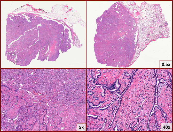
Low magnification (top panels) photomicrographs of H&E stained sections show a well-circumscribed mass that is predominantly extrapulmonary with a fibrous capsule, and focally appears connected with lung parenchyma. On high magnification (bottom panels) a biphasic histology with 2 cell populations is evident: epithelial cuboidal lining cells with minimal atypia and a variably cellular underlying stromal population. There is a papillary configuration with some large pseudopapillae and solid areas.

A panel of immunohistochemical stains show that the lining epithelial cells are positive for Cam5.2 (cytokeratin), TTF-1, and Napsin-A. The stromal cells are positive for smooth muscle actin (SMA) and desmin.
References
-
- Carter B.W., Benveniste M.F., Madan R., Godoy M.C., de Groot P.M., Truong M.T. ITMIG classification of mediastinal compartments and multidisciplinary approach to mediastinal masses. Radiographics. 2017;37(2):413–436. - PubMed
-
- Pinkus G.S., Kurtin P.J. Epithelial membrane antigen—a diagnostic discriminant in surgical pathology: immunohistochemical profile in epithelial, mesenchymal, and hematopoietic neoplasms using paraffin sections and monoclonal antibodies. Hum Pathol. 1985;16(9):929–940. - PubMed
-
- Turner B.M., Cagle P.T., Sainz I.M., Fukuoka J., Shen S.S., Jagirdar J. Napsin A, a new marker for lung adenocarcinoma, is complementary and more sensitive and specific than thyroid transcription factor 1 in the differential diagnosis of primary pulmonary carcinoma: evaluation of 1674 cases by tissue microarray. Arch Pathol Lab Med. 2012;136(2):163–171. - PubMed
Publication types
Grants and funding
LinkOut - more resources
Full Text Sources
