

Case Reports
doi: 10.1089/ped.2019.1065.
Epub 2019 Dec 11.
Endobronchial Glomus Tumor in a Child
Affiliations
- PMID: 32140287
- PMCID: PMC7057049
- DOI: 10.1089/ped.2019.1065
Item in Clipboard
Case Reports
Endobronchial Glomus Tumor in a Child
Pediatr Allergy Immunol Pulmonol.
.
Abstract
Glomus tumors (GTs) are rare, usually benign, mesenchymal neoplasms typically located in the cutaneous tissues of the extremities. Visceral locations have been reported in ∼5% of cases. The average age at diagnosis is 42 years. GTs originating in the respiratory tract of pediatric patients are exceedingly rare. We report a 16-year-old male with a GT of the right lower lobe bronchus.
Keywords: glomus tumor; histology; lung; pediatric; soft tissue tumors.
Copyright 2019, Mary Ann Liebert, Inc., publishers.
Conflict of interest statement
No competing financial interests exist.
Figures
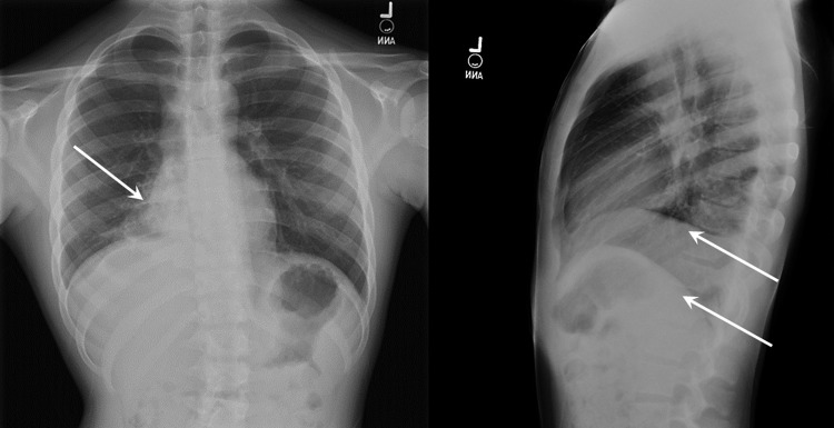
Chest radiographs demonstrating consolidative process involving portions of right middle and lower lobes (single arrow). A sub-pulmonic process on the right suggests a pleural effusion or pleural reactivity (double arrows).

Axial computed tomography image (A) demonstrates superior margin of enhancing intraluminal mass distal to the bronchus intermedius (arrow). (B) Demonstrates 1.4 × 1.6 × 2.2 mass completely obstructing lower lobe bronchus (arrow) with postobstructive pneumonia and bronchiectasis.

(A) Tumor within the lumen and wall of the bronchus. Normal bronchial mucosa is on the left. H&E; 20 × . (B) Tumor is lobulated with sheaths of uniform cells with distinct membranes, moderate amphophilic cytoplasm, round nuclei with dispersed chromatin; some cells have visible small nucleoli. H&E; 200 × . (C) Smooth muscle actin shows positive cytoplasmic staining by immunohistochemistry; 400 × . (D) CD34 stain; showing richly vascular tumor; 100 × . H&E, hematoxylin and eosin.
References
-
- Goldblum JR, Folpe AL, Weiss SW. Perivascular tumors. In: Goldblum JR, Folpe AL, Wess SW, eds. Enzinger and Weiss's soft tissue tumors, sixth ed., Philadelphia, PA: Elsevier, 2014, pp. 749–765
-
- Gombos Z, Zhang PJ. Glomus tumor. Arch Pathol Lab Med 2008; 132:1448–1452 - PubMed
-
- Folpe AL, Fanburg-Smith JC, Miettinen M, et al. . Atypical and malignant glomus tumors: analysis of 52 cases, with a proposal for the reclassification of glomus tumors. Am J Surg Pathol 2001; 25:1–12 - PubMed
-
- Chou T, Pan SC, Shieh SJ, et al. . Glomus tumor: twenty-year experience and literature review. Ann Plast Surg 2016; 76 Suppl 1:S35–S40 - PubMed
-
- Gaertner EM, Steinberg DM, Huber M, et al. . Pulmonary and mediastinal glomus tumors—report of five cases including a pulmonary glomangiosarcoma: a clinicopathologic study with literature review. Am J Surg Pathol 2000; 24:1105–1114 - PubMed
Publication types
LinkOut - more resources
Full Text Sources
Miscellaneous
