

Review
doi: 10.7759/cureus.6813.
A Review of Benign Hepatic Tumors and Their Imaging Characteristics
Affiliations
- PMID: 32140369
- PMCID: PMC7047931
- DOI: 10.7759/cureus.6813
Item in Clipboard
Review
A Review of Benign Hepatic Tumors and Their Imaging Characteristics
Cureus.
.
Abstract
This paper concisely reviews the benign hepatic tumors most commonly encountered by clinicians. It includes the epidemiology, pathology, and imaging characteristics of hepatic hemangiomas, focal nodular hyperplasia (FNH), and hepatic adenomas (HAs).
Keywords: focal nodular hyperplasia; hepatic adenoma; hepatic hemangioma; hepatic tumor.
Copyright © 2020, Patacsil et al.
Conflict of interest statement
The authors have declared that no competing interests exist.
Figures

A. Sagittal ultrasound image of the liver demonstrating a hyperechoic mass within the liver with increased posterior through transmission. B. Doppler evaluation of this mass reveals no significant internal flow

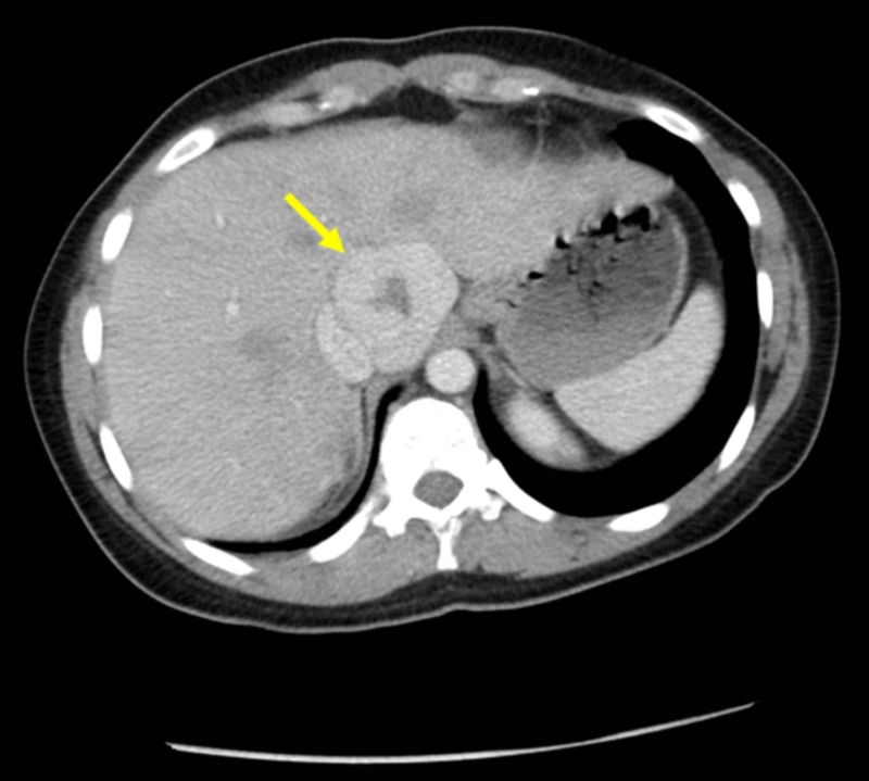
CT: computed tomography A. Late arterial phase axial CT image in the same patient demonstrates discontinuous peripheral nodular enhancement (arrow) – classic for hepatic cavernous hemangiomas. B. Portal venous phase axial CT image at a more superior slice more clearly demonstrates the enhancement pattern (arrow). C. Venous phase axial CT image demonstrates slow centripetal filling (arrow). D. Portal venous phase coronal CT image provided to demonstrate location of hemangioma (arrow)
CT: computed tomography Contrast-enhanced CT in portal venous phase demonstrates an enhancing mass with central non-enhancement. Imaging characteristics favor focal nodular hyperplasia

MRI: magnetic resonance imaging Contrast-enhanced (hepatobiliary agent) MRI of the same patient. A. T1 axial MR image demonstrates T1 isointense signal peripherally with a central focus of T1 hypointensity. B. T2 axial MRI demonstrates central T2 hyperintensity with relatively decreased signal in the periphery. C. Contrast-enhanced T1 MRI in bolus phase demonstrates peripheral enhancement of the mass. D. Contrast-enhanced T1 MR image in delayed/hepatobiliary phase demonstrates persistent contrast enhancement. Imaging characteristics favor FNH and confirm CT findings

CT: computed tomography CT image of a 38-year-old female on oral contraceptives. Portal venous phase contrast-enhanced axial CT image demonstrates a multilobulated mass with heterogeneous enhancement. This was a biopsy-proven adenoma

MRI: magnetic resonance imaging Contrast-enhanced (hepatobiliary agent) MRI from the same patient as in Figure 5. A. The mass demonstrates T1 hypointensity (arrow). B. There are scattered regions of T2 hyperintensity within the mass. C. Contrast-enhanced T1 bolus phase demonstrates early arterial enhancement. D. Contrast-enhanced T1 hepatobiliary phase image demonstrates relative wash-out of the mass compared to surrounding hepatic parenchyma. Enhancement pattern is consistent with a benign adenoma in this young female
References
-
- Prevalence and clinical outcome of hepatic haemangioma with specific reference to the risk of rupture: a large retrospective cross-sectional study. Mocchegiani F, Vincenzi P, Coletta M, et al. Dig Liver Dis. 2016;48:309–314. - PubMed
-
- Liver haemangioma: common and uncommon findings and how to improve the differential diagnosis. Caseiro-Alves F, Brito J, Araujo AE, et al. Eur Radiol. 2007;17:1544–1554. - PubMed
-
- Prevalence of hepatic hemangioma in patients with focal nodular hyperplasia: MR imaging analysis. Vilgrain V, Uzan F, Brancatelli G, Federle MP, Zappa M, Menu Y. Radiology. 2003;229:75–79. - PubMed
Publication types
LinkOut - more resources
Full Text Sources