

mTOR pathway somatic variants and the molecular pathogenesis of hemimegalencephaly
- PMID: 32140648
- PMCID: PMC7049797
- DOI: 10.1002/epi4.12377
mTOR pathway somatic variants and the molecular pathogenesis of hemimegalencephaly
Abstract
Objectives: Recently, defects in the protein kinase mTOR (mammalian target of rapamycin) and its associated pathway have been correlated with hemimegalencephaly (HME). mTOR acts as a central regulator of important physiological cellular functions such as growth and proliferation, metabolism, autophagy, death, and survival. This study was aimed at identifying specific variants in mTOR signaling pathway genes in patients diagnosed with HME.
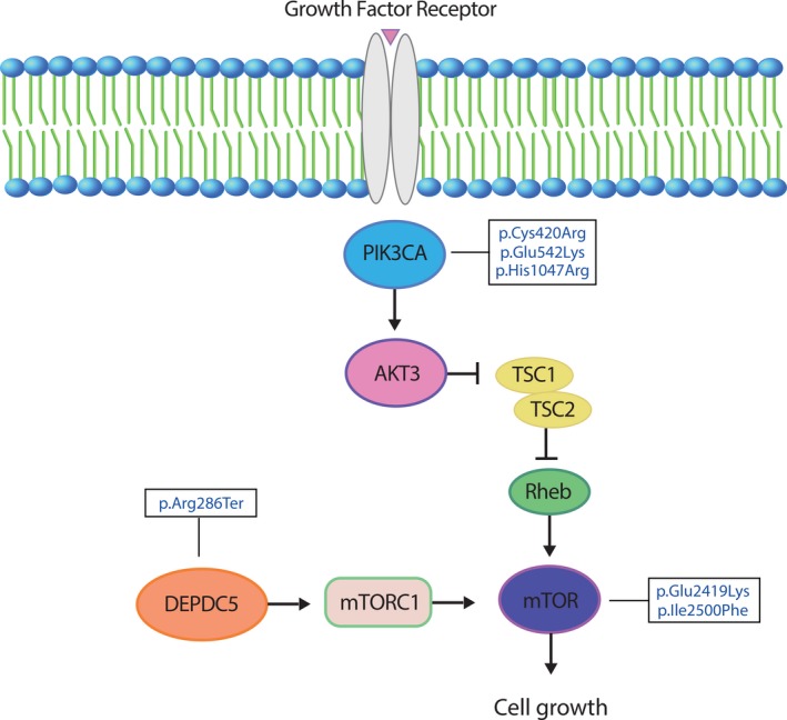
Methods: Using amplicon and whole exome sequencing (WES) of resected brain and paired blood samples from five HME patients, we were able to identify pathogenic mosaic variants in the mTOR pathway genes MTOR, PIK3CA, and DEPDC5.
Results: These results strengthen the hypothesis that somatic variants in PI3K-Akt-mTOR pathway genes contribute to HME. We also describe one patient presenting with a pathogenic variant on DEPDC5 gene, which reinforces the role of DEPDC5 on cortical structural changes due to mTORC1 hyperactivation. These findings also provide insights into when in brain development these variants occurred. An early developmental variant is expected to affect a larger number of cells and to result in a larger malformation, whereas the same variant occurring later in development would cause a minor malformation.
Significance: In the future, numerous somatic variants in known or new genes will undoubtedly be revealed in resected brain samples, making it possible to draw correlations between genotypes and phenotypes and allow for a genetic clinical diagnosis that may help to predict a given patient's outcome.
Keywords: epilepsy; hemimegalencephaly; mTOR.
© 2020 The Authors. Epilepsia Open published by Wiley Periodicals Inc. on behalf of International League Against Epilepsy.
Conflict of interest statement
We thank the support provided by the Research Support Foundation (FAEPA) of the University Hospital of Ribeirão Preto Medical School and the Center of Genomic Medicine, University Hospital, Ribeirão Preto Medical School (CMG/HCFMRP) for patient recruitment and sample preparation. This work was supported by grants from the Howard Hughes Medical Institute (to JGG), and Camila B. Garcia was recipient of a scholarship from the National Support Program for the Health Care of Persons with Disabilities (PRONAS/PCD) and Coordination of Improvement of Higher Education Personnel‐Brazil (CAPES). The authors confirm that they have read the journal's position on issues involved in ethical publication and affirm that this report is consistent with those guidelines.
Figures
References
-
- Di Rocco C, Battaglia D, Pietrini D, Piastra M, Massimi L. Hemimegalencephaly: clinical implications and surgical treatment. Childs Nerv Syst. 2006;22:852–66. - PubMed
-
- Becker AJ, Urbach H, Scheffler B, Al‐Falaki R, Cronshaw M, Parker S, et al. Focal cortical dysplasia of Taylor's balloon cell type: mutational analysis of the TSC1 gene indicates a pathogenic relationship to tuberous sclerosis. Ann Neurol. 2002;52:29–37. - PubMed
LinkOut - more resources
Full Text Sources
Molecular Biology Databases
Miscellaneous
