. 2020 Jul;107(1):31-40.
doi: 10.1007/s00223-020-00682-7. Epub 2020 Mar 5.

Wnt16 Overexpression in Osteoblasts Increases the Subchondral Bone Mass but has no Impact on Osteoarthritis in Young Adult Female Mice

Affiliations

Wnt16 Overexpression in Osteoblasts Increases the Subchondral Bone Mass but has no Impact on Osteoarthritis in Young Adult Female Mice

Anna E Törnqvist et al. Calcif Tissue Int. 2020 Jul.

Abstract

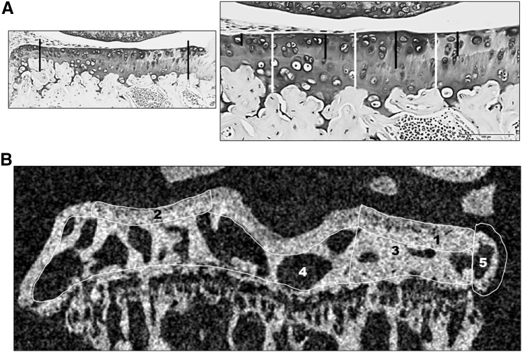
Epidemiological studies have shown that high bone mineral density (BMD) is associated with an increased risk of osteoarthritis (OA), but the causality of this relationship remains unclear. Both bone mass and OA have been associated with the WNT signaling pathway in genetic studies, there is thus an interest in studying molecular partners of the WNT signaling pathway and OA. Female mice overexpressing WNT16 in osteoblasts (Obl-Wnt16 mice) have an increased bone mass. We aimed to evaluate if the high bone mass in Obl-Wnt16 mice leads to a more severe experimental OA development than in WT control mice. We induced experimental OA in female Obl-Wnt16 and WT control mice by destabilizing the medial meniscus (DMM). The Obl-Wnt16 mice displayed thicker medial and lateral subchondral bone plates as well as increased subchondral trabecular bone volume/tissue volume (BV/TV) but un-altered thickness of articular cartilage compared to WT mice. After DMM surgery, there was no difference in OA severity in the articular cartilage in the knee joint between the Obl-Wnt16 and WT mice. Both the Obl-Wnt16 and WT mice developed osteophytes in the DMM-operated tibia to a similar extent. We conclude that although the Obl-Wnt16 female mice have a high subchondral bone mass due to increased WNT signaling, they do not exhibit a more severe OA phenotype than their WT controls. This demonstrates that high bone mass does not result in an increased risk of OA per se.

Keywords: Cartilage; DMM; Mouse model; Osteoarthritis; WNT16.


Conflict of interest statement

Anna E. Törnqvist, Louise Grahnemo, Karin H. Nilsson, Thomas Funck-Brentano, Claes Ohlsson and Sofia Movérare-Skrtic declare that they have no conflict of interest.

Figures

Fig. 1
Fig. 1
Measurements of articular cartilage and subchondral bone in tibia. a Photos of the articular cartilage. The left photo shows two black lines between which the measurements were made (magnification × 10). The right photo (magnification × 20, black scale bar 100 µm) shows how the measurements of the un-calcified cartilage (black lines) and the total cartilage thickness (white lines) were made. b Image in the coronal plane of a right DMM-operated tibia from the µCT. The regions of interest are the medial subchondral bone plate (1), the lateral subchondral bone plate (2), the medial subchondral trabecular bone (3), the total subchondral trabecular bone (3 + 4), and the osteophyte (5)
Fig. 2
Fig. 2
The subchondral bone. A mixed-design two-way repeated-measures ANOVA was used to assess the effects of genotype (WT vs. Obl-Wnt16) and surgery (un-operated [Ctrl] vs. surgery destabilizing the medial meniscus [DMM]) on the subchondral bone. a The subchondral bone plate thickness in the medial and lateral tibia was affected by the genotype, where Obl-Wnt16 mice had thicker bone plates than the WT mice. The DMM surgery had an effect on the medial, but not the lateral, subchondral bone plate, where the surgery led to increased bone plate thickness. b The medial and total subchondral bone volume/tissue volume (BV/TV) was affected both by genotype and by surgery. The Obl-Wnt16 mice had higher BV/TV than WT mice and the surgery led to increased BV/TV. c The medial subchondral trabecular number (Tb.N) was affected both by genotype and by surgery, whereas the total subchondral Tb.N was only affected by genotype. The Obl-Wnt16 mice had a higher medial and total subchondral Tb.N than the WT mice and the DMM surgery led to a higher Tb.N on the medial side. d The medial and total subchondral trabecular thickness (Tb.Th) was decreased after DMM surgery, while the genotype had no effect. e Micro-CT reconstructions show subchondral bone, mainly in the tibia, of the left un-operated control knees (control) and the right knees that underwent DMM surgery. The arrows point at osteophytes. Data are shown as scatter plots where the bars show the mean (N = 15/group)
Fig. 3
Fig. 3
OARSI scores for un-operated control knees and knees that underwent DMM surgery. A mixed-design two-way repeated-measures ANOVA was used to assess the effects of genotype (WT vs. Obl-Wnt16) and surgery (un-operated [Ctrl] vs. surgery destabilizing the medial meniscus [DMM]) on OARSI scores. a The summed OARSI scores for each compartment (medial = medial tibial plateau [MTP] + medial femoral condyle [MFC]; lateral = lateral tibial plateau [LTP] + lateral femoral condyle [LFC]), where the maximum score for each compartment is 12. The surgery but not the genotype affected the summed medial and lateral OARSI score. b The surgery but not the genotype affected the OARSI score in the MTP and in the MFC. The maximum score for each region is 6. c The surgery but not the genotype affected the OARSI score in the LFC. The maximum score for each region is 6. d Representative microphotographs of the MTP and MFC in un-operated control (Ctrl) and DMM-operated knees in Obl-Wnt16 and WT mice. Especially the MTP in the DMM-operated knees show severe cartilage damage both in the Obl-Wnt16 and WT mice. Data are shown as scatter plots where the bars show the mean (summed lateral compartment and LFC in Obl-Wnt16 mice N = 11; LTP in Obl-Wnt16 mice N = 14; summed lateral compartment and LFC in WT mice N = 13; all other parameters N = 15/group)

References

    1. Bergink AP, Rivadeneira F, Bierma-Zeinstra SM, et al. Are Bone Mineral density and fractures related to the incidence and progression of radiographic osteoarthritis of the knee, hip, and hand in elderly men and women? The Rotterdam study. Arthritis Rheumatol. 2019;71:361–369. doi: 10.1002/art.40735. - DOI - PubMed
    1. Funck-Brentano T, Nethander M, Moverare-Skrtic S, et al. Causal factors for knee, hip and hand osteoarthritis: a Mendelian randomization study in the UK Biobank. Arthritis Rheumatol. 2019;71(10):1634–1641. doi: 10.1002/art.40928. - DOI - PMC - PubMed
    1. Hart DJ, Cronin C, Daniels M, et al. The relationship of bone density and fracture to incident and progressive radiographic osteoarthritis of the knee: the Chingford Study. Arthritis Rheum. 2002;46:92–99. doi: 10.1002/1529-0131(200201)46:1<92::AID-ART10057>3.0.CO;2-#. - DOI - PubMed
    1. Nevitt MC, Zhang Y, Javaid MK, et al. High systemic bone mineral density increases the risk of incident knee OA and joint space narrowing, but not radiographic progression of existing knee OA: the MOST study. Ann Rheum Dis. 2010;69:163–168. doi: 10.1136/ard.2008.099531. - DOI - PMC - PubMed
    1. Baron R, Kneissel M. WNT signaling in bone homeostasis and disease: from human mutations to treatments. Nat Med. 2013;19:179–192. doi: 10.1038/nm.3074. - DOI - PubMed

Publication types