

Bilateral spontaneous thrombosis of the pampiniform plexus mimicking incarcerated inguinal hernia: case report of a rare condition and literature review
- PMID: 32140844
- PMCID: PMC7058780
- DOI: 10.1186/s40792-020-00810-3
Bilateral spontaneous thrombosis of the pampiniform plexus mimicking incarcerated inguinal hernia: case report of a rare condition and literature review
Erratum in
-
Correction to: Bilateral spontaneous thrombosis of the pampiniform plexus mimicking incarcerated inguinal hernia: case report of a rare condition and literature review.Surg Case Rep. 2020 Mar 23;6(1):56. doi: 10.1186/s40792-020-00821-0. Surg Case Rep. 2020. PMID: 32206942 Free PMC article.
Abstract
Background: Pampiniform plexus thrombosis is a very rare disease (only less than 25 published cases are available till date), and it is a diagnostic dilemma. The present case is an unusual condition of an elderly gentleman who was finally diagnosed as a case of spontaneous thrombosis of bilateral pampiniform plexus and was managed conservatively. Literature was reviewed to explore potential etiologies and therapeutic strategies.
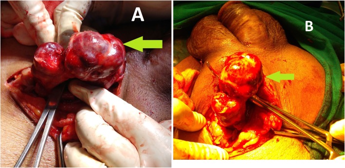
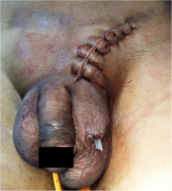
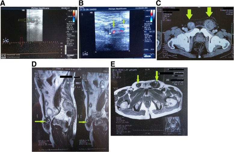
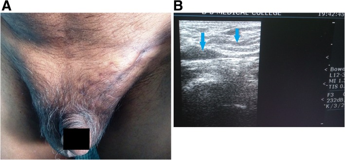
Case presentation: A 65-year-old afebrile gentleman, laborer (in brick industry), and non-smoker with no previous major health problems was admitted with swelling in the bilateral inguinal region. The swelling had started one and half months ago. He had developed severe pain over the swelling for last 1 day with tenderness and indurations. Neither he had history of previous surgeries, chronic cough, dysuria, prostatism, and trauma nor he presented any thrombogenic factors. There was no history of vomiting, abdominal pain, and obstipation. Physical examination revealed normotensive person with BMI of 22.5, was significant only for one tender, movable, and firm to hard 10 cm × 3 cm mass extending from the left deep inguinal ring up to the upper pole of the testis in the scrotum. Another 5 cm × 3 cm mass of similar characteristics was found extending from deep inguinal ring up to the root of the scrotum on right side. The testes and prostate were normal on palpation. On the contrary to preoperative USG, which clinched suspicion of incarcerated inguinal hernia, a thrombosed pampiniform plexus without any evidence of hernia sac was found on the left side during inguino-scrotal exploration. Wound was closed without doing any further procedure. Contralateral inguino-scrotal exploration was spared considering same nature of disease. Postoperative Doppler ultrasonography confirmed the diagnosis of bilateral thrombosed pampiniform plexus. MDCT of whole abdomen revealed no abnormality other than bilateral spermatic cord thrombosis. Blood thrombophilia screening came normal. The subject had an uneventful postoperative hospital course. With 2 years of follow-up, the gentleman is doing well, remaining asymptomatic and had returned to his usual life.
Conclusions: Due to extreme rarity, spontaneous thrombosis of the pampiniform plexus may be a diagnostic dilemma and requires a high index of suspicion. Doppler ultrasound is the initial investigation of choice. In the absence of other concomitant disease, beginning the treatment conservatively instead of excising the thrombosed segment is more suitable.
Keywords: Incarcerated inguinal hernia; Pampiniform plexus; Thrombosis.
Conflict of interest statement
The author declares that he has no competing interests.
Figures
References
-
- L. Hashimoto Brett Vibeto. Spontaneous thrombosis of the pampiniform plexus. Scand J Urol Nephrol, 40 (2006), pp. 252–254. - PubMed
-
- Kleinclauss F, Della Negra E, Martin M, et al. Spontaneous thrombosis of left varicocele. Prog Urol. 2001;11:95–96. - PubMed
-
- Campagnola S, Flessati P, Fasoli L, et al. A rare case of acute scrotum. Thrombophlebitis from ectasia of the left pampiniforme plexus. Minerva Uroll Nephrol. 1999;51:163–165. - PubMed
LinkOut - more resources
Full Text Sources
