. 2020 May;29(5):1172-1185.
doi: 10.1002/pro.3850. Epub 2020 Mar 25.

Ab initio folding of a trefoil-fold motif reveals structural similarity with a β-propeller blade motif

Affiliations

Ab initio folding of a trefoil-fold motif reveals structural similarity with a β-propeller blade motif

Connie A Tenorio et al. Protein Sci. 2020 May.

Abstract

Many protein architectures exhibit evidence of internal rotational symmetry postulated to be the result of gene duplication/fusion events involving a primordial polypeptide motif. A common feature of such structures is a domain-swapped arrangement at the interface of the N- and C-termini motifs and postulated to provide cooperative interactions that promote folding and stability. De novo designed symmetric protein architectures have demonstrated an ability to accommodate circular permutation of the N- and C-termini in the overall architecture; however, the folding requirement of the primordial motif is poorly understood, and tolerance to circular permutation is essentially unknown. The β-trefoil protein fold is a threefold-symmetric architecture where the repeating ~42-mer "trefoil-fold" motif assembles via a domain-swapped arrangement. The trefoil-fold structure in isolation exposes considerable hydrophobic area that is otherwise buried in the intact β-trefoil trimeric assembly. The trefoil-fold sequence is not predicted to adopt the trefoil-fold architecture in ab initio folding studies; rather, the predicted fold is closely related to a compact "blade" motif from the β-propeller architecture. Expression of a trefoil-fold sequence and circular permutants shows that only the wild-type N-terminal motif definition yields an intact β-trefoil trimeric assembly, while permutants yield monomers. The results elucidate the folding requirements of the primordial trefoil-fold motif, and also suggest that this motif may sample a compact conformation that limits hydrophobic residue exposure, contains key trefoil-fold structural features, but is more structurally homologous to a β-propeller blade motif.

Keywords: domain swapping; folding pathway; protein evolution; protein symmetry.


Conflict of interest statement

M.B. is a cofounder and has equity ownership in Trefoil Therapeutics, Inc.

Figures

Figure 1
Figure 1
The β‐trefoil and Monofoil trefoil‐fold structural features. (a) Ribbon diagram of the Symfoil‐4P de novo designed symmetric β‐trefoil protein (research collaboratory for structural bioinformatics (RCSB) accession http://firstglance.jmol.org/fg.htm?mol=304D). The view is down the threefold axis of rotational symmetry (indicated by triangle), and the repeating motif is termed a “trefoil‐fold”. (b) A similar representation for the homotrimer assembly of the Monofoil trefoil‐fold polypeptide (RCSB accession http://firstglance.jmol.org/fg.htm?mol=3OL0). The individual Monofoil polypeptides are indicated by different colors. (c) The Monofoil trimer as in panel b, but with a view normal to the axis of symmetry. (d) An isolated Monofoil polypeptide “outside face” (i.e., a side view as in panel c). (e) A space‐filling representation of d, and colored to indicate side chain properties (gray is hydrophobic, red is acidic, blue is basic, orange is polar). (f) A view of the Monofoil polypeptide “inside face” (i.e., panel d from the opposite direction). (g) Space‐filling representation of panel f (note the extensive hydrophobic character of the buried inside face)
Figure 2
Figure 2
Size exclusion chromatography (SEC) of Symfoil‐4P, Monofoil and permutant P1 and P3 polypeptides. Analytical concentrations of proteins were loaded onto a Superdex 75 column and eluted with Pi buffer. Mass standards included bovine serum albumin (BSA) (66.5 kDa), carbonic anhydrase (29.0 kDa) and cytochrome C (12.3 kDa). Monofoil was also resolved in Pi buffer with 6 M GuHCl denaturant. GuHCl, guanidine hydrochloride
Figure 3
Figure 3
Isothermal equilibrium denaturation (IED) of Monofoil and permutant P3 polypeptides. Upper panel: the IED data for Monofoil showing cooperative unfolding with indicated thermodynamic parameters. Lower panel: the IED data for permutant P3 showing non‐cooperative unfolding behavior, additionally, unlike Monofoil the permutant P3 polypeptide exhibits increased fluorescence signal with increasing denaturant concentration
Figure 4
Figure 4
The primary structure of Symfoil‐4P, Monofoil and Monofoil P1, P2, and P3 circular permutants. The three repeating trefoil‐fold motifs in Symfoil‐4P are indicated by color shading (single letter amino acid code is used). Locations of β‐strand secondary structure are indicated with underline. The single Monofoil trefoil‐fold was constructed by introducing a stop codon at position 53 in Symfoil‐4P (indicated by gray shading). Circular permutants P1, P2, and P3 of the Monofoil sequence were constructed at positions within surface turn positions and outside the four β‐strand regions (following a previous convention 17). Their primary structure relationship to Monofoil is indicated by color shading
Figure 5
Figure 5
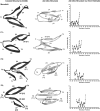
Monofoil, P1, P2, and P3 crystal structure, ab initio predicted structure, and ab initio Cα error estimates. Left column: Ribbon diagram (side view) of the Cα coordinates of residue positions 10–52 (Monofoil), 19–60 (P1), 28–69 (P2), and 38–78 (P3) from the Symfoil‐4P crystal structure (research collaboratory for structural bioinformatics (RCSB) accession 3O4D) (see Figure 4). Regions of β‐strand secondary structure are indicated by an arrow. Locations of the N‐ and C‐termini, β‐strands, and turns are also indicated. Central column: a similar ribbon diagram for the predicted ab initio structure for each polypeptide. Right column: Cα error estimate for the respective ab initio structure. The location of the individual β‐strands is indicated. Due to the primary structure symmetry β5 = β1, β6 = β2, and β7 = β3 in the circular permutations (see Figure 4)
Figure 6
Figure 6
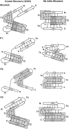
Regions of structural similarity between the ab initio and crystal structures of Monofoil and permutant sequences. The diagram shows the general secondary structure arrangement for the Monofoil and permutant sequences based upon the Symfoil‐4P crystal structure (left column) and the ab initio structure (right column). β‐Strands are indicated by arrows and interstrand H‐bonds by dashed lines. Due to the exact primary structure symmetry turns T1 and T5 are equivalent, turns T1 and T6 are equivalent, and so on. The shaded regions indicate subdomains that overlay with rmsd <1.5 Å for the set of Cα positions (see Section 2)
Figure 7
Figure 7
Monofoil and Monofoil P3 ab initio structures overlaid onto http://firstglance.jmol.org/fg.htm?mol=1UTC β‐propeller subdomains. (a) A ribbon diagram of the Monofoil ab initio structure (blue) overlaid onto the fifth blade motif of clathrin terminal domain (research collaboratory for structural bioinformatics (RCSB) accession http://firstglance.jmol.org/fg.htm?mol=1UTC). (b) A ribbon diagram of the Monofoil P3 ab initio structure (green) overlaid onto the second blade motif of clathrin terminal domain (RCSB accession http://firstglance.jmol.org/fg.htm?mol=1UTC). (c) A ribbon diagram of http://firstglance.jmol.org/fg.htm?mol=1UTC clathrin terminal domain (view down the pseudoaxis of rotational symmetry) with the locations of the overlays of Monfoil and Monofoil P3 ab initio structures (same color scheme as in panels a and b). RCSB, Research Collaboratory for Structural Bioinformatics

Similar articles

Cited by

References

    1. Lang D, Thoma R, Henn‐Sax M, Sterner R, Wilmanns M. Structural evidence for evolution of the beta/alpha barrel scaffold by gene duplication and fusion. Science. 2000;289:1546–1550. - PubMed
    1. Richter M, Bosnali M, Carstensen L, et al. Computational and experimental evidence for the evolution of a (βα)8‐barrel protein from an ancestral quarter‐barrel stabilized by disulfide bonds. J Mol Biol. 2010;398:763–773. - PubMed
    1. Fülöp V, Jones DT. β propellers: Structural rigidity and functional diversity. Curr Opin Struct Biol. 1999;9:715–721. - PubMed
    1. Chaudhuri I, Soding J, Lupas AN. Evolution of the β‐propeller fold. Proteins. 2008;71:795–803. - PubMed
    1. Voet ARD, Noguchi H, Addy C, et al. Computational design of a self‐assembling symmetrical β‐propeller protein. Proc Natl Acad Sci U S A. 2014;111:15102–15107. - PMC - PubMed

Publication types

Substances

LinkOut - more resources