Review
. 2020 Jun;11(3):619-635.
doi: 10.1002/jcsm.12528. Epub 2020 Mar 6.

Cancer cachexia and its pathophysiology: links with sarcopenia, anorexia and asthenia

Affiliations
Review

Cancer cachexia and its pathophysiology: links with sarcopenia, anorexia and asthenia

Sara Peixoto da Silva et al. J Cachexia Sarcopenia Muscle. 2020 Jun.

Abstract

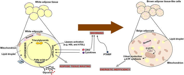
Cancer cachexia is a multifactorial syndrome characterized by a progressive loss of skeletal muscle mass, along with adipose tissue wasting, systemic inflammation and other metabolic abnormalities leading to functional impairment. Cancer cachexia has long been recognized as a direct cause of complications in cancer patients, reducing quality of life and worsening disease outcomes. Some related conditions, like sarcopenia (age-related muscle wasting), anorexia (appetite loss) and asthenia (reduced muscular strength and fatigue), share some key features with cancer cachexia, such as weakness and systemic inflammation. Understanding the interplay and the differences between these conditions is critical to advance basic and translational research in this field, improving the accuracy of diagnosis and contributing to finally achieve effective therapies for affected patients.

Keywords: Anorexia; Asthenia; Cachexia; Cancer; Muscle wasting; Sarcopenia.


Conflict of interest statement

None declared.

The manuscript does not contain clinical studies or patient data.

Figures

Figure 1
Figure 1
Molecular signalling involved in muscle wasting during cancer cachexia. Inflammatory mediators, such as pro‐inflammatory cytokines (interleukin‐1 and tumour necrosis factor‐α) and myostatin, and proteolysis‐inducing factor (PIF), derived from the tumour and/or immune cells, activate intracellular signals. Cytokines and PIF, through nuclear factor‐kappa B (NF‐κB), activate forkhead box O (FOXO) leading to increased transcription of ubiquitin ligase genes—Atrogin 1 and muscle RING finger‐containing protein 1 (MURF1)—that promote muscle protein degradation. The activation of p38 and Janus kinase/mitogen‐activated protein kinase (JAK/MAPK) cascades by PIF, cytokines and myostatin, leads to apoptosis mediated by caspases. Myostatin can also activate protein degradation through FOXOs. Additionally, myostatin may decrease protein synthesis, inhibiting protein kinase B (AKT) through SMAD. Insulin‐like growth factor‐1 (IGF‐1) is decreased during muscle wasting, suppressing the IGF‐1 pathway (dashed lines) and therefore inhibiting protein synthesis. Peroxisome proliferator‐activated receptor‐γ co‐activator 1α (PGC1α) increases uncoupling protein (UCP) expression, leading to mitochondrial dysfunction. The consumption of high levels of amino acids, such as glutamine, by the tumour increases protein breakdown in skeletal muscle, contributing to cancer cachexia. PIFR, PIF receptor; ACTRIIB, activin receptor type IIB; IGF1R, insulin‐like growth factor‐1 receptor; PI3K, phosphatidylinositol 3‐kinase; mTOR, mammalian target of rapamycin; UPR, ubiquitin‐mediated proteasome degradation; REE, resting energy expenditure
Figure 2
Figure 2
Impaired regeneration capacity during cancer cachexia. Satellite cells are dysregulated during cancer cachexia: although they are able to be activated and proliferate, they cannot complete their differentiation process, because of persistent expression of Paired box 7 (PAX7), via nuclear factor‐kappa B (NF‐κB) activation. PAX7 negatively regulates MyoD and myogenin, which mediate differentiation
Figure 3
Figure 3
Adipose tissue lipolysis and browning during cancer cachexia. In cancer cachexia, adipose tissue wasting is observed. High levels of circulating free fatty acids (FFA) and glycerol are observed, because of a massive lipolysis in white adipose tissue (WAT), promoted by lipases activation, zinc‐α2‐glycoprotein (ZAG) and cytokines. Additionally, these high levels of FFA may also result from lipoprotein lipase (LPL) decreased activity that reduces lipogenesis (inhibition represented by the dashed line). Moreover, WAT can acquire features of brown adipose cells, a process called ‘WAT browning’. In these beige adipocytes, uncoupling protein 1 (UCP1) is expressed, promoting uncoupling mitochondrial respiration. This results in heat production and less ATP synthesis, leading to an energetic inefficiency. This browning can be promoted by cytokines, ZAG and tumoural‐derived compounds such as parathyroid‐hormone‐related protein (PTHRP). HSL, hormone‐sensitive lipase; ATGL, adipose triglycerides lipase
Figure 4
Figure 4
Links and overlaps between related conditions. Inflammation, weakness and fatigue are features of cachexia, sarcopenia, anorexia and asthenia. *Cachexia can occur with or without loss of appetite and fat mass

References

    1. Katz AM, Katz PB. Diseases of the heart in the works of Hippocrates. Br Heart J. 1962;24:257–264. - PMC - PubMed
    1. Fearon K, Strasser F, Anker SD, Bosaeus I, Bruera E, Fainsinger RL, et al. Definition and classification of cancer cachexia: an international consensus. Lancet Oncol. 2011;12:489–495. - PubMed
    1. Argiles JM, Busquets S, Stemmler B, Lopez‐Soriano FJ. Cancer cachexia: understanding the molecular basis. Nat Rev Cancer. 2014;14:754–762. - PubMed
    1. Schmidt SF, Rohm M, Herzig S, Berriel DM. Cancer Cachexia: More Than Skeletal Muscle Wasting. Trends Cancer. 2018;4:849–860. - PubMed
    1. Tisdale MJ. Cachexia in cancer patients. Nat Rev Cancer. 2002;2:862–871. - PubMed

Publication types