. 2020 May 13;12(19):21192-21200.
doi: 10.1021/acsami.9b22621. Epub 2020 Mar 6.

Submicrometer-Sized Roughness Suppresses Bacteria Adhesion

Affiliations

Submicrometer-Sized Roughness Suppresses Bacteria Adhesion

Noemí Encinas et al. ACS Appl Mater Interfaces. .

Abstract

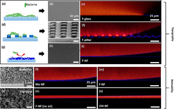
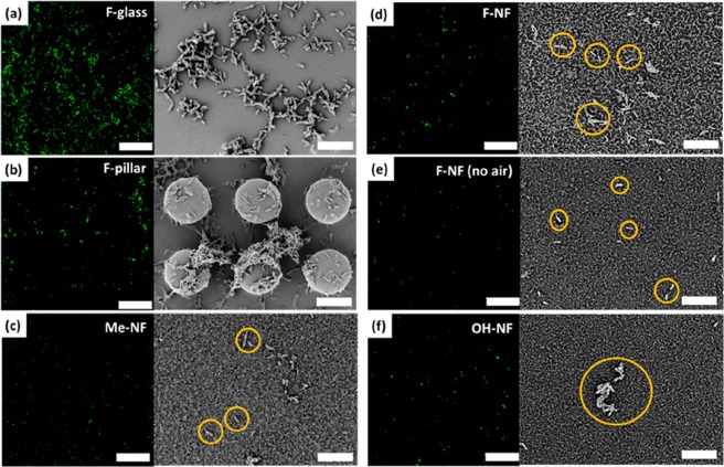
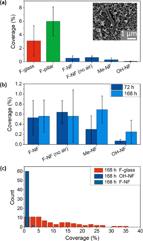
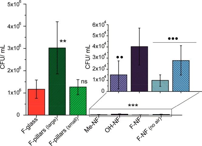
Biofilm formation is most commonly combatted with antibiotics or biocides. However, proven toxicity and increasing resistance of bacteria increase the need for alternative strategies to prevent adhesion of bacteria to surfaces. Chemical modification of the surfaces by tethering of functional polymer brushes or films provides a route toward antifouling coatings. Furthermore, nanorough or superhydrophobic surfaces can delay biofilm formation. Here we show that submicrometer-sized roughness can outweigh surface chemistry by testing the adhesion of E. coli to surfaces of different topography and wettability over long exposure times (>7 days). Gram-negative and positive bacterial strains are tested for comparison. We show that an irregular three-dimensional layer of silicone nanofilaments suppresses bacterial adhesion, both in the presence and absence of an air cushion. We hypothesize that a 3D topography can delay biofilm formation (i) if bacteria do not fit into the pores of the coating or (ii) if bending of the bacteria is required to adhere. Thus, such a 3D topography offers an underestimated possibility to design antibacterial surfaces that do not require biocides or antibiotics.

Keywords: antifouling; bacterial size; biofouling; roughness; silicone nanofilaments.


Conflict of interest statement

The authors declare no competing financial interest.

Figures

Figure 1
Figure 1
(a, d, g) Schemes of bacteria (green) attaching on (a) flat surface, (d) SU-8 micropillar arrays, and (g) surface coated with silicone nanofilaments. For simplicity, the bacteria are drawn straight. (b, e, h) Corresponding scanning electron microscope (SEM) micrographs of the surfaces are displayed in gray (scale bar, 10 μm). (c, f, i, l–o) Wetting properties were investigated by laser scanning confocal microscopy (LSCM) using an inverted microscope (Leica TCS SP8 SMD) and a 40× water immersion objective. Red, dyed medium for bacteria cultivation; blue reflection of light from the glass, culture medium; and air, culture medium interface; black, air, glass substrate, or coating (scale bar, 25 μm). (j, k) Cross-section SEM image and top view of a nanofilament-coated sample (dcale bar: 1 μm).
Figure 2
Figure 2
Biofilm formation investigated by LSCM and SEM after 72 h of incubation at 37 °C. The bacterial suspension was incubated on the following surfaces: (a) fluorinated flat glass, (b) fluorinated micropillar array with 5 μm height and 7 μm of spacing and 13 μm of diameter (image shows the coverage with bacteria (green spots) close to the pillars’ top surface (see Figures S2 and S3 for enlarged versions of the confocal images), (c) methyl-terminated nanofilaments (d) fluorinated nanofilaments, (e) fluorinated nanofilament coating where the air cushion was removed prior to incubation, and (f) plasma-activated nanofilament coating (some of the bacterial cells on the silicone nanofilaments in c–f are highlighted in yellow circles to enhance the contrast with the surface background) The E. coli were exposed to l-arabinose for expression of the green fluorescent protein, which can be excited at 488 nm. Scale bar of LSCM, 50 μm; scale bar of SEM, 10 μm.
Figure 3
Figure 3
(a) Coverage area based on SEM images (Table S2, Figures S4 and S8a) of different surfaces after 72 h of incubation with E. coli. F-glass, fluorinated glass surfaces, F-pillar, superhydrophobic micropillar arrays, F-NF, fluorinated nanofilaments in the presence of an air cushion; F-NF (no-air), fully wetted fluorinated filaments; Me-NF, methyl-terminated nanofilaments; OH-NF, plasma-activated nanofilaments. The inset SEM image shows E. coli bacterial cells attached to a surface coated with nanofilaments (Figure S8b). (b) Comparison of coverage of adhered bacteria on incubation duration for surfaces coated with nanofilaments showing different surface functionalities in the presence and absence of an air cushion. (c) Histogram based on the number of evaluated SEM images, showing the coverage on three surfaces: fluorinated glass, fully wetted fluorinated nanofilament-coated surface, and superhydrophilic nanofilament-coated surface (see also Figure S9). The y-axis stands for the number of bacteria aggregates of a certain size.
Figure 4
Figure 4
CFU/mL of E. coli for differently treated surfaces. The sample surfaces were incubated for 168 h at 37 °C. The inset shows the CFU/mL for the surfaces coated with silicone nanofilaments at enlarged magnification. F-pillar: diameter = 13 μm, height = 5 μm, pillar–pillar distance = 20 μm. F-pillar 2: diameter = 5 μm, height = 5 μm, pillar–pillar distance = 10 μm. Six independent samples were prepared. Results, standard deviations, and statistical paired t test (95, 99, and 99.9% confidence levels) are calculated from independent experiments, using as calculated probabilities (p): *p < 0.05; **p < 0.01, and ***p < 0.001 as significant differences; ns corresponds to no difference. Asterisks denote comparison between F-glass, pillars, and all the nanofilament samples (main plot), whereas circles mark the comparison between OH-NF and the rest of the nanofilaments (inset).
Figure 5
Figure 5
SEM images after incubating with Micrococcus luteus a for 3 days: (a) fluorinated glass (F-glass), (b) fluorinated nanofilament coating (F-NF), (c) methyl-terminated nanofilament (Me-NF), and (d) plasma-activated nanofilament (OH-NF). Scale bar, 10 μm; scale bar of inset SEM image, 1 μm.

References

    1. Klevens R. M.; Edwards J. R.; Richards C. L.; Horan T. C.; Gaynes R. P.; Pollock D. A.; Cardo D. M. Estimating Health Care-Associated Infections and Deaths in U.S. Hospitals, 2002. Public Health Rep. 2007, 122, 160–166. 10.1177/003335490712200205. - DOI - PMC - PubMed
    1. Chmielewski R. A. N.; Frank J. F. Biofilm Formation and Control in Food Processing Facilities. Compr. Rev. Food Sci. Food Saf. 2003, 2, 22–32. 10.1111/j.1541-4337.2003.tb00012.x. - DOI - PubMed
    1. Dinh H. T.; Kuever J.; Mußmann M.; Hassel A. W.; Stratmann M.; Widdel F. Iron Corrosion by Novel Anaerobic Microorganisms. Nature 2004, 427, 829–832. 10.1038/nature02321. - DOI - PubMed
    1. Enning D.; Venzlaff H.; Garrelfs J.; Dinh H. T.; Meyer V.; Mayrhofer K.; Hassel A. W.; Stratmann M.; Widdel F. Marine Sulfate-reducing Bacteria Cause Serious Corrosion of Iron under Electroconductive Biogenic Mineral Crust. Environ. Microbiol. 2012, 14, 1772–1787. 10.1111/j.1462-2920.2012.02778.x. - DOI - PMC - PubMed
    1. Hall-Stoodley L.; Costerton J. W.; Stoodley P. Bacterial Biofilms: from the Natural Environment to Infectious Diseases. Nat. Rev. Microbiol. 2004, 2, 95–108. 10.1038/nrmicro821. - DOI - PubMed

MeSH terms

LinkOut - more resources