

A Benchtop Automated Sputum-to-Genotype System Using a Lab-on-a-Film Assembly for Detection of Multidrug-Resistant Mycobacterium tuberculosis
- PMID: 32142258
- PMCID: PMC7354060
- DOI: 10.1021/acs.analchem.9b05853
A Benchtop Automated Sputum-to-Genotype System Using a Lab-on-a-Film Assembly for Detection of Multidrug-Resistant Mycobacterium tuberculosis
Abstract
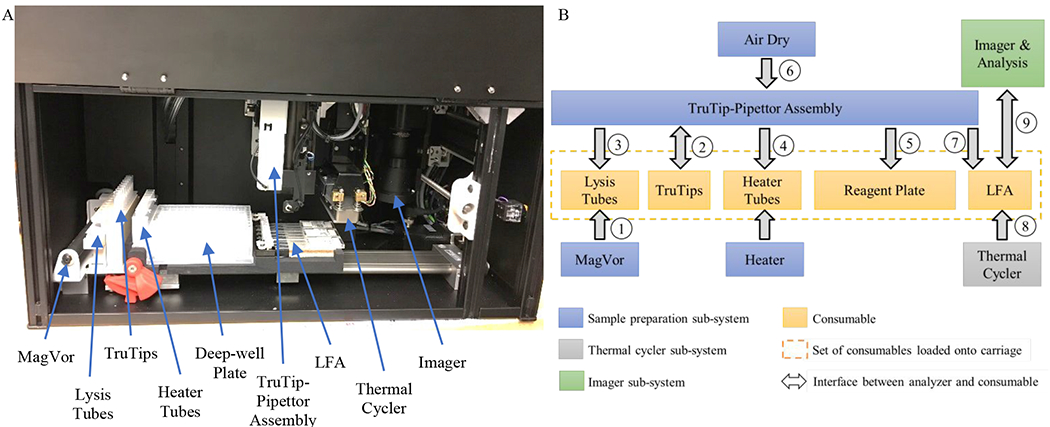
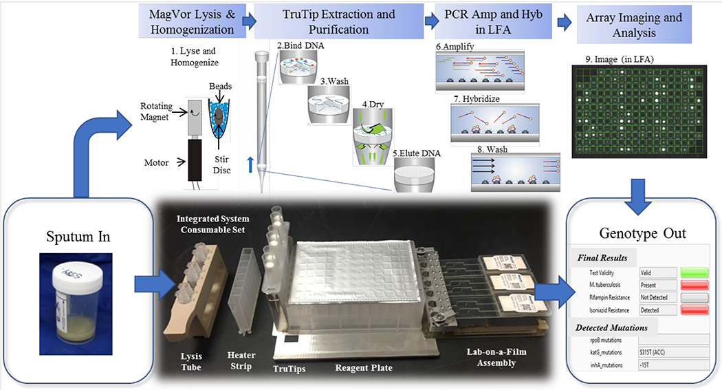
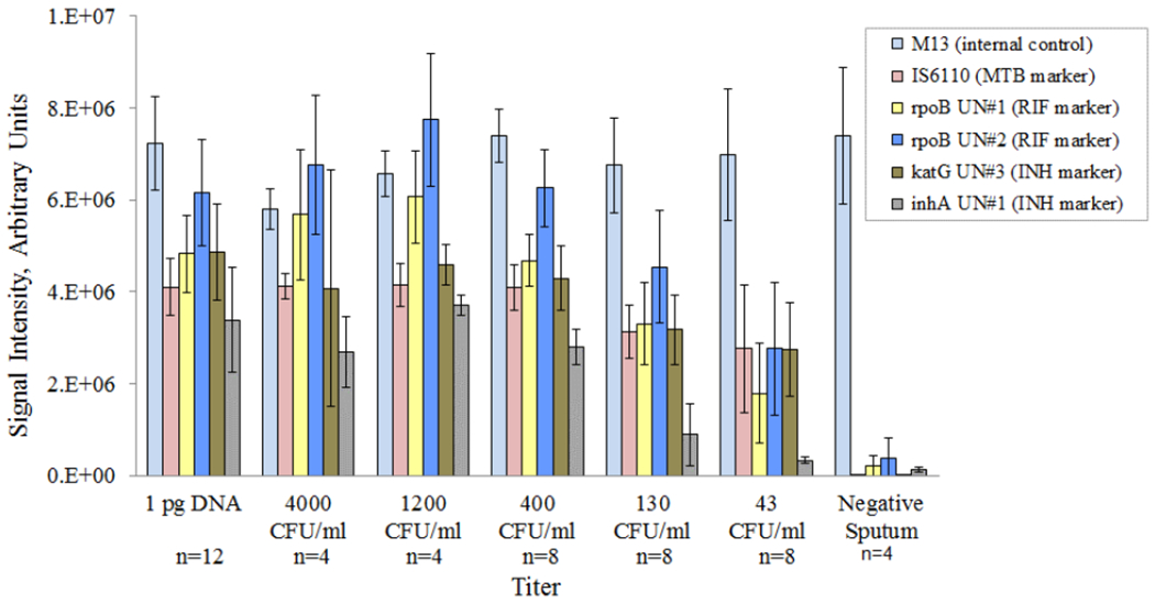
Automated genotyping of drug-resistant Mycobacterium tuberculosis (MTB) directly from sputum is challenging for three primary reasons. First, the sample matrix, sputum, is highly viscous and heterogeneous, posing a challenge for sample processing. Second, acid-fast MTB bacilli are difficult to lyse. And third, there are hundreds of MTB mutations that confer drug resistance. An additional constraint is that MTB is most prevalent where test affordability is paramount. We address the challenge of sample homogenization and cell lysis using magnetic rotation of an external magnet, at high (5000) rpm, to induce the rotation of a disposable stir disc that causes chaotic mixing of glass beads ("MagVor"). Nucleic acid is purified using a pipet tip with an embedded matrix that isolates nucleic acid ("TruTip"). We address the challenge of cost and genotyping multiple mutations using 203 porous three-dimensional gel elements printed on a film substrate and enclosed in a microfluidic laminate assembly ("Lab-on-a-Film"). This Lab-on-a-Film assembly (LFA) serves as a platform for amplification, hybridization, washing, and fluorescent imaging, while maintaining a closed format to prevent amplicon contamination of the workspace. We integrated and automated MagVor homogenization, TruTip purification, and LFA amplification in a multisample, sputum-to-genotype system. Using this system, we report detection down to 43 cfu/mL of MTB bacilli from raw sputum.
Conflict of interest statement
CONFLICT OF INTEREST DISCLOSURE
AVK, RN, AB, PQ, DPC, RCH and CGC were employed by Akonni when this work was conducted. AVK, DPC, RCH and CGC are also shareholders at Akonni.
Figures
Similar articles
-
Lab-on-a-Film disposable for genotyping multidrug-resistant Mycobacterium tuberculosis from sputum extracts.Lab Chip. 2019 Mar 27;19(7):1217-1225. doi: 10.1039/c8lc01404c. Lab Chip. 2019. PMID: 30801596 Free PMC article.
-
Automated TruTip nucleic acid extraction and purification from raw sputum.PLoS One. 2018 Jul 5;13(7):e0199869. doi: 10.1371/journal.pone.0199869. eCollection 2018. PLoS One. 2018. PMID: 29975759 Free PMC article.
-
Genotyping Multidrug-Resistant Mycobacterium tuberculosis from Primary Sputum and Decontaminated Sediment with an Integrated Microfluidic Amplification Microarray Test.J Clin Microbiol. 2018 Feb 22;56(3):e01652-17. doi: 10.1128/JCM.01652-17. Print 2018 Mar. J Clin Microbiol. 2018. PMID: 29305543 Free PMC article.
-
Xpert MTB/RIF and Xpert MTB/RIF Ultra assays for active tuberculosis and rifampicin resistance in children.Cochrane Database Syst Rev. 2020 Aug 27;8(8):CD013359. doi: 10.1002/14651858.CD013359.pub2. Cochrane Database Syst Rev. 2020. Update in: Cochrane Database Syst Rev. 2022 Sep 6;9:CD013359. doi: 10.1002/14651858.CD013359.pub3. PMID: 32853411 Free PMC article. Updated.
-
[Tuberculosis in 2015: From diagnosis to the detection of multiresistant cases].Rev Mal Respir. 2015 Oct;32(8):784-90. doi: 10.1016/j.rmr.2015.06.008. Epub 2015 Jul 10. Rev Mal Respir. 2015. PMID: 26169235 Review. French.
Cited by
-
Clinical Evaluation of the XDR-LFC Assay for the Molecular Detection of Isoniazid, Rifampin, Fluoroquinolone, Kanamycin, Capreomycin, and Amikacin Drug Resistance in a Prospective Cohort.J Clin Microbiol. 2023 Mar 23;61(3):e0147822. doi: 10.1128/jcm.01478-22. Epub 2023 Feb 9. J Clin Microbiol. 2023. PMID: 36757183 Free PMC article.
-
Detecting rifampin and isoniazid resistance in Mycobacterium tuberculosis direct from patient sputum using an automated integrated system.J Clin Tuberc Other Mycobact Dis. 2022 Feb 22;27:100304. doi: 10.1016/j.jctube.2022.100304. eCollection 2022 May. J Clin Tuberc Other Mycobact Dis. 2022. PMID: 35252594 Free PMC article.
-
Point-of-care diagnostics for infectious diseases: From methods to devices.Nano Today. 2021 Apr;37:101092. doi: 10.1016/j.nantod.2021.101092. Epub 2021 Feb 6. Nano Today. 2021. PMID: 33584847 Free PMC article. Review.
-
Microfluidics as a Novel Technique for Tuberculosis: From Diagnostics to Drug Discovery.Microorganisms. 2021 Nov 11;9(11):2330. doi: 10.3390/microorganisms9112330. Microorganisms. 2021. PMID: 34835455 Free PMC article. Review.
References
Publication types
MeSH terms
Grants and funding
LinkOut - more resources
Full Text Sources
Other Literature Sources
Miscellaneous
