. 2020 May 4;217(5):e20192282.
doi: 10.1084/jem.20192282.

Intratumoral accumulation of gut microbiota facilitates CD47-based immunotherapy via STING signaling

Affiliations

Intratumoral accumulation of gut microbiota facilitates CD47-based immunotherapy via STING signaling

Yaoyao Shi et al. J Exp Med. .

Abstract

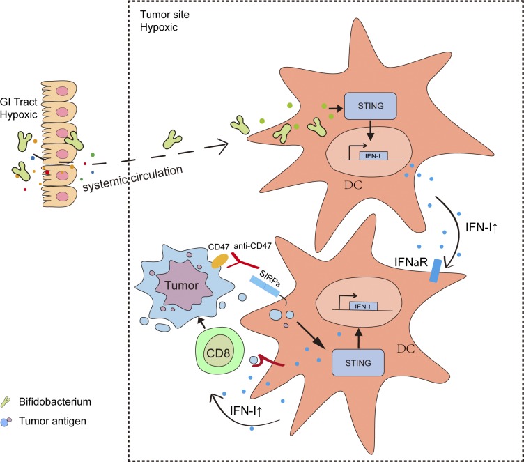
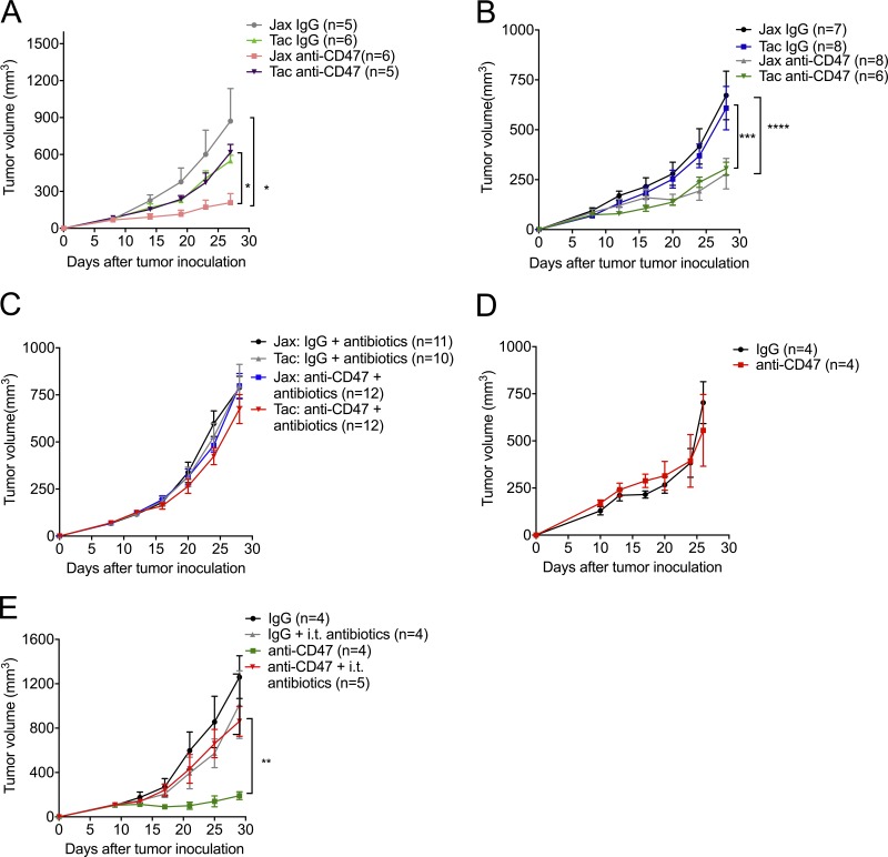
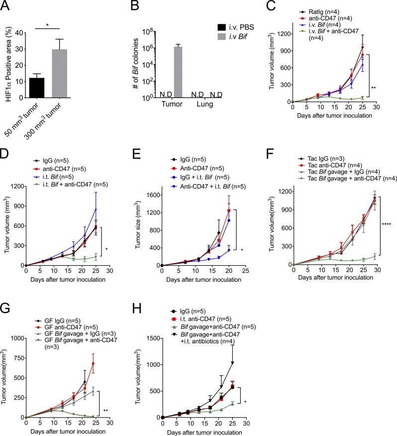
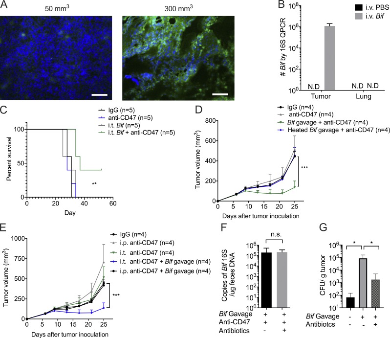
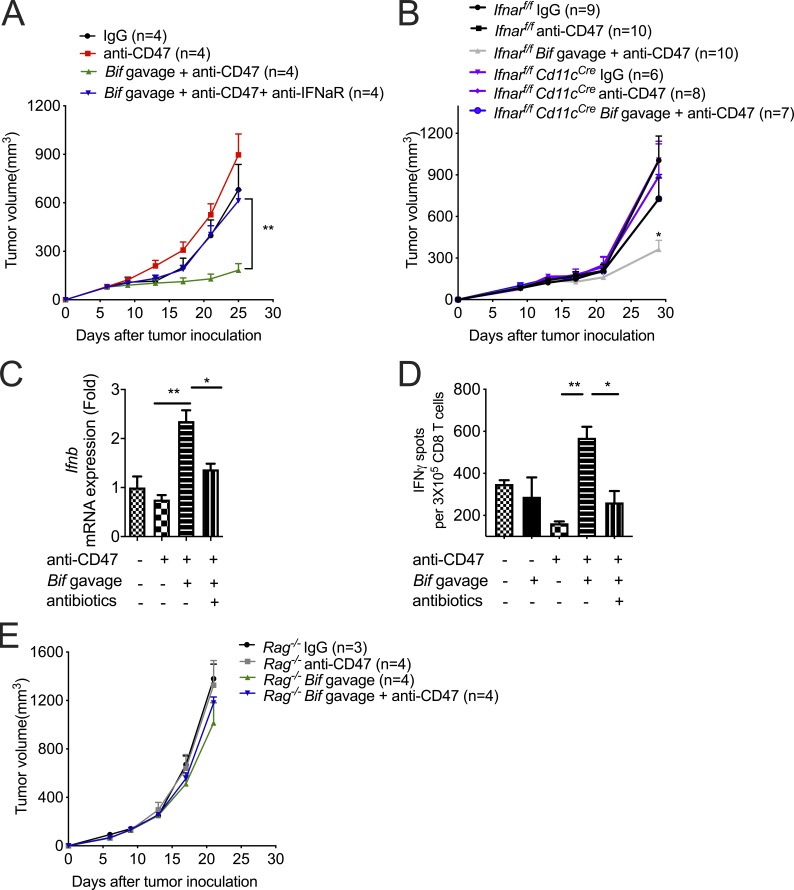
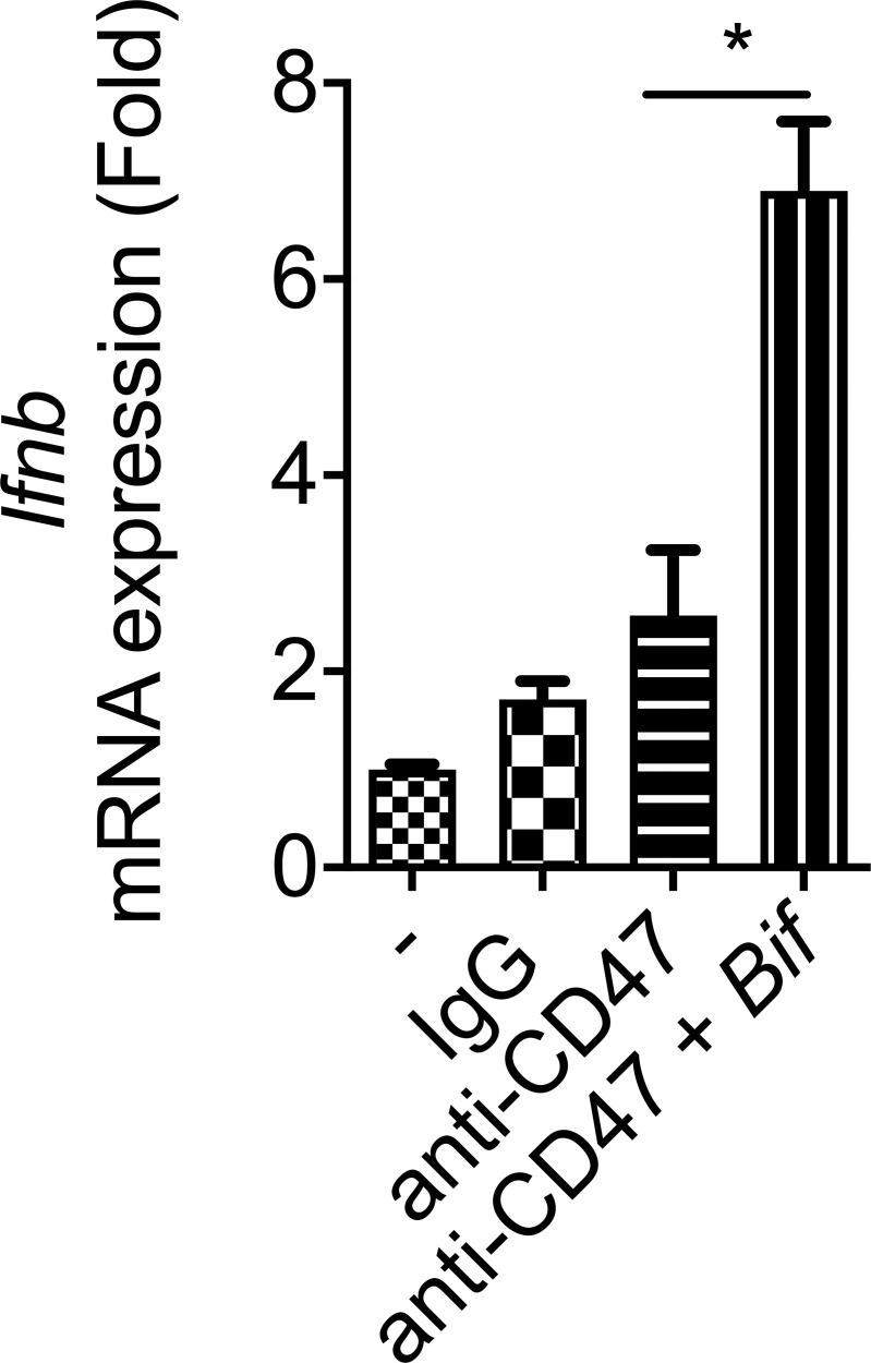
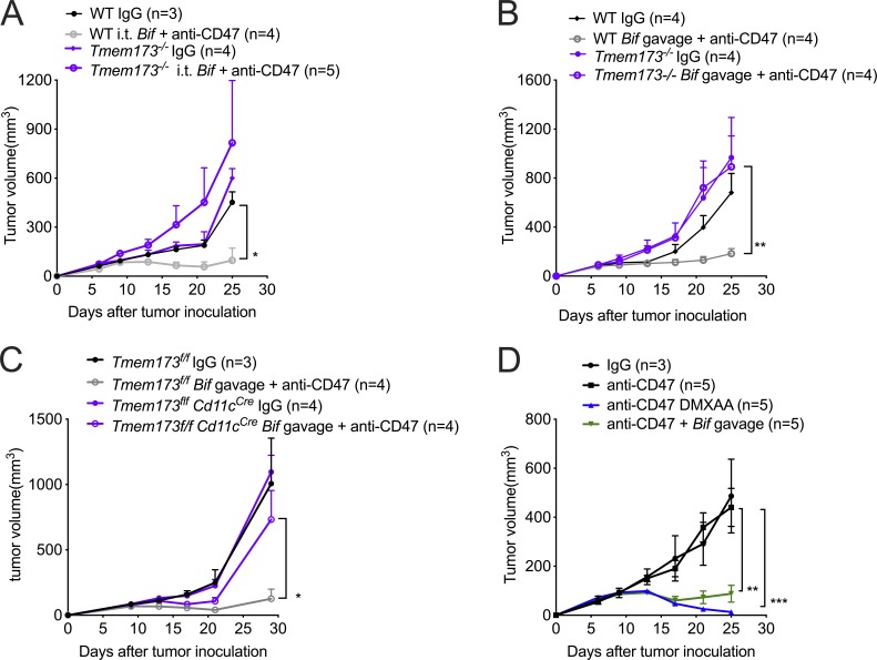
Most studies focus on how intestinal microbiota influence cancer immunotherapy through activating gut immunity. However, immunotherapies related to innate responses such as CD47 blockade rely on the rapid immune responses within the tumor microenvironment. Using one defined anaerobic gut microbiota to track whether microbiota interact with host immunity, we observed that Bifidobacterium facilitates local anti-CD47 immunotherapy on tumor tissues through the capacity to accumulate within the tumor microenvironment. Systemic administration of Bifidobacterium leads to its accumulation within the tumor and converts the nonresponder mice into responders to anti-CD47 immunotherapy in a stimulator of interferon genes (STING)- and interferon-dependent fashion. Local delivery of Bifidobacterium potently stimulates STING signaling and increases cross-priming of dendritic cells after anti-CD47 treatment. Our study identifies the mechanism by which gut microbiota preferentially colonize in tumor sites and facilitate immunotherapy via STING signaling.


Conflict of interest statement

Disclosures: The authors declare no competing interests exist.

Figures

None
Graphical abstract
Figure 1.
Figure 1.
The antitumor responses of CD47 blockade rely on gut microbiota accumulated outside the GI tract. (A–D) C57BL/6 mice were injected subcutaneously with 5 × 10⁵ MC38 cells and treated i.t. with 50 µg anti-CD47 antibody (Ab) or rat IgG on days 10 and 14 after tumor inoculation. Tumor volume was measured at indicated time points. (A) MC38 tumor growth kinetics in newly arrived Jax and Tac mice treated with anti-CD47 Ab or rat IgG. (B) Jax and Tac mice were cohoused for 3 wk before MC38 tumor inoculation and anti-CD47 Ab treatments. (C) Jax and Tac mice were given an oral antibiotic cocktail solution (0.5 mg/ml ampicillin, 0.5 mg/ml gentamicin, 0.5 mg/ml metronidazole, 0.5 mg/ml neomycin, and 0.25 mg/ml vancomycin) 3 wk before MC38 tumor inoculation and anti-CD47 Ab treatment and the oral feeding of antibiotics was stopped at the end of the experiments. (D) MC38 tumor growth kinetics in germ-free mice treated with anti-CD47 Ab or rat IgG. (E) Newly arrived Jax C57BL/6 mice were s.c. injected with 5 × 10⁵ MC38 cells. Each mouse was i.t. injected with 70 µg anti-CD47 Ab or 70 µg rat IgG on day 9 or day 13, respectively. The mice in antibiotics groups were i.t. injected with 100 µl antibiotic cocktail solution every other day. Tumor volume was measured at indicated time points. One representative experiment (A andD) is depicted from at least two experiments yielding similar results. Each group contains at least four mice. Results in C were pooled based on two experiments yielding similar results. Each group contains ≥10 mice. Results in B and E were from one experiment; each group contains at least six (B) and four (E) samples. Presented as mean ± SEM. Two-way ANOVA was used to analyze data in A–E. *, P < 0.05; **, P < 0.01; ***, P < 0.001; ****, P < 0.0001.
Figure 2.
Figure 2.
Administration of Bifidobacterium, a tumor-targeting member of the microbiota, sufficiently recovers the antitumor efficacy of anti-CD47 immunotherapy in nonresponders. (A) Statistical analysis of the percentage of HIF1α-positive area on different-volume tumor tissue slides. (B) Detection of Bifidobacterium in tumor tissues and lung sites after treatment by anaerobic culture. MC38 tumor–bearing Tac mice were intravenously injected with 1.5 × 10⁷ CFU activated Bifidobacterium three times when tumor sizes approached 200 mm3. 7 d after the final injection, tumor tissues were collected and homogenized for anaerobic culture of Bifidobacterium. N.D., not detected. (C and D) MC38 tumor–bearing Tac mice were i.t. injected with 70 µg anti-CD47 antibody (Ab) or 70 µg rat IgG on days 9 and 13, respectively. (C) Tumor growth kinetics of intravenous injection of Bifidobacterium. MC38 tumor-bearing Tac mice were treated with 70 µg anti-CD47 Ab or 70 µg rat IgG on days 9 and 13. For i.v. injection of Bifidobacterium, mice were intravenously injected with 1.5 × 10⁷ CFU Bifidobacterium or PBS on the same days on days 6, 9, and 13. (D) For i.t. injection of Bifidobacterium, mice were i.t. injected with 2.5 × 10⁶ CFU Bifidobacterium or PBS on the same days as i.v. injection of Bifidobacterium. The i.t. Bif + anti-CD47 group were compared with the IgG group. (E) EG7 tumor–bearing TAC mice were treated with 70 µg anti-CD47 Ab or 70 µg rat IgG on days 11 and 13. I.t. injection of 2.5 × 10⁶ CFU Bifidobacterium or PBS was administered on days 8, 11, and 13. Tumor volume was measured at the indicated time points. (F–H) Newly arrived Tac C57BL/6 mice (F and H) and germ-free (GF) mice (G) were s.c. injected with 5 × 10⁵ MC38 cells. Each mouse was i.t. injected with 70 µg anti-CD47 Ab or 70 µg rat IgG on days 9 and 13, respectively. For oral administration of Bifidobacterium (Bif), mice were gavaged with 5 × 10⁸ CFU Bifidobacterium or PBS on days 6, 9, and 13, respectively. The Tac Bif gavage + anti-CD47 group was compared with the Tac IgG group. (H) Mice were i.t. injected with 100 µl antibiotic cocktail solution or PBS every other day. Tumor volume was measured at the indicated time points. One representative experiment from at least two experiments yielding similar results is depicted in A–H. Each group in A and B contains at least three samples. The number of mice in each group is depicted in the panels C–H. Presented as mean ± SEM. *, P < 0.05 (nonpaired Student’s t test; A); *, P < 0.05; **, P < 0.01; ****, P < 0.0001 (two-way ANOVA; C–H).
Figure S1.
Figure S1.
Gut microbiota could survive in the hypoxic TME and prolong mouse survival. Related to Fig. 2. (A) Representative image of HIF1α stain of different volumes of tumors. Immunofluorescence analysis of hypoxia: HIF1α (green) expression on different-volume tumor tissue slides. Tumor volume of the left: 50 mm3; tumor volume of the right: 300 mm3. Scale bars, 20 µm. One representative experiment is depicted from two experiments yielding similar results. (B) Accumulation of Bifidobacterium inside tumors but not lung sites after treatments. MC38 tumor–bearing Tac mice were i.v. injected with 1.5 × 10⁷ CFU Bifidobacterium or PBS three times when tumor size approaches 200 mm3. 7 d later, tumor tissues were collected for qPCR detection of Bifidobacterium. One representative experiment from two experiments yielding similar results is depicted. N.D., not detected. (C) Survival curves of tumor-burden mice with different treatments. MC38 tumor-bearing Tac mice were i.t. injected with 70 µg anti-CD47 antibody (Ab) or 70 µg rat IgG on days 9 and 13, respectively. For i.t. injection of Bifidobacterium, mice were i.t. injected with 2.5 × 10⁶ CFU Bifidobacterium or PBS on days 6, 9, and 13. Mice were euthanized when the tumor size approached end points. The anti-CD47+ i.t. Bifidobacterium group is compared with the IgG group. One representative experiment from at least two experiments yielding similar results is depicted. **, P < 0.01 (log-rank test). (D) Heat-killed Bifidobacterium failed to facilitate CD47-based immunotherapy. Newly arrived Tac C57BL/6 mice were subcutaneously injected with 5 × 10⁵ MC38 cells. Each mouse was i.t. injected with 70 µg anti-CD47 Ab or 70 µg rat IgG on days 9 and 13, respectively. The mice with Bifidobacterium administration were gavaged with 5 × 10⁸ Bifidobacterium or heat-inactivated Bifidobacterium on days 6, 9, and 13. Tumor volume was measured at the indicated time points. ***, P < 0.001 (two-way ANOVA). (E) Oral administration of Bifidobacterium failed to facilitate antitumor efficacy after systemic administration of anti-CD47 antibody. Newly arrived Tac C57BL/6 mice were subcutaneously injected with 5 × 10⁵ MC38 cells. Each mouse was i.t. injected with 70 µg anti-CD47 Ab or 70 µg rat IgG on days 9 and 13 or i.p. injected with 500 µg anti-CD47 or rat IgG on days 9 and 13, respectively. The mice with Bifidobacterium administration were gavaged with 5 × 10⁸ CFU Bifidobacterium on days 6, 9, and 13, respectively. Tumor volume was measured at indicated time points. ***, P < 0.001 (two-way ANOVA). (F) I.t. antibiotics therapy does not reduce the Bifidobacterium copies in mouse feces. qPCR detection of Bifidobacterium in fresh feces collected from mice with indicated treatments on day 14 (also see Fig. 2 H; nonpaired Student’s t test). (G) Detection of Bifidobacterium inside tumor tissues after Bifidobacterium gavage and i.t. antibiotics treatment. Newly arrived Tac C57BL/6 mice were subcutaneously injected with 5 × 10⁵ MC38 cells. For oral administration of Bifidobacterium (Bif), mice were gavaged with 5 × 10⁸ CFU Bifidobacterium or PBS on day 7, 10, and 13. Mice were i.t. injected with 100 µl antibiotic cocktail solution or PBS every other day from day 6. Tumor tissues were collected on day 17 and homogenized for anaerobic culture of Bifidobacterium. Presented as mean ± SEM. *, P < 0.05 (Mann–Whitney U test of log scale). n.s., not significant.
Figure 3.
Figure 3.
I.t. type I IFN signaling and T cells are essential for Bifidobacterium-mediated tumor control. (A and B) MC38 tumor–bearing TAC mice (A) and Ifnarf/f or Ifnarf/fCd11cCre mice (B) were i.t. injected with 70 µg CD47 antibody or 70 µg rat IgG on days 9 and 13. For gavage of Bifidobacterium, mice were gavaged with 5 × 10⁸ Bifidobacterium on days 6, 9, and 13. Tumor volume was measured at the indicated time points. (A) To block type I IFN signaling, mice were i.t. injected with 200 µg anti-IFNAR or rat IgG on days 6, 8, 10, 12, and 14. The Bif gavage + anti-CD47 group is compared with the Bif gavage + anti-CD47 + anti-IFNaR group. (B) The Ifnarf/fCd11cCre Bif gavage + anti-CD47 group is compared with the Ifnarf/f Bif gavage + anti-CD47 group. (C) qPCR of Ifnb1 mRNA levels in tumor-infiltrating DCs. MC38 tumors of TAC mice were injected with 70 µg CD47 antibody or 70 µg rat IgG on day 9. Mouse tumors were injected with 100 µl antibiotic cocktail solution or PBS every other day. For gavage of Bifidobacterium, mice were gavaged with 5 × 10⁸ CFU Bifidobacterium on days 6 and 9, respectively. On day 13, tumor tissues were collected for flow cytometric sorting of tumor-infiltrating DCs. Ifnb1 mRNA levels were quantified by qPCR. (D) Number of ELISPOT IFNγ spots of 3 × 10⁵ sorted OTI-TCR CD8+ T Cells cocultured with DCs from tumor tissues in Tac mice. Tac mice were subcutaneously injected with 2 × 10⁶ MC38-OTI tumor cells and i.t. injected with 70 µg CD47 antibody or 70 µg rat IgG on days 9 and 12. Mice were i.t. injected with 100 µl antibiotic cocktail solution or PBS every other day from day 6. 2 d after the final injection of antibody, tumor tissues were collected for tumor DC sorting. For gavage of Bifidobacterium, mice were gavaged with 5 × 10⁸ CFU Bifidobacterium on days 6, 9, and 12. (E) MC38 tumors of Rag−/− mice were injected with 70 µg CD47 antibody or 70 µg rat IgG on days 9 and 13. For gavage of Bifidobacterium, mice were gavaged with 5 × 10⁸ CFU Bifidobacterium on days 6, 9, and 13. Tumor volume was measured at the indicated time points. One representative experiment from at least two experiments yielding similar results is depicted in A, C, D, and E, and each group contains four mice (A and E). The results in B were based on data pooled from two independent experiments yielding similar results, and each group contains at least six mice. Presented as mean ± SEM. **, P < 0.01 (two-way ANOVA; A and E). *, P < 0.05; **, P < 0.01 (nonpaired Student’s t test; B, C, and D).
Figure S2.
Figure S2.
Type I IFN could be up-regulated in bone marrow–derived DCs cocultured with tumor cells and Bifidobacterium after CD47 blockade. Related to Fig. 3. qPCR of Ifnb mRNA levels in bone marrow–derived DCs. Bone marrow–derived DCs (2 × 10⁶) were cocultured with MC38 cells (2 × 10⁶) with 10 µg/ml anti-CD47 or 10 µg/ml rat IgG in the presence or absence of 2 × 10⁶ Bifidobacterium for 8 h. DCs were then sorted out for Ifnb quantification by qPCR. Presented as mean ± SEM. *, P < 0.05 (nonpaired Student’s t test).
Figure 4.
Figure 4.
I.t. Bifidobacterium facilitates CD47-based immunotherapy via STING signaling. (A–D) Mice were injected subcutaneously with 5 × 10⁵ MC38 cells and treated i.t. with 70 µg CD47 antibody or rat IgG on days 9 and 13 after tumor inoculation. Tumor volume was measured at indicated time points. (A) Tumor growth kinetics in Tmem173−/− mice treated with anti-CD47 antibody (Ab) or rat IgG and i.t. administration of Bifidobacterium or PBS. (B) Tumor growth kinetics in Tmem173−/− mice treated with anti-CD47 Ab or rat IgG and oral administration of Bifidobacterium or PBS. The WT Bif gavage + anti-CD47 group is compared with the Tmem173−/− Bif gavage + anti-CD47 group. (C) Tumor growth kinetics in Tmem173f/f and Tmem173f/fCd11cCre mice treated with anti-CD47 Ab or rat IgG and oral administration of Bifidobacterium or PBS. The Tmem173f/f Bif gavage + anti-CD47 group is compared with the Tmem173f/f Cd11cCre Bif gavage + anti-CD47 group. (D) Tumor growth kinetics in Tac C57BL/6 mice treated with anti-CD47 Ab and/or oral administration of Bifidobacterium. 20 µg DMXAA were i.t. injected on days 9 and 13. For A–D, one representative experiment from at least two experiments yielding similar results is depicted, and the number of mice per group is depicted in the panels. Presented as mean ± SEM. *, P < 0.05; **, P < 0.01; ***, P < 0.001 (two-way ANOVA; A–D).

References

    1. Advani R., Flinn I., Popplewell L., Forero A., Bartlett N.L., Ghosh N., Kline J., Roschewski M., LaCasce A., Collins G.P., et al. . 2018. CD47 Blockade by Hu5F9-G4 and Rituximab in Non-Hodgkin’s Lymphoma. N. Engl. J. Med. 379:1711–1721. 10.1056/NEJMoa1807315 - DOI - PMC - PubMed
    1. Arch Oncology 2019. AO-176 in Multiple Solid Tumor Malignancies. Available at: https://ichgcp.net/clinical-trials-registry/NCT03834948.
    1. Bullman S., Pedamallu C.S., Sicinska E., Clancy T.E., Zhang X., Cai D., Neuberg D., Huang K., Guevara F., Nelson T., et al. . 2017. Analysis of Fusobacterium persistence and antibiotic response in colorectal cancer. Science. 358:1443–1448. 10.1126/science.aal5240 - DOI - PMC - PubMed
    1. Cani P.D., and de Vos W.M.. 2017. Next-Generation Beneficial Microbes: The Case of Akkermansia muciniphila. Front. Microbiol. 8:1765 10.3389/fmicb.2017.01765 - DOI - PMC - PubMed
    1. Chao M.P., Alizadeh A.A., Tang C., Myklebust J.H., Varghese B., Gill S., Jan M., Cha A.C., Chan C.K., Tan B.T., et al. . 2010. Anti-CD47 antibody synergizes with rituximab to promote phagocytosis and eradicate non-Hodgkin lymphoma. Cell. 142:699–713. 10.1016/j.cell.2010.07.044 - DOI - PMC - PubMed

Publication types