. 2020 Mar 5;106(3):412-421.
doi: 10.1016/j.ajhg.2020.02.007.

Bi-allelic JAM2 Variants Lead to Early-Onset Recessive Primary Familial Brain Calcification

Collaborators, Affiliations

Bi-allelic JAM2 Variants Lead to Early-Onset Recessive Primary Familial Brain Calcification

Lucia V Schottlaender et al. Am J Hum Genet. .

Abstract

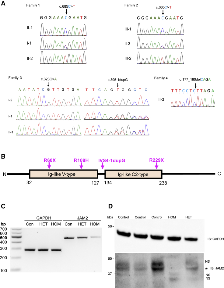
Primary familial brain calcification (PFBC) is a rare neurodegenerative disorder characterized by a combination of neurological, psychiatric, and cognitive decline associated with calcium deposition on brain imaging. To date, mutations in five genes have been linked to PFBC. However, more than 50% of individuals affected by PFBC have no molecular diagnosis. We report four unrelated families presenting with initial learning difficulties and seizures and later psychiatric symptoms, cerebellar ataxia, extrapyramidal signs, and extensive calcifications on brain imaging. Through a combination of homozygosity mapping and exome sequencing, we mapped this phenotype to chromosome 21q21.3 and identified bi-allelic variants in JAM2. JAM2 encodes for the junctional-adhesion-molecule-2, a key tight-junction protein in blood-brain-barrier permeability. We show that JAM2 variants lead to reduction of JAM2 mRNA expression and absence of JAM2 protein in patient's fibroblasts, consistent with a loss-of-function mechanism. We show that the human phenotype is replicated in the jam2 complete knockout mouse (jam2 KO). Furthermore, neuropathology of jam2 KO mouse showed prominent vacuolation in the cerebral cortex, thalamus, and cerebellum and particularly widespread vacuolation in the midbrain with reactive astrogliosis and neuronal density reduction. The regions of the human brain affected on neuroimaging are similar to the affected brain areas in the myorg PFBC null mouse. Along with JAM3 and OCLN, JAM2 is the third tight-junction gene in which bi-allelic variants are associated with brain calcification, suggesting that defective cell-to-cell adhesion and dysfunction of the movement of solutes through the paracellular spaces in the neurovascular unit is a key mechanism in CNS calcification.

Keywords: Fahr disease; JAM2; JAM3; MYORG; OCLN; SLC20A2; familial idiopathic basal ganglia calcification; knock out mouse model; primary familial brain calcification; recessive brain calcification.


Conflict of interest statement

The authors declare that A. Begtrup and E.T. are employees of GeneDx, Inc., USA, and S.K. and C.B. are employees of CENTOGENE AG, Rostock, Germany. The other authors declare no competing interests.

Figures

Figure 1
Figure 1
Clinical and Neuroimaging Features of JAM2-Related Disease (A) Pedigrees of the four families with bi-allelic JAM2 mutations. (B) Brain images of JAM2-related disease. B1 to B3 are CT scans acquired from case subject F1-II:2 from family 1 and B4 to B6 are CT acquired from case subject F2-III:3 from family 2. In both individuals there is extensive, symmetrical, bilateral calcification involving the basal ganglia, deep cortical gray matter and cerebellum. B7, 8, and 9 are CT from case subject F3-II:1 demonstrating calcification in the basal ganglia and cortical gyri but not in the cerebellum. B10, 11, and 12 are axial MRI scans from individual F3-II:1 from family 3 showing basal ganglia and frontal calcification (B10 is T1 MRI, B11 is T2 MRI, and B12 is Axial Ven Bold reconstruction MRI that is sensitive to calcium shown as hypointense regions). Asterisk () areas of calcification on MRI.
Figure 2
Figure 2
Validation of JAM2 Variants Identified in This Study (A) Validation by Sanger sequencing of the stop variant c.685C>T (p.Arg229Ter) in families 1 and 2, the bi-allelic variants in family 3 (IVS4-1dupG and p.Arg108His), and the homozygous change in family 4 c.177_180delCAGA (p.Arg60Ter). (B) Protein structure of JAM2 showing the 2 main domains (Ig-like V-type and Ig-like C2-type) as well as the variants identified in the 4 families. (C) Total RNA and protein extracts were prepared from fibroblast cell lines isolated from the proband of family 1 (HOM), his unaffected mother (HET), and unrelated control subjects (Con) to assess the c.685C>T (p.Arg229Ter) JAM2 variant. Agarose gel separation of RT-PCR products shows a reduction in JAM2 mRNA expression in the homozygous proband (HOM) compared to the proband’s mother (HET) and unrelated control subject (Con). GAPDH RT-PCR served as a loading control. (D) Western blot analysis of total protein lysates using anti-JAM2 and GAPDH (loading control) antibodies revealed presence of JAM2 protein in control (Control) and unaffected carrier (HET) cells but complete loss of JAM2 protein in the homozygous proband of family 1 (HOM). NS, non-specific band; asterisk, JAM2.
Figure 3
Figure 3
Behavioral Study on jam2 KO Mice and Wild-Type (WT) (A) Representative images of wild-type (left) and jam2 KO strides (right) in the gait test. Forepaw (blue) and hindpaw (red). Altered gait of the jam2 KO mice was analyzed in stance length, stride, and sway compared to wild-type (∗∗∗p < 0.0001, ∗∗p = 0.002; means ± SEM; n = 6 mice per genotype). (B and C) Walking beam performance on test day, showing elevated latency to cross the beam in jam2 KO (p = 0.017; means ± SEM; n = 5 wild-type, n = 6 jam2 KO). See Video S2 for example beam walking test of a jam2 KO mouse and a control (wild-type).
Figure 4
Figure 4
Brain Pathology in Control and jam2 KO Mice of Young (6 Months Old) and Old Age (18 Months Old) Littermate controls (wild-type) (a, a1–a6) show no significant pathology on (H&E) stained sections (a, cortex; a1, cerebellum; a2, midbrain) and on immunohistochemistry for astrocytes (GFAP) (a3, midbrain), microglia (Iba1) (a4, midbrain), neuropil (APP) (a5, midbrain), and neurones (NeuN) (a6, midbrain). In age-matched jam2 KO mice, occasional cortical vacuolation is evident in the cerebral (b) and cerebellar (b1) cortex and widespread prominent vacuolation is seen in the midbrain (b2). In the midbrain region there is also marked reactive astrocytosis (b3) and mild microglial activation (b4). APP immunostaining highlights vacuolar change in the neuropil (b5) and NeuN shows mild reduction in the neuronal density (b6). Aged wild-type mice show occasional vacuolation in the cerebral cortex (c), cerebellar cortex (c1), and midbrain (c2) (H&E). There is mild patchy astrogliosis in the midbrain (c3) and mild microglial activation (c4). No significant disruption of the cytoarchitecture is seen on APP immunostaining (c5), but NeuN highlights some degree of neuronal loss (c6). In age-matched old jam2 KO mice, there is increasingly prominent vacuolation in the cerebral cortex (d) and in the cerebellar white matter (d1), midbrain (d2), and thalamus (not shown). Similar to young jam2 KO mice, there is widespread astrogliosis in the midbrain (d3), but microglial activation remains mildly increased (d4). Frequent vacuolation is highlighted in the neuropil with APP immunostaining (d5). Similar to age-matched littermate controls, there is reduction of the neuronal density in the midbrain of jam2 KO mouse (d6). Scale bar: 100 μm in a–d, a2–a6, b2–b6, c2–c6, d2–d6; 200 μm in a1–d1.

Comment in

References

    1. Batla A., Tai X.Y., Schottlaender L., Erro R., Balint B., Bhatia K.P. Deconstructing Fahr’s disease/syndrome of brain calcification in the era of new genes. Parkinsonism Relat. Disord. 2017;37:1–10. - PubMed
    1. Taglia I., Bonifati V., Mignarri A., Dotti M.T., Federico A. Primary familial brain calcification: update on molecular genetics. Neurol. Sci. 2015;36:787–794. - PubMed
    1. Legati A., Giovannini D., Nicolas G., López-Sánchez U., Quintáns B., Oliveira J.R.M., Sears R.L., Ramos E.M., Spiteri E., Sobrido M.-J. Mutations in XPR1 cause primary familial brain calcification associated with altered phosphate export. Nat. Genet. 2015;47:579–581. - PMC - PubMed
    1. Yao X.-P., Cheng X., Wang C., Zhao M., Guo X.-X., Su H.-Z., Lai L.-L., Zou X.-H., Chen X.-J., Zhao Y. Biallelic Mutations in MYORG Cause Autosomal Recessive Primary Familial Brain Calcification. Neuron. 2018;98:1116–1123.e5. - PubMed
    1. Mochida G.H., Ganesh V.S., Felie J.M., Gleason D., Hill R.S., Clapham K.R., Rakiec D., Tan W.-H., Akawi N., Al-Saffar M. A homozygous mutation in the tight-junction protein JAM3 causes hemorrhagic destruction of the brain, subependymal calcification, and congenital cataracts. Am. J. Hum. Genet. 2010;87:882–889. - PMC - PubMed

Publication types

Substances

LinkOut - more resources