. 2020 May:241:119903.
doi: 10.1016/j.biomaterials.2020.119903. Epub 2020 Feb 24.

Enabling sublingual peptide immunization with molecular self-assemblies

Affiliations

Enabling sublingual peptide immunization with molecular self-assemblies

Sean H Kelly et al. Biomaterials. 2020 May.

Abstract

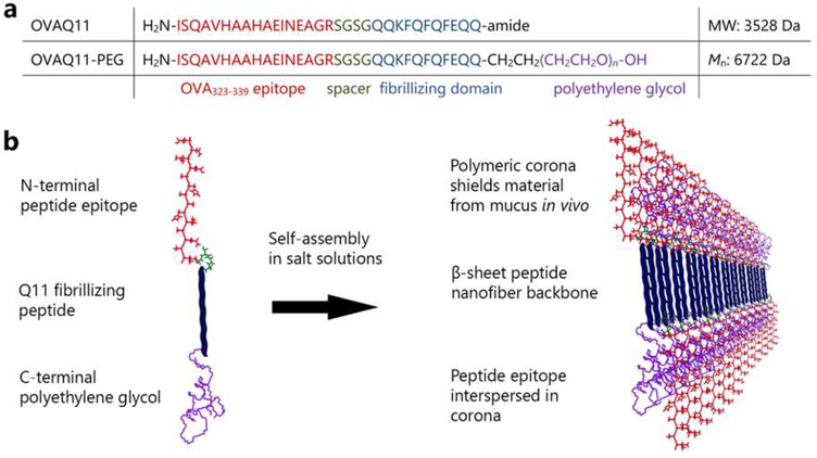
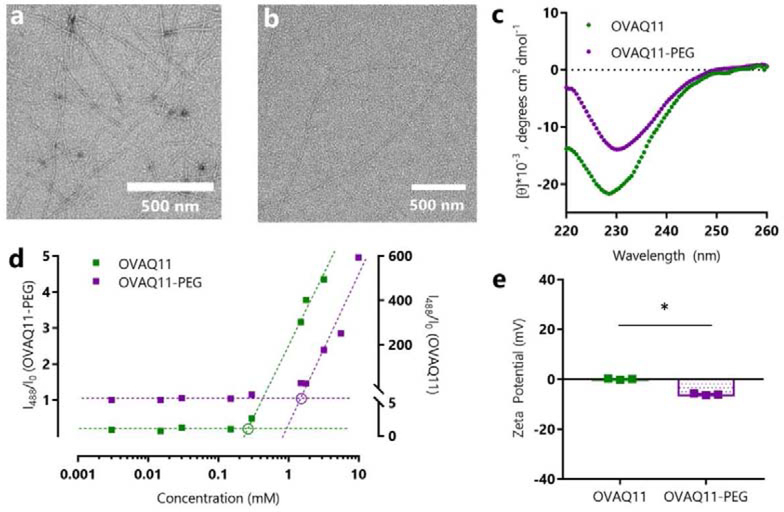
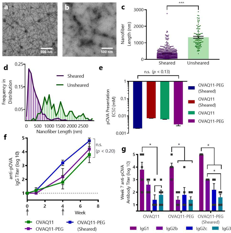
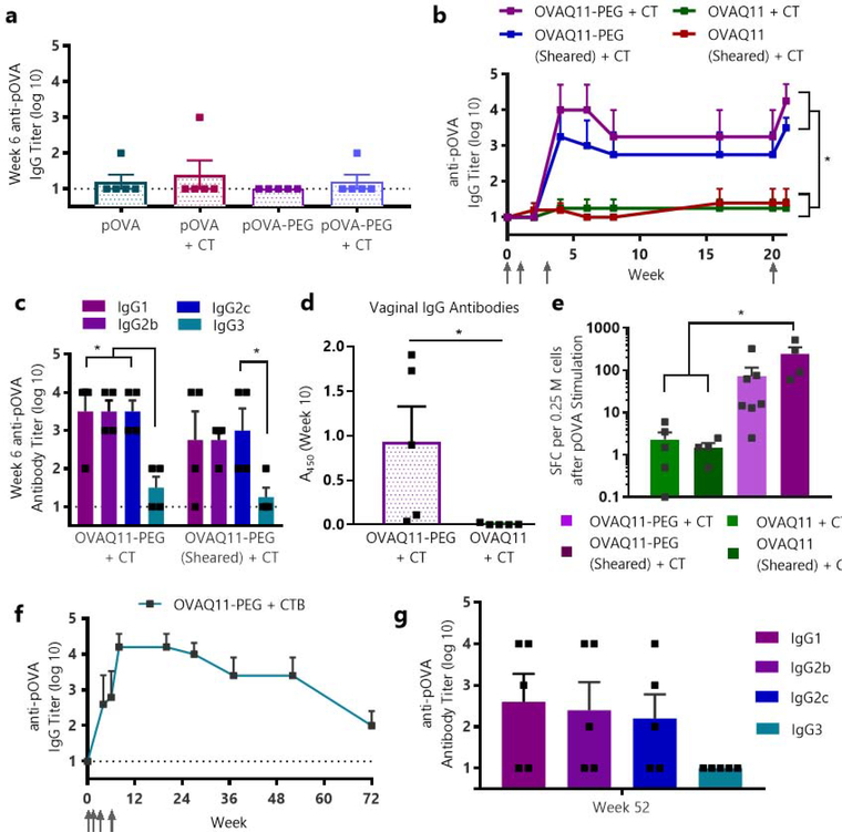
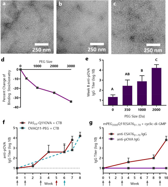
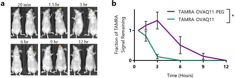
Short peptides are poorly immunogenic when delivered sublingually - under the tongue. Nanomaterial delivery of peptides could be utilized to improve immunogenicity towards designed sublingual vaccines, but nanomaterials have not been widely successful in sublingual vaccines owing to the challenges of transport through the sublingual mucosa. Here, we report that the sublingual immunogenicity of peptides is negligible, even in the presence of sublingual adjuvants or when PEGylated, but can be dramatically enhanced by assembly into supramolecular polymer-peptide nanofibers bearing low-molecular weight PEG, optimally between 2000 and 3000 Da. Neither PEGylation nor a sublingual adjuvant were capable of rendering peptides immunogenic without assembly into nanofibers. We found that PEG decreased nanofiber interactions with mucin and promoted longer residence time at the sublingual immunization site. Parallel investigations with shortened nanofibers indicated that the size of the assemblies had a surprisingly negligible influence over sublingual immunogenicity. In mice, optimized formulations were capable of raising strong and highly durable systemic antibody responses, antibodies in the upper respiratory and reproductive tracts, and systemic antigen-specific T-cell responses. These nanofiber-based sublingual vaccines were effective with both protein and nucleotide adjuvants and raised responses against both a model peptide epitope and a peptide epitope from M. tuberculosis. Further, PASylation (modification of nanofibers with peptide sequences rich in Pro, Ala, and Ser) could be substituted for PEGylation to also achieve sublingual immunogenicity. These findings indicated that surface properties supersede nanomaterial size in modulating sublingual nanomaterial immunogenicity, having important implications for the design of synthetic sublingual vaccines.

Keywords: Mucosal; Nanofiber; Self-assembly; Sublingual; Supramolecular; Vaccine.


Conflict of interest statement

Declaration of competing interest The authors declare the following financial interests/personal relationships which may be considered as potential competing interests: JHC and SHK are listed as inventors on a patent application associated with the technology described.

Figures

Figure 1.
Figure 1.. Design of supramolecular peptide-polymer nanofibers.
(a) Primary sequences of OVAQ11 and OVAQ11-PEG. (b) Peptide-polymers self-assemble to form supramolecular nanofibers. The hypothesized structure was produced using previously published knowledge regarding the anti-parallel β strand architecture in Q11 nanofibers and the propensity of similar self-assembling PEG-conjugated β-sheet peptides to form peptide cores with PEG coronas.,
Figure 2.
Figure 2.
OVAQ11-PEG self-assembled into β-sheet nanofibers, as indicated by negative stained TEM images of (a) OVAQ11 and (b) OVAQ11-PEG nanofibers assembled from 2 mM peptide and (c) Circular dichroism of peptides assembled at 3 mM in PBS and diluted to 0.1 mM in potassium fluoride immediately prior to analyzing. (d) β-sheet structure was further confirmed using Thioflavin T. Following the method of Hamley and coworkers, the graphical estimates of critical aggregation concentration correspond to the intersection of the pre- and post-assembly tangent lines (circled). (e) Zeta-potentials of OVAQ11-PEG and OVAQ11 indicated that surface charge was minimally altered by PEGylation. Peptides were prepared at 2 mM in 1X PBS and diluted to 0.2 mM in 1X PBS prior to measurement at 25 °C. * p < 0.05, unpaired, two-tailed T-test
Figure 3.
Figure 3.. Shearing of OVAQ11 nanofibers reduced nanofiber lengths, and neither PEGylation nor shearing diminished immunogenicity via traditional subcutaneous immunization.
Ten TEM images of OVAQ11 were obtained before and after shearing through a 100 nm track-etched polycarbonate membrane. ImageJ was used to determine the length of 450 individual sheared fibers and 96 non-sheared fibers. Representative images of unsheared (a) and sheared (b) OVAQ11 fibers, and (c) individual lengths of each nanofiber. ***p < 0.001, unpaired, two-tailed t-test. (d) Histogram showing the frequency distribution of fiber lengths before and after shearing. All TEM images and corresponding nanofiber traces are shown in Fig. S4. (e) Neither shearing nor PEGylation of nanofibers significantly affected presentation of pOVA in MHC class II molecules, as measured by DOBW reporter cells, which secrete IL-2 upon encountering DCs with pOVA-loaded MHC II. IL-2 concentration in the supernatant was measured by ELISA. EC50 corresponds to the concentration of material that gives the half-maximal antigen presentation; n.s. (p < 0.13), one-way ANOVA, n=3/group. (f) Neither shearing nor PEGylation disrupted the subcutaneous immunogenicity of OVAQ11 nanofibers. Mice were immunized subcutaneously on weeks 0 and 4 with two 50 μL injections of 2 mM peptide and serum was analyzed by ELISA; ns (p < 0.20); one-way ANOVA, n=5/group. Arrows indicate timepoints of immunizations. (g) Neither PEGylation nor shearing significantly altered the dominant subclasses of IgG raised by nanofibers. Shown is week 7 serum of mice from (f). *p < 0.05, two-way ANOVA with Tukey’s multiple comparisons test, n=5/group.
Figure 4.
Figure 4.. Supramolecular PEG-peptide nanofibers enabled sublingual immunization against peptide epitopes.
(a) Adjuvant plus soluble OVA peptide (pOVA) alone fails to elicit sublingual antibody responses, even when the peptide is PEGylated. Mice were primed and boosted at weeks 1 and 3 with 7 μL of 5.6 mM peptide with or without 2 μg of cholera toxin (CT) adjuvant, and serum pOVA-specific IgG was measured by ELISA at week 6, n=5/group. (b) PEGylated nanofibers were immunogenic sublingually with CT, regardless of whether they were sheared. Nanofibers lacking PEG were non-immunogenic sublingually. Mice were primed and boosted at weeks 1, 3, and 20 with 7 μL of 5.6 mM peptide with 2 μg of cholera toxin per mouse, and pOVA-specific IgG was measured by ELISA. *p < 0.05, one-way ANOVA, Tukey’s multiple comparisons test, n=4/group. Arrows indicate timepoints of immunizations. (c) Antibody subclasses at week 6 were balanced between IgG1, IgG2b, and IgG2c. *p < 0.05, two-way ANOVA, n=4/group. (d) Sublingually delivered OVAQ11-PEG nanofibers also raised IgG antibodies in the reproductive tract at week 10. *p<0.05, unpaired, two-tailed t-test, n=5/group. (e) PEGylated nanofibers elicited T cell responses, measured by splenocyte ELISPOT at week 21 (same mouse groups as shown in (b) and Fig. S7). SFC (spot-forming cells) after pOVA stimulation are shown. *p < 0.05, one-way ANOVA, Tukey’s multiple comparisons test, n = 4/group (OVAQ11 + CT, OVAQ11-PEG + CT), n = 7 (OVAQ11-PEG (Sheared) + CT), or n = 5 (OVAQ11 (Sheared) + CT). (f) OVA-Q11-PEG nanofibers remained strongly and durably immunogenic using the more clinically translatable adjuvant CTB. n=5 mice, immunized sublingually at weeks 0, 1, 3, and 6 with 7 μL of 5.6 mM OVAQ11-PEG with 10 μg of cholera toxin subunit B, pOVA-specific serum IgG measured by ELISA. (g) At week 52 for the mice immunized with CTB-adjuvanted nanofibers shown in (e), Ig responses were balanced between IgG1, IgG2b, and IgG2c.
Figure 5.
Figure 5.. Sublingual immune responses and mucin binding to OVAQ11 were dependent on the length of conjugated PEG.
Q11OVA with N-terminal mPEG blocks of 350, 1000, and 2000 average molecular weight formed nanofibers, as evidenced by negative stained TEM: (a) mPEG350-Q11OVA (b) mPEG1000-Q11OVA (c) mPEG2000Q11OVA. (d) Isothermal titration calorimetry indicated that increasing PEG length diminished interactions with mucin. * p < 0.05, R2 = 0.94 by linear regression of PEG size vs. percent change in binding stoichiometry. (e) Increasing PEG length improved OVA-specific titers raised against OVAQ11-PEG nanofibers. Mice were immunized sublingually with 7 μL of 5 mM peptide with 2 μg of cholera toxin per mouse and boosted with the same dose at weeks 1, 3, and 6. IgG against the pOVA epitope was measured at week 8 by ELISA. A, B, C: Groups in (e) that do not share a letter are statistically different (p < 0.05) by 2-way ANOVA with Tukey’s multiple comparisons test, n=6 (0 PEG) or n=8/group from two independent experiments. Complete titer data is shown in Fig. S11. (f) Proline-Alanine-Serine modification (PASylation) had a similar effect as PEGylation, enabling sublingual immunization against PAS20-Q11OVA nanofibers in a fully peptidic formulation. Mice were immunized with 8 μL of 5 mM peptide and 10 μg CTB at weeks 0, 1, 3, and 5. Data from OVAQ11-PEG + CTB immunizations are re-presented from Figure 4f as a comparison. Arrows indicate timepoints of immunizations. (g) Sublingual immunization was achieved against the ESAT651–70 epitope from M. tuberculosis using cyclic-di-AMP adjuvant. CBA/J mice (n=5) were immunized at weeks 0, 1, 3, 7, and 9 with 8 μL of 5 mM mPEG2000-Q11ESAT651–70 with 10 μg of cyclic-di-AMP adjuvant per mouse; IgG against the ESAT651–70 and pOVA epitopes were measured by ELISA.
Figure 6:
Figure 6:. PEG conjugation prolonged nanofiber residence at the sublingual immunization site.
(a) SKH-1 Elite mice (n=3/group) were administered 7 μL of 5 mM TAMRA-OVAQ11 or TAMRA-OVAQ11-PEG, and the radiant efficiency of the immunization site was monitored using an IVIS Lumina XR. Shown in (a) are representative images of one mouse given TAMRA-OVAQ11 (left in each image) and TAMRA-OVAQ11-PEG (right in each image). Images for all mice are shown in Fig. S12. (b) Quantification of IVIS imaging results. The radiant efficiency at each timepoint for each mouse was normalized to the value at the first timepoint (20 minutes) to determine the fraction of signal remaining. *p < 0.05, 2-way ANOVA.

References

    1. Bookstaver ML; Tsai SJ; Bromberg JS; Jewell CM, Improving Vaccine and Immunotherapy Design Using Biomaterials. Trends in Immunology 2017. - PMC - PubMed
    1. Kelly SH; Shores LS; Votaw NL; Collier JH, Biomaterial strategies for generating therapeutic immune responses. Advanced Drug Delivery Reviews 2017, 114, 3–18. - PMC - PubMed
    1. Hotaling NA; Tang L; Irvine DJ; Babensee JE, Biomaterial strategies for immunomodulation. Annual Review of Biomedical Engineering 2015, 17, 317–349. - PMC - PubMed
    1. Chen J; Pompano RR; Santiago FW; Maillat L; Sciammas R; Sun T; Han H; Topham DJ; Chong AS; Collier JH, The use of self-adjuvanting nanofiber vaccines to elicit high-affinity B cell responses to peptide antigens without inflammation. Biomaterials 2013, 34 (34), 8776–8785. - PMC - PubMed
    1. Mora-Solano C; Wen Y; Han H; Chen J; Chong AS; Miller ML; Pompano RR; Collier JH, Active immunotherapy for TNF-mediated inflammation using self-assembled peptide nanofibers. Biomaterials 2017, 149, 1–11. - PMC - PubMed

Publication types