

Basal forebrain atrophy in frontotemporal dementia
- PMID: 32143137
- PMCID: PMC7058403
- DOI: 10.1016/j.nicl.2020.102210
Basal forebrain atrophy in frontotemporal dementia
Abstract
Background: The basal forebrain is a subcortical structure that plays an important role in learning, attention, and memory. Despite the known subcortical involvement in frontotemporal dementia (FTD), there is little research into the role of the basal forebrain in this disease. We aimed to investigate differences in basal forebrain volumes between clinical, genetic, and pathological diagnoses of FTD.
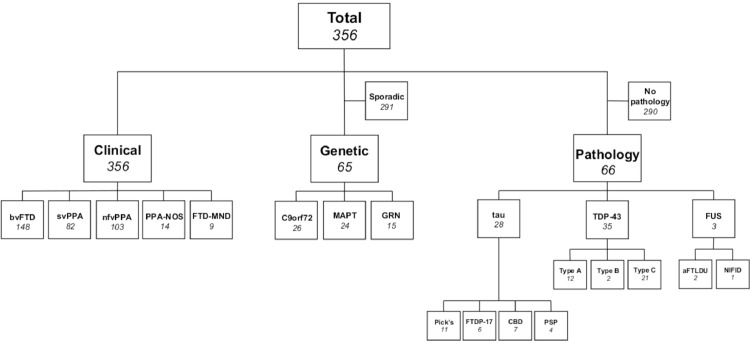
Methods: 356 patients with FTD were recruited from the UCL Dementia Research Centre and matched on age and gender with 83 cognitively normal controls. All subjects had a T1-weighted MR scan suitable for analysis. Basal forebrain volumes were calculated using the Geodesic Information Flow (GIF) parcellation method and were compared between clinical (148 bvFTD, 82 svPPA, 103 nfvPPA, 14 PPA-NOS, 9 FTD-MND), genetic (24 MAPT, 15 GRN, 26 C9orf72) and pathological groups (28 tau, 3 FUS, 35 TDP-43) and controls. A subanalysis was also performed comparing pathological subgroups of tau (11 Pick's disease, 6 FTDP-17, 7 CBD, 4 PSP) and TDP-43 (12 type A, 2 type B, 21 type C).
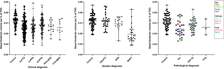
Results: All clinical subtypes of FTD showed significantly smaller volumes than controls (p ≤ 0.010, ANCOVA), with svPPA (10% volumetric difference) and bvFTD (9%) displaying the smallest volumes. Reduced basal forebrain volumes were also seen in MAPT mutations (18%, p < 0.0005) and in individuals with pathologically confirmed FTDP-17 (17%), Pick's disease (12%), and TDP-43 type C (8%) (p < 0.001).
Conclusion: Involvement of the basal forebrain is a common feature in FTD, although the extent of volume reduction differs between clinical, genetic, and pathological diagnoses. Tauopathies, particularly those with MAPT mutations, had the smallest volumes. However, atrophy was also seen in those with TDP-43 type C pathology (most of whom have svPPA clinically). This suggests that the basal forebrain is vulnerable to multiple types of FTD-associated protein inclusions.
Keywords: Frontotemporal dementia; MRI, Basal forebrain; Volumetry.
Copyright © 2020 The Authors. Published by Elsevier Inc. All rights reserved.
Figures
References
-
- Baker M., Mackenzie I.R., Pickering-Brown S.M., Gass J., Rademakers R., Lindholm C., Snowden J., Adamson J., Sadovnick A.D., Rollinson S., Cannon A., Dwosh E., Neary D., Melquist S., Richardson A., Dickson D., Berger Z., Eriksen J., Robinson T., Zehr C., Dickey C.A., Crook R., McGowan E., Mann D., Boeve B., Feldman H., Hutton M. Mutations in progranulin cause tau-negative frontotemporal dementia linked to chromosome 17. Nature. 2006;442:916–919. - PubMed
-
- Baxter M.G., Chiba A.A. Cognitive functions of the basal forebrain. Curr. Opin. Neurobiol. 1999;9:178–183. - PubMed
-
- Bergeron D., Gorno-Tempini M.L., Rabinovici G.D., Santos-Santos M.A., Seeley W., Miller B.L., Pijnenburg Y., Keulen M.A., Groot C., van Berckel B.N.M., van der Flier W.M., Scheltens P., Rohrer J.D., Warren J.D., Schott J.M., Fox N.C., Sanchez-Valle R., Grau-Rivera O., Gelpi E., Seelaar H., Papma J.M., van Swieten J.C., Hodges J.R., Leyton C.E., Piguet O., Rogalski E.J., Mesulam M.M., Koric L., Nora K., Pariente J., Dickerson B., Mackenzie I.R., Hsiung G.-Y.R., Belliard S., Irwin D.J., Wolk D.A., Grossman M., Jones M., Harris J., Mann D., Snowden J.S., Chrem-Mendez P., Calandri I.L., Amengual A.A., Miguet-Alfonsi C., Magnin E., Magnani G., Santangelo R., Deramecourt V., Pasquier F., Mattsson N., Nilsson C., Hansson O., Keith J., Masellis M., Black S.E., Matías-Guiu J.A., Cabrera-Martin M.-.N., Paquet C., Dumurgier J., Teichmann M., Sarazin M., Bottlaender M., Dubois B., Rowe C.C., Villemagne V.L., Vandenberghe R., Granadillo E., Teng E., Mendez M., Meyer P.T., Frings L., Lleó A., Blesa R., Fortea J., Seo S.W., Diehl-Schmid J., Grimmer T., Frederiksen K.S., Sánchez-Juan P., Chételat G., Jansen W., Bouchard R.W., Laforce R.J., Visser P.J., Ossenkoppele R. Prevalence of amyloid-β pathology in distinct variants of primary progressive aphasia. Ann. Neurol. 2018;84:729–740. - PMC - PubMed
-
- Bierer L.M., Haroutunian V., Gabriel S., Knott P.J., Carlin L.S., Purohit D.P., Perl D.P., Schmeidler J., Kanof P., Davis K.L. Neurochemical correlates of dementia severity in Alzheimer’s disease: relative importance of the cholinergic deficits. J. Neurochem. 2002;64:749–760. - PubMed
Publication types
MeSH terms
Grants and funding
LinkOut - more resources
Full Text Sources
Miscellaneous
