. 2020 Mar 4;21(5):1759.
doi: 10.3390/ijms21051759.

A Novel Way of Preventing Postoperative Pancreatic Fistula by Directly Injecting Profibrogenic Materials into the Pancreatic Parenchyma

Affiliations

A Novel Way of Preventing Postoperative Pancreatic Fistula by Directly Injecting Profibrogenic Materials into the Pancreatic Parenchyma

Sang Chul Lee et al. Int J Mol Sci. .

Abstract

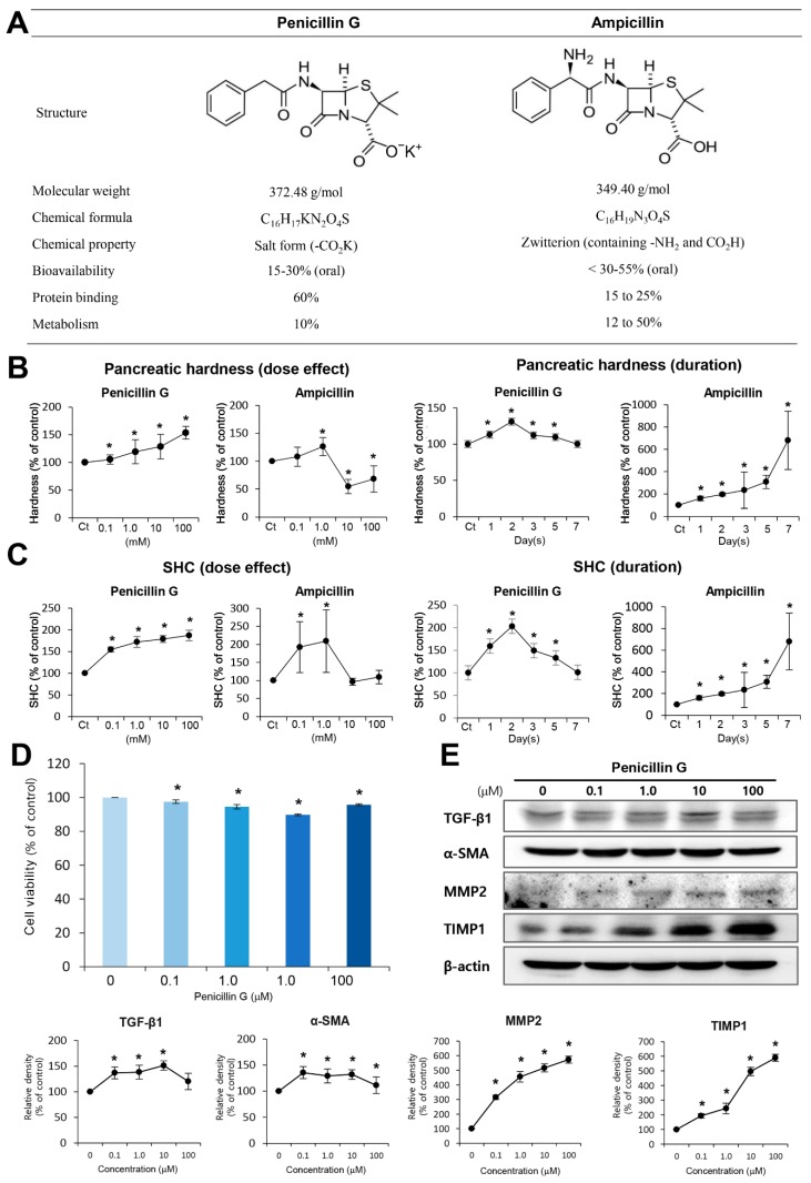
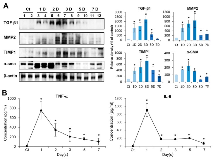
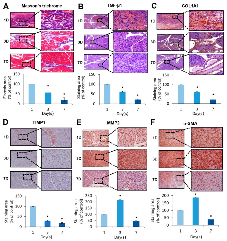
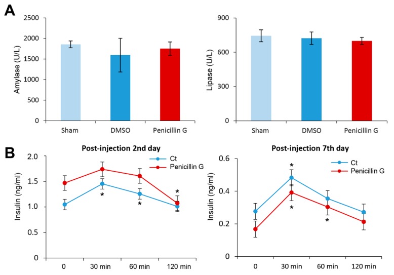
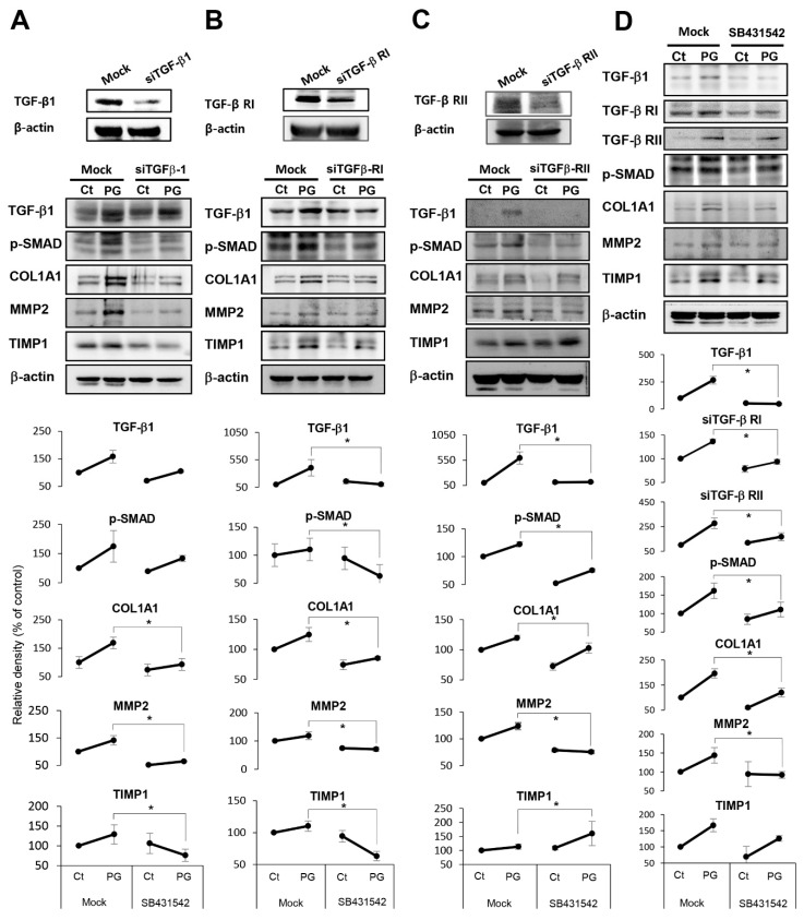
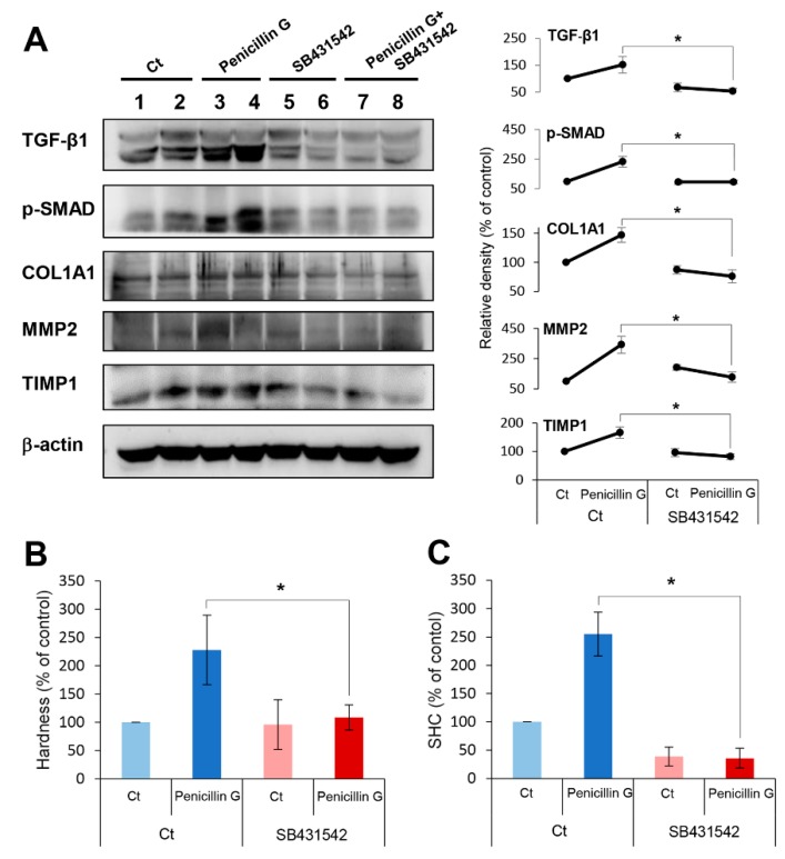
This paper aims to validate if intrapancreatic injection of penicillin G can enhance hardness and suture holding capacity (SHC) of the pancreas through prompting the fibrosis process. Soft pancreatic texture is constantly mentioned as one of the most contributory predictors of postoperative pancreatic fistula (POPF). Soft pancreas has poor SHC and higher incidence of parenchymal tearing, frequently leading to POPF. From a library of 114 antibiotic compounds, we identified that penicillin G substantially enhanced pancreatic hardness and SHC in experimental mice. Specifically, we injected penicillin G directly into the pancreas. On determined dates, we measured the pancreatic hardness and SHC, respectively, and performed molecular and histological examinations for estimation of the degree of fibrosis. The intrapancreatic injection of penicillin G activated human pancreatic stellate cells (HPSCs) to produce various fibrotic materials such as transforming growth factor-β1 (TGF-β1) and metalloproteinases-2. The pancreatic hardness and SHC were increased to the maximum at the second day after injection and then it gradually subsided demonstrating its reversibility. Pretreatment of mice with SB431542, an inhibitor of the TGF-β1 receptor, before injecting penicillin G intrapancreatically, significantly abrogated the increase of both pancreatic hardness and SHC caused by penicillin G. This suggested that penicillin G promotes pancreatic fibrosis through the TGF-β1 signaling pathway. Intrapancreatic injection of penicillin G promotes pancreatic hardness and SHC by enhancing pancreatic fibrosis. We thus think that penicillin G could be utilized to prevent and minimize POPF, after validating its actual effectiveness and safety by further studies.

Keywords: fibrosis; pancreas texture; pancreatic stellate cells; penicillin G; postoperative pancreatic fistula (POPF); transforming growth factor-β1.


Conflict of interest statement

The authors declare no conflict of interest.

Figures

Figure 1
Figure 1
Comparison of pancreas-hardening capacity and SHC between penicillin G and ampicillin. (A) Molecular structure of penicillin G (Left) and ampicillin (Right). Ampicillin differs from penicillin G only by the presence of an amino group. (B) Pancreas-hardening capacity between penicillin G and ampicillin. [Top] Dose-dependent effects of penicillin G (Left) and ampicillin (Right) on hardening pancreas. Whereas penicillin G increased the pancreatic hardness dose-dependently, ampicillin showed the pattern of increasing the pancreatic hardness, especially at a certain concentration (1 mM). [Bottom] Alterations of pancreatic hardness over time between ampicillin-injected (Left) and penicillin G-injected (Right) mice. While the pancreatic hardness increased to the maximum at the second day in the penicillin G-injected mice, the hardness of the pancreas gradually increased over time until the seventh post-injection day in the ampicillin-injected mice. (C) Suture-holding capacity (SHC) of the pancreas between varying concentrations of penicillin G and ampicillin [Top] Dose-dependent effects of SHC of the pancreas between penicillin G [Left] and ampicillin [Right]. Whereas penicillin G increased the SHC of the pancreas dose-dependently, ampicillin increased it at certain concentrations (0.1 and 1.0 mM) only. [Bottom] Alterations of SHC of the pancreas over time between ampicillin-injected [Left] and penicillin G-injected [Right] mice. Whereas the SHC of the pancreas increased to the maximum at the second day after injection in the penicillin G-injected mice, it gradually increased over time until the seventh post-injection day in the ampicillin-injected mice. (D) Cell viability assay of penicillin G using human pancreatic stellate cells (HPSCs). Penicillin G did not decrease the viability of HPSCs within the tested concentrations (100 nM–100 μM) (E) Western blot analysis of HPSCs after treatment of penicillin G. Penicillin G increased the expression of fibrosis-related proteins, such as TGFβ-1, α-SMA, MMP2, and TIMP1, dose-dependently. Values are presented as mean ± standard deviation of three independent experiments. * p < 0.05. Abbreviations: α-SMA, alpha smooth muscle actin; DMSO, dimethyl sulfoxide; MMP-2, metalloproteinases-2; HPSCs, pancreatic stellate cells; SHC; suture-holding capacity; TGF-β1, transforming growth factor-β1; TIMP1, tissue inhibitor of metalloproteinases-1.
Figure 2
Figure 2
In vivo determination of profibrotic property of penicillin G. (A) Western blot analysis of mouse pancreas after direct injection of penicillin G into mouse pancreas over time. The expression of TGF-β1, MMP2, and TIMP1 in the pancreas gradually increased and peaked after three days of injection with penicillin, and then decreased gradually. The expression of α-SMA peaked at the second day after injection and gradually decreased thereafter. (B) Enzyme linked immunosorbent assay (ELISA) of serum pro-inflammatory cytokines (IL-6 and TNF-α) after injection of penicillin G over time. The levels of IL-6 and TNF-α peaked at day 1 post-injection with penicillin, and almost subsided by seven days post-injection. Values are presented as mean ± standard deviation of three independent experiments. * p < 0.05. Abbreviations: α-SMA, alpha smooth muscle actin; MMP-2, metalloproteinases-2; TGF-β1, transforming growth factor-β1, TIMP-1, tissue inhibitor of metalloproteinases-1, TNF-α, tumor necrosis factor-α.
Figure 3
Figure 3
Histological changes induced by the injection of penicillin G. (A) Masson-trichrome stains of mouse pancreas after injection of penicillin G. The degree of fibrosis was highest at the first day after injection and decreased gradually thereafter until seven days after injection. (BF) TGF-β1, COL1A1, TIMP1, MMP2, and α-SMA immunohistochemistry of mouse pancreas after injecting penicillin G. In TGF-β1, COL1A1, and TIMP1 immunohistochemistry, the expression of the markers was the highest on the first day after injection, and gradually decreased thereafter until seven days after injection. In MMP2 and α-SMA immunohistochemistry, the expression of the markers was highest at three days after injection and decreased thereafter until seven days after injection. Values are presented as mean ± standard deviation of three independent experiments. * p < 0.05. Abbreviations: α-SMA, alpha smooth muscle actin; COL1A1, collagen type 1 alpha 1; MMP-2, metalloproteinases-2; TGF-β1, transforming growth factor-β1, TIMP-1, tissue inhibitor of metalloproteinases-1.
Figure 4
Figure 4
The effects of intrapancreatic penicillin G injection on pancreatic function. (A) Serum levels of amylase and lipase after intrapancreatic penicillin G injection. Injecting penicillin G into the pancreas did not significantly alter the serum levels of amylase or lipase. (B) Serial measurement of serum levels of insulin at certain time intervals by enzyme-linked immunosorbent assay (ELISA) after glucose infusion on the second and seventh day of penicillin G injection. Penicillin G-injected mice had slightly less insulin secretion than the control group although it was not statistically significant. Values are presented as mean ± standard deviation of three independent experiments. * p < 0.05. Abbreviation: DMSO, dimethyl sulfoxide.
Figure 5
Figure 5
In vitro demonstration of mechanism leading to pancreatic fibrosis by penicillin G. (A) Western blot analysis of HPSCs after treating penicillin G followed by addition of TGF-β1 siRNA. The addition of TGF-β1 siRNA significantly abrogated profibrotic effects of penicillin G, demonstrated by the lower expression of profibrotic markers, such as collagen, MMP2, and p-SMAD. (B) Western blot analysis of HPSCs after treatment with penicillin G followed by addition of TGF-β receptor I (TGF-β RI) siRNA. The addition of TGF-β1 siRNA significantly abrogated profibrotic effects of penicillin G. (C) Western blot analysis of HPSCs after injecting penicillin G followed by addition of TGF-β receptor II (TGF-β RI) siRNA. The addition of TGF-β1 RI siRNA also significantly abrogated profibrotic effects of penicillin (D) Alterations in fibrosis-related factors after treatment of pancreatic tissue with Penicillin G combined with SB431542, an inhibitor of the TGF-β1 receptor. Treatment with SB431542 significantly abrogated the expression of all fibrosis-related factors that had been increased by Penicillin G. * p < 0.05.
Figure 6
Figure 6
In vivo demonstration of mechanism leading to pancreatic fibrosis by penicillin G. Mice were injected with SB431542, an inhibitor of the TGF-β1 receptor intraperitoneally, followed by an intra-pancreatic injection of penicillin G. (A) Western blot analysis showing the expression of fibrosis-related markers in the mouse pancreas. Pretreatment with SB431542 in mice injected with intrapancreatic penicillin G significantly reduced the expression of fibrosis-related markers, such as TGF-β1, p-SMAD, collagen, MMP2, and TIMP1, in comparison with mice without the pretreatment (p < 0.05). (B) Measurement of pancreatic hardness. Pretreatment of SB431542 significantly abrogated the pancreatic hardening capacity by penicillin G in the mouse pancreas. (C) Measurement of SHC of the pancreas. Pretreatment of SB431542 significantly abrogated the enhanced SHC induced by penicillin G in the mouse pancreas. Values are presented as mean ± standard deviation of three independent experiments. * p < 0.05. Abbreviations: Ct, control; MMP-2, metalloproteinases-2; p-SMAD2, phosphorylated Smad2; SHC, suture holding capacity; TGF-β1, transforming growth factor-β1; TIMP-1, tissue inhibitor of metalloproteinases-1.

Similar articles

Cited by

References

    1. Bassi C., Dervenis C., Butturini G., Fingerhut A., Yeo C., Izbicki J., Neoptolemos J., Sarr M., Traverso W., Buchler M., et al. Postoperative pancreatic fistula: An international study group (ISGPF) definition. Surgery. 2005;138:8–13. doi: 10.1016/j.surg.2005.05.001. - DOI - PubMed
    1. Schafer M., Mullhaupt B., Clavien P.A. Evidence-based pancreatic head resection for pancreatic cancer and chronic pancreatitis. Ann. Surg. 2002;236:137–148. doi: 10.1097/00000658-200208000-00001. - DOI - PMC - PubMed
    1. Buchler M.W., Friess H., Wagner M., Kulli C., Wagener V., Z’Graggen K. Pancreatic fistula after pancreatic head resection. Br. J. Surg. 2000;87:883–889. doi: 10.1046/j.1365-2168.2000.01465.x. - DOI - PubMed
    1. Cullen J.J., Sarr M.G., Ilstrup D.M. Pancreatic anastomotic leak after pancreaticoduodenectomy: Incidence, significance, and management. Am. J. Surg. 1994;168:295–298. doi: 10.1016/S0002-9610(05)80151-5. - DOI - PubMed
    1. Miedema B.W., Sarr M.G., van Heerden J.A., Nagorney D.M., McIlrath D.C., Ilstrup D. Complications following pancreaticoduodenectomy. Current management. Arch. Surg. 1992;127:945–949. doi: 10.1001/archsurg.1992.01420080079012. - DOI - PubMed

MeSH terms

Substances