. 2020 Mar 6;20(1):73.
doi: 10.1186/s12906-020-2849-1.

Inhibition of alpha-synuclein seeded fibril formation and toxicity by herbal medicinal extracts

Affiliations

Inhibition of alpha-synuclein seeded fibril formation and toxicity by herbal medicinal extracts

Mustafa T Ardah et al. BMC Complement Med Ther. .

Retraction in

Abstract

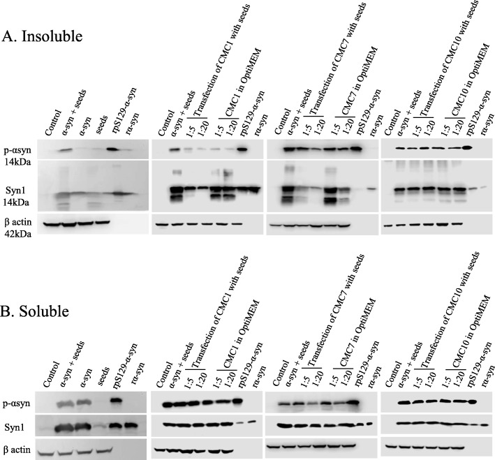
Background: Recent studies indicated that seeded fibril formation and toxicity of α-synuclein (α-syn) play a main role in the pathogenesis of certain diseases including Parkinson's disease (PD), multiple system atrophy, and dementia with Lewy bodies. Therefore, examination of compounds that abolish the process of seeding is considered a key step towards therapy of several synucleinopathies.

Methods: Using biophysical, biochemical and cell-culture-based assays, assessment of eleven compounds, extracted from Chinese medicinal herbs, was performed in this study for their effect on α-syn fibril formation and toxicity caused by the seeding process.

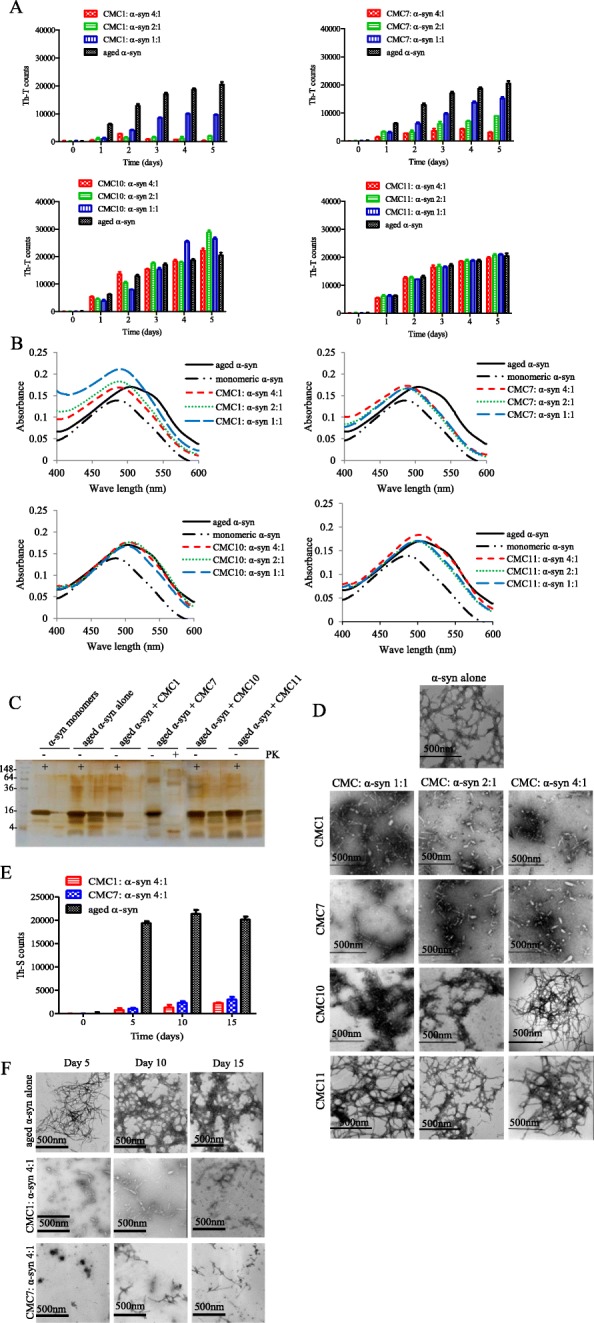
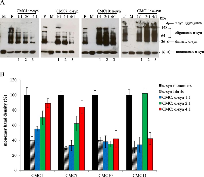
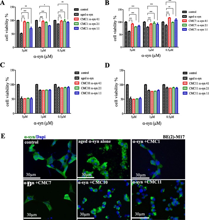
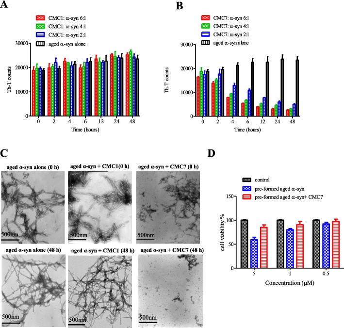
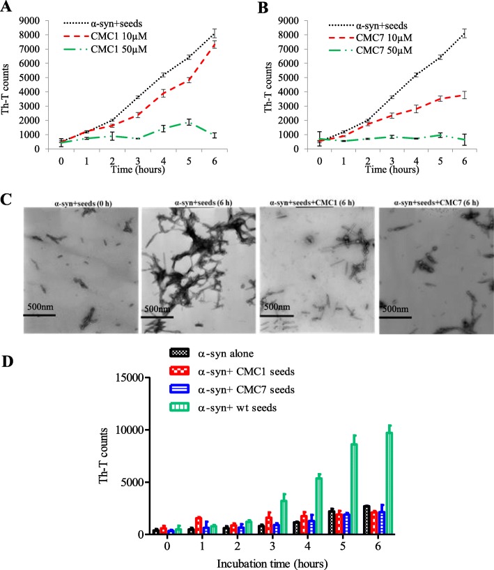
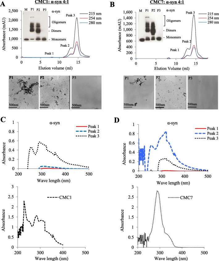
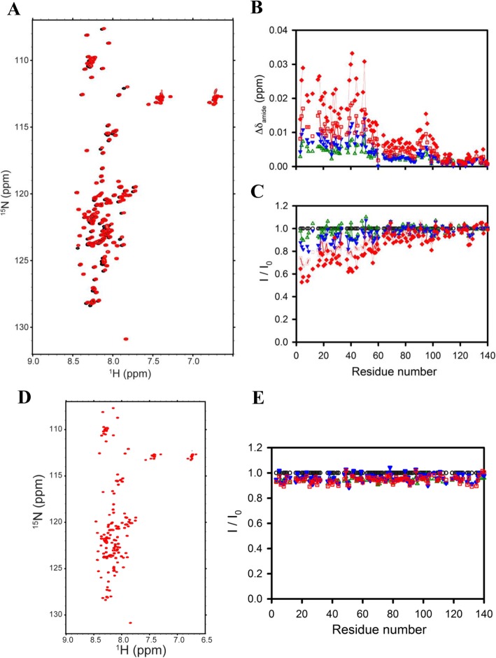
Results: Salvianolic acid B and dihydromyricetin were the two compounds that strongly inhibited the fibril growth and neurotoxicity of α-syn. In an in-vitro cell model, these compounds decreased the insoluble phosphorylated α-syn and aggregation. Also, in primary neuronal cells, these compounds showed a reduction in α-syn aggregates. Both compounds inhibited the seeded fibril growth with dihydromyricetin having the ability to disaggregate preformed α-syn fibrils. In order to investigate the inhibitory mechanisms of these two compounds towards fibril formation, we demonstrated that salvianolic acid B binds predominantly to monomers, while dihydromyricetin binds to oligomeric species and to a lower extent to monomers. Remarkably, these two compounds stabilized the soluble non-toxic oligomers lacking β-sheet content after subjecting them to proteinase K digestion.

Conclusions: Eleven compounds were tested but only two showed inhibition of α-syn aggregation, seeded fibril formation and toxicity in vitro. These findings highlight an essential beginning for development of new molecules in the field of synucleinopathies treatment.

Keywords: Amyloid fibrils; Dihydromyricetin; Parkinson’s disease; Salvianolic acid B; Seeded fibril formation; α-Synuclein.


Conflict of interest statement

The authors declare that they have no competing interests.

Figures

Fig. 1
Fig. 1
Chemical structures of eleven naturally derived Chinese medicinal compounds used to test the effect of α-syn fibril formation. These compounds are designated as CMC 1–11
Fig. 2
Fig. 2
Effect of CMCs on α-syn fibril formation. a and b Fibril content formation analysis. α-syn protein (25 μM) was incubated for 5 days at 37 °C with continuous shaking in the absence (aged alone) or presence of CMC1, CMC7, CMC10, and CMC11 using different molar ratios (CMC: α-syn at 1:1, 2:1, and 4:1). Fibril content for each sample was then measured using a Thioflavin-T (Th-T) and b Congo red binding assays. Means ± standard deviations are from triplicates of one experiment. c Silver staining for SDS-PAGE of α-syn monomers, α-syn aged alone or in the presence of CMC1, CMC7, CMC10 and CMC11 at molar ratio 1:1 after Protein Kinase (PK) digestion. d Negative stain electron microscopy images showing fibril formation of α-syn aged alone or in the presence of the indicated CMCs (CMC: α-syn molar ratios of 1:1, 2:1, and 4:1). Scale bar, 500 nm. e Th-T binding assay for α-syn aged alone or with CMC1 or CMC7 for 5, 10 and 15 days at a molar ratio of 4:1 (CMC: α-syn). Means ± standard deviations are from the triplicates of one experiment. f Negative stain electron microscopy images showing fibril formation of α-syn aged alone or in the presence of CMC1 and CMC7 using molar ratio of 4:1 (CMC: α-syn) for 5, 10 and 15 days. Scale bar, 500 nm
Fig. 3
Fig. 3
Effect of CMCs on α-syn oligomerization. a Immunoblot analysis of different species of α-syn (monomeric, dimeric, oligomeric, and aggregates). Different species of α-syn were detected in samples collected from α-syn aged alone or in the presence of CMC1, CMC7, CMC10 and CMC11 at CMC: α-syn molar ratios of 1:1, 2:1 and 4:1 for 5 days. F: α-syn fibrils, M: monomeric α-syn, Lane 1, CMC: α-syn 1:1, lane 2, CMC: α-syn 2:1, lane 3, CMC: α-syn 4:1, b Band density representing the amount of monomeric α-syn was quantified for each sample using ImageJ software
Fig. 4
Fig. 4
Effect of CMCs on cell toxicity of α-syn aggregates. a-d MTT cell viability analysis. The viability of BE(2)-M17 human cells treated with either α-syn aged alone or with a CMC1, b CMC7, c CMC110, and d CMC11 was tested by MTT assay. Cells were treated with the indicated concentrations of α-syn and CMCs for 48 h prior to the addition of MTT. The results are expressed as percentages of the average of the control (i.e., untreated cells). Means ± standard deviations are from the average of 3 wells. Statistical analysis was performed using a two-tailed unpaired t-test. ***, p < 0.001; **, p < 0.01; *, p < 0.05. e Immunocytochemistry against α-syn (green) and DAPI (blue) in BE(2)-M17 cells treated with aggregated α-syn. The cells were either non-treated or treated for 48 h with 5 μM of α-syn aged alone or in the presence of CMC1, CMC7, CMC10, and CMC11at a molar ratio of 1:4. Scale bar 30 μm
Fig. 5
Fig. 5
Effect of CMC1 and CMC7 on performed α-syn fibrils. a and b Th-T binding assay used to measure the fibril content resulted from the incubation of aggregated α-syn for the indicated times in the absence or presence of a CMC1 and b CMC7 using different molar ratios (CMC: α-syn at 6:1, 4:1, and 2:1). The assays were performed in triplicates (Means ± standard deviations are from the average of the triplicates). c Electron microscopy images of negatively stained samples of aged α-syn incubated alone or in the presence of CMC1 and CMC7 (CMC: α-syn at 6:1) for 0 h (upper panels) or 48 h (lower panels) with continuous shaking at 37 °C. Scale bar, 500 nm. d Cell viability of BE(2)-M17 cells was tested using MTT assay. Cells were treated for 48 h with pre-formed α-syn fibrils or with the pre-formed fibrils incubated with CMC7 prior to the addition of MTT. Results are expressed as percentages of the average of the control (i.e., untreated cells). Means ± standard deviations are from the average of 3 wells
Fig. 6
Fig. 6
Effect of CMC1 and CMC7 on the seeding of α-syn monomers with fibrils. α-syn monomers (100 μM) were seeded with 2 μM sonicated α-syn fibrils, which were then incubated in the presence or absence of a CMC1 and b CMC7 at different concentrations (10 and 50 μM) for 6 h with continuous shaking at 37 °C. The extent of fibril formation was estimated by Th-T binding assay. The assays were performed in triplicate (average of triplicate measurements ± standard deviations). c Electron microscopy images of negatively stained samples of α-syn incubated with seeds alone or in the presence of CMC1 or CMC7 in a concentration of 50 μM for the indicated times with continuous shaking at 37 °C. Scale bar, 500 nm. d Th-T binding assay for seeding the aggregation of α-syn monomers by either 2 μM of untreated seeds (wt seeds) or seeds generated from the incubation of CMC1 or CMC7 with α-syn fibrils for 5 days at a molar ratio of 4:1
Fig. 7
Fig. 7
CMC1 and CMC7 binding activity to α-syn monomers and oligomers. a and b Upper panels: Gel filtration profiles of 100 μM α-syn sample incubated with a CMC1 and b CMC7 at 4:1 M ratio (CMC1: α-syn) for 5 days as described in Materials and Methods using Superdex 200 SE column. Peak 1 (P1) and Peak 2 (P2) represent the oligomers species while Peak 3 (P3) represents the monomeric species. The elution was monitored at absorbance wavelengths of A215,A254 and A280. The inset shows immunoblot analysis of different α-syn species (monomers, dimers, and oligomers) separated from the pooled fractions of P1, P2 and P3. Lower panels: Electron microscopy images showing α-syn species resulted from the indicated peaks. c UV absorbance of α-syn P1, P2 and P3 samples collected from the gel filtration chromatography resulted from the incubation of α-syn with CMC1 (left panel) or CMC7 (right panel). d UV absorbance of CMC1 alone (left panel) and CMC7 (right panel). Samples were placed in a10 mm quartz cuvette and the UV absorbance spectra were recorded from 200 nm to 600 nm
Fig. 8
Fig. 8
Analysis of CMC1 and CMC7 binding to monomeric α-syn by NMR spectroscopy. a Proton-nitrogen correlation (HSQC) spectra b averaged NMR chemical shift perturbation of 1H/15N resonances, and c Residue-specific NMR signal intensity ratios of α-syn in the absence and presence of increasing stoichiometries of CMC1 (CMC1: α-syn of 0:1, 1:1, 2:1, 4:1, 6:1, 10:1, and 20:1). d Proton-nitrogen correlation (HSQC) spectra and e Residue-specific NMR signal intensity ratios of α-syn in the absence and presence of increasing stoichiometries of CMC7 (CMC7: α-syn of 0:1, 1:1, 2:1, 4:1, 6:1, and 10:1)
Fig. 9
Fig. 9
Effects of CMC1, CMC7, and CMC10 on insoluble pS129-α-syn and aggregation. Insoluble pS129 and aggregation of α-syn were assessed in 10 μg and 15 μg of insoluble a and soluble b proteins from cell lysates of untransfected (control) and transfected HEK cells by immunoblotting proteins using antibodies specific to pS129-α-syn and total α-syn (Syn1). While one group of wt α-syn transfected HEK cells were simultaneously transfected with seeds and CMC at different molar ratios (1:5 and 1:20) after an incubation at 37 °C for 1 h, the other group was transfected with seeds to be followed by CMC treatment at different molar ratios (1:5 and 1:20) in OptiMEM for 48 h. Recombinant pS129-α-syn (rpS129-α-syn) and recombinant α-syn (r-α-syn) proteins were loaded (50 ng) as positive controls. Re-immunoblotting with β-actin antibody was performed to normalize the amount of loaded proteins

References

    1. Obeso JA, Rodriguez-Oroz MC, Benitez-Temino B, Blesa FJ, Guridi J, Marin C, et al. Functional organization of the basal ganglia: therapeutic implications for Parkinson’s disease. Mov Disord. 2008;23(Suppl 3):S548–S559. doi: 10.1002/mds.22062. - DOI - PubMed
    1. Galvin JE, Lee VM, Schmidt ML, Tu PH, Iwatsubo T, Trojanowski JQ. Pathobiology of the Lewy body. Adv Neurol. 1999;80:313–324. - PubMed
    1. Spillantini MG, Crowther RA, Jakes R, Hasegawa M, Goedert M. Alpha-Synuclein in filamentous inclusions of Lewy bodies from Parkinson's disease and dementia with lewy bodies. Proc Natl Acad Sci U S A. 1998;95(11):6469–6473. doi: 10.1073/pnas.95.11.6469. - DOI - PMC - PubMed
    1. Goedert M, Spillantini MG, Del Tredici K, Braak H. 100 years of Lewy pathology. Nat Rev Neurol. 2013;9(1):13–24. doi: 10.1038/nrneurol.2012.242. - DOI - PubMed
    1. Kim WS, Kågedal K, Halliday GM. Alpha-synuclein biology in Lewy body diseases. Alzheimers Res Ther. 2014;6(5):73. doi: 10.1186/s13195-014-0073-2. - DOI - PMC - PubMed

Publication types

LinkOut - more resources