. 2020 Mar 6;49(1):13.
doi: 10.1186/s40463-020-00408-7.

Endoscopic cartilage myringoplasty with the removal of a small rim of the external auditory canal to repair marginal perforations

Affiliations

Endoscopic cartilage myringoplasty with the removal of a small rim of the external auditory canal to repair marginal perforations

Zheng-Cai Lou. J Otolaryngol Head Neck Surg. .

Abstract

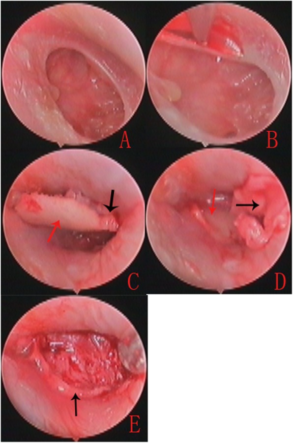
Objective: To evaluate the graft success rate and postoperative hearing gain for marginal perforations using endoscopic cartilage myringoplasty with the removal of a small rim of the external auditory canal (EAC).

Study design: Prospective case series.

Materials and methods: We performed a prospective study in 41 patients with marginal perforations who underwent endoscopic cartilage myringoplasty with the removal of a small rim of EAC. Patients were followed up for 6 months.

Results: Of the 41 patients with unilateral marginal perforation included in this study, the graft success rate was 100% (41/41). The mean ABG improved from 11.31 ± 9.71 dB preoperatively to 7.31 ± 2.32 dB postoperatively for small-and medium-sized perforations (P = 0.13); the mean ABG improved from 21.46 ± 8.39 dB preoperatively to 9.84 ± 2.41 dB postoperatively for large perforations (P < 0.05); the mean ABG improved from 28.79 ± 6.74 dB preoperatively to 10.13 ± 3.56 dB postoperatively for subtotal and total perforations (P < 0.05). There were no cases of graft lateralization or significant blunting or atelectasis or graft adhesions. Three patients developed postoperative otorrhoea and five patients had mild myringitis.

Conclusions: Endoscopic cartilage myringoplasty with the removal of a small rim of the EAC is simple and feasible, showing a high graft success rate and minimal complications for repairing marginal perforations.

Keywords: Cartilage myringoplasty; Endoscopy; Lateralization; Tympanic membrane perforation; Tympanomeatal flap.


Conflict of interest statement

The author declares that he has no competing interests.

Figures

Fig. 1
Fig. 1
Diagram of the marginal perforations not involving the malleus. Tympanic membrane perforation (a); the perforation edges were refreshed and annulus was de-epithelialized, a small rim of EAC was removed (b); the perichondrium was listed off of the cartilage over 2 mm on one end (c); the uncover cartilage was partially removed (d); the cartilage covered by perichondrium was placed medial to the remnant TM, the free perichondrium was placed lateral to the annulus and exposed EAC (e)
Fig. 2
Fig. 2
Diagram of the marginal perforations involving the malleus. Tympanic membrane perforation (a); the perforation edges were refreshed and annulus was de-epithelialized, a small rim of EAC was removed (b); the lateral perichondrium was lifted off of the superior and inferior end, a notch was made for the malleus (c and d); the cartilage graft was placed medial to the remnant TM and the annulus, a notch of cartilage was clipped into the malleus, the free perichondrium was placed lateral to the malleus, the annulus and exposed EAC (e). TM: tympanic membrane; TMP: tympanic membrane perforation; EAC: external auditory canal; MA: malleus handle; PE: perichondrium; CA: cartilage; CAP: cartilage with perichondrium. The black shadow region indicates the excision of a small rim of EAC. Red dotted line indicates the fresh perforation edges. The section E: Black hidden line indicated the underlay cartilage. Pink shadow indicates the overlay perichondrium
Fig. 3
Fig. 3
The operative photos of right large TM perforation in a 32-year-old female patient. Preoperative total TMP (a). The perforation edges were refreshed and annulus was de-epithelialized (b). Cartilage graft was pushed into the EAC and middle ear; the cartilage notch was made for the malleus (c). The cartilage graft was placed medial to the annulus and remanent TM (d). The free perichondrium graft was placed lateral to the malleus, the annulus and exposed EAC (e).Black arrows indicates the perichondrium, red arrows indicates the cartilage and notch
Fig. 4
Fig. 4
Photographs showing the perforation before surgery (a), and at 3 weeks (b), 5 weeks (c), and 6 weeks (d) post-surgery. Please note, this is the same patient as in Fig. 3. The irregular curve indicates the overlay perichondrium area

References

    1. Sharp JF, Terzis TF, Robinson J. Myringoplasty for anterior perforation: experience with the Kerr flap. J Laryngol Otol. 1992;106:14–16. doi: 10.1017/S002221510011847X. - DOI - PubMed
    1. Telian Steven A., Kemink John L. Lateral technique tympanoplasty. Operative Techniques in Otolaryngology-Head and Neck Surgery. 1992;3(4):214–219. doi: 10.1016/S1043-1810(10)80117-4. - DOI
    1. Farrior JB. Sandwich graft tympanoplasty: experience, results, and complications. Laryngoscope. 1989;99:213–217. doi: 10.1288/00005537-198902000-00015. - DOI - PubMed
    1. Kartush JM, Michaelides EM, Becvarovski Z, LaRouere MJ. Over-under tympanoplasty. Laryngoscope. 2002;112:802–807. doi: 10.1097/00005537-200205000-00007. - DOI - PubMed
    1. Jung TT, Park SK. Mediolateral graft tympanoplasty for anterior or subtotal tympanic membrane perforation. Otolaryngol Head Neck Surg. 2005;132:532–536. doi: 10.1016/j.otohns.2004.10.018. - DOI - PubMed