. 2020 Jun;26(6):724-738.
doi: 10.1261/rna.073114.119. Epub 2020 Mar 6.

Reciprocal monoallelic expression of ASAR lncRNA genes controls replication timing of human chromosome 6

Affiliations

Reciprocal monoallelic expression of ASAR lncRNA genes controls replication timing of human chromosome 6

Michael B Heskett et al. RNA. 2020 Jun.

Abstract

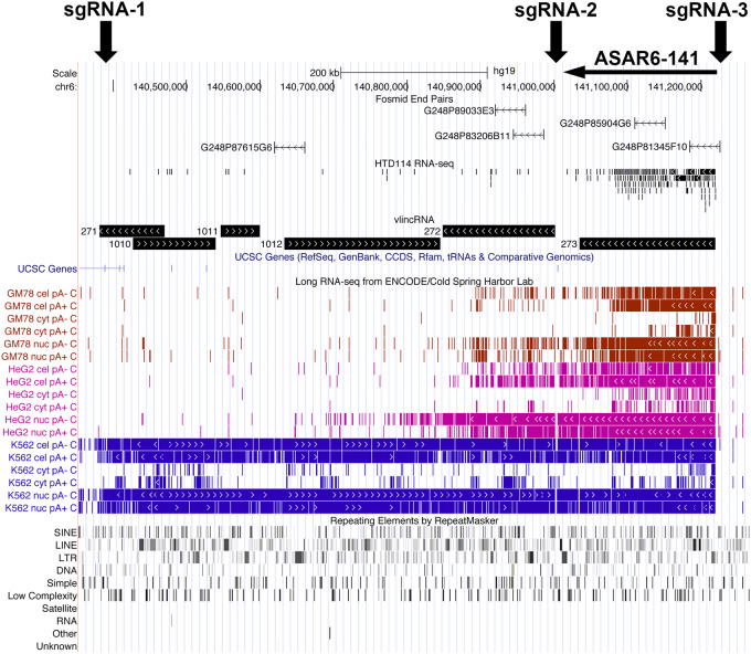
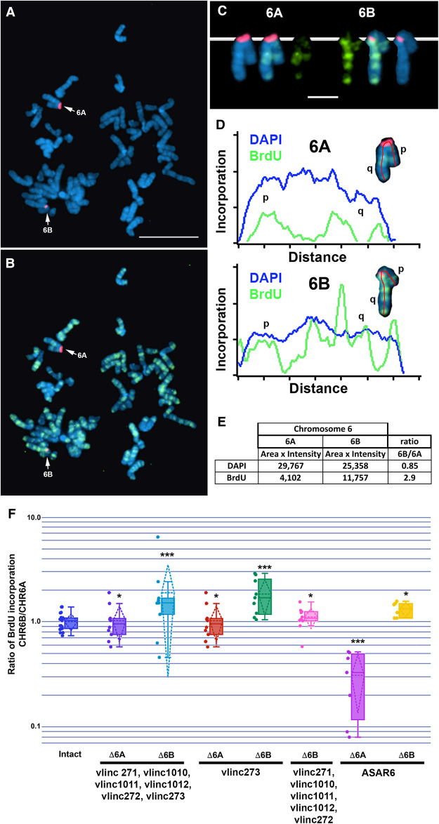
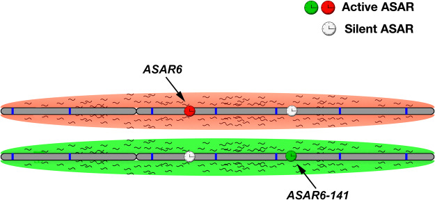
DNA replication occurs on mammalian chromosomes in a cell-type distinctive temporal order known as the replication timing program. We previously found that disruption of the noncanonical lncRNA genes ASAR6 and ASAR15 results in delayed replication timing and delayed mitotic chromosome condensation of human chromosomes 6 and 15, respectively. ASAR6 and ASAR15 display random monoallelic expression and display asynchronous replication between alleles that is coordinated with other random monoallelic genes on their respective chromosomes. Disruption of the expressed allele, but not the silent allele, of ASAR6 leads to delayed replication, activation of the previously silent alleles of linked monoallelic genes, and structural instability of human chromosome 6. In this report, we describe a second lncRNA gene (ASAR6-141) on human chromosome 6 that when disrupted results in delayed replication timing in cisASAR6-141 is subject to random monoallelic expression and asynchronous replication and is expressed from the opposite chromosome 6 homolog as ASAR6 ASAR6-141 RNA, like ASAR6 and ASAR15 RNAs, contains a high L1 content and remains associated with the chromosome territory where it is transcribed. Three classes of cis-acting elements control proper chromosome function in mammals: origins of replication, centromeres, and telomeres, which are responsible for replication, segregation, and stability of all chromosomes. Our work supports a fourth type of essential chromosomal element, the "Inactivation/Stability Center," which expresses ASAR lncRNAs responsible for proper replication timing, monoallelic expression, and structural stability of each chromosome.

Keywords: cis-acting element; noncoding RNA; replication timing.


Figures

FIGURE 1.
FIGURE 1.
UCSC Genome Browser view of the vlinc cluster on chromosome 6 between 140.3 and 141.3 Mb. The genomic locations of vlinc271, vlinc1010, vlinc1011, vlinc1012, vlinc272, and vlinc273 are illustrated using the UCSC Genome Browser (see Supplemental Table S4 for genomic locations). RNA-seq data from nuclear RNA isolated from HTD114 is shown (HTD114 RNA-seq). Long RNA-seq data from the ENCODE Project (Cold Spring Harbor Laboratory) is shown using the Contigs view. Expression from the human cell lines GM12878 (red), HepG2 (magenta), and K562 (blue) are shown. RNA from total cellular Poly A+ (cel pA+), total cellular Poly A− (cel pA−), nuclear Poly A+ (nuc pA+), nuclear Poly A− (nuc pA−), cytoplasmic Poly A+ (cyt pA+), and cytoplasmic Poly A− (cyt pA−) are shown. Also shown are the repeating elements using the RepeatMasker track. The location of five RNA FISH probes (Fosmids) that were used to detect the expression of vlinc1012 (G248P87615G6), vlinc272 (G248P89033E3 and G248P83206B11), and vlinc273 (G248P85904G6 and G248P81345F10) are shown.
FIGURE 2.
FIGURE 2.
Monoallelic expression and nuclear retention of vlinc273 in HTD114 cells. (A) DNA sequencing traces from PCR products designed to detect SNPs rs989613623 and rs2328092 (see Supplemental Fig. S1A; Supplemental Table S2). PCRs were carried out on genomic DNAs isolated from HTD114, two monochromosomal hybrids containing the two different chromosome 6s from HTD114 [L(Hyg)-1 contains chromosome 6A (CHR6A) and expresses ASAR6, and L(Neo)-38 contains chromosome 6B (CHR6B) and is silent for ASAR6 (Stoffregen et al. 2011)]. The top and bottom panels also show the sequencing traces from HTD114 cDNA (RNA). The asterisks mark the location of the heterozygous SNPs. (BF) RNA–DNA FISH to detect vlinc273 expression in HTD114 cells. Fosmid G248P81345F10 was used as probe to detect vlinc273 RNA (green), and a chromosome 6 centromeric probe was used to detect chromosome 6 DNA (red). The nuclear DNA was stained with DAPI. Bars are 2.5 µM. (GJ) RNA–DNA FISH to detect vlinc273 and ASAR6 expression in HTD114 cells. Fosmid G248P81345F10 was used as probe to detect vlinc273 RNA (green), Fosmid G248P86031A6 was used as probe to detect ASAR6 RNA (red), and a chromosome 6 paint was used to detect chromosome 6 DNA (magenta). The nuclear DNA was stained with DAPI. Bars are 2.5 µM. Quantitation of the number of RNA FISH signals is shown in Supplemental Figure S2A and B.
FIGURE 3.
FIGURE 3.
Monoallelic expression and nuclear retention of vlinc273 in EBV transformed lymphoblasts and PBLs. (AE) RNA–DNA FISH to detect vlinc273 expression in GM12878 EBV transformed lymphocytes. Fosmid G248P81345F10 was used to detect vlinc273 RNA (green), and a chromosome 6 centromeric probe (CHR6 cen) was used to detect chromosome 6 DNA (red). (FJ) RNA FISH to detect the coordinated expression of vlinc273 and KCNQ5, a known random monoallelic gene, in PBLs. Fosmid G248P81345F10 was used to detect vlinc273 RNA (green), and Fosmid G248P80791F6 was used to detect the expression of the first intron of KCNQ5. (K,L) RNA FISH to detect the expression of vlinc273 and XIST RNAs, in female PBLs. The nuclear DNA was stained with DAPI. Bars are 2.5 µM. Quantitation of the number of RNA FISH signals per nucleus and the cis versus trans expression is shown in Supplemental Figure S2B–D.
FIGURE 4.
FIGURE 4.
Coordinated asynchronous replication timing on chromosome 6. (A) Schematic representation of the ReTiSH assay. Cells were exposed to BrdU during the entire length of S phase (14 h) or only during late S phase (5 h). The ReTiSH assay can distinguish between alleles that replicate early (E) and late (L) in S phase. (BD) Mitotic spreads from human PBLs were processed for ReTiSH and hybridized with three different FISH probes. First, each hybridization included a centromeric probe to chromosome 6 (magenta). Arrows mark the centromeric signals in panels B (5 h) and C (14 h). Each assay also included BAC probes representing ASAR6 (RP11-374I15; green) and vlinc273 (RP11-715D3; red). Panel D shows the two chromosome 6s, from both the 5 and 14 h time points, aligned at their centromeres. The ASAR6 BAC and the vlinc273 BAC show hybridization signals on the same chromosome 6 at the 5-h time point, and as expected hybridized to both chromosome 6s at the 14-h time point. The chromosomal DNA was stained with DAPI. (EG) ReTiSH assay on HTD114 cells. Each ReTiSH assay included a centromeric probe to chromosome 6 (magenta). Arrows mark the centromeric signals in panels E (5 h) and F (14 h). Each assay also included BAC probes for ASAR6 (RP11-374I15; green) and vlinc273 (RP11-715D3; red). The chromosomal DNA was stained with DAPI. The ASAR6 BAC and the vlinc273 BAC show hybridization signals to one chromosome 6 homolog (CHR6A) at the 5-h time point, and as expected hybridized to both chromosome 6s at the 14-h time point.
FIGURE 5.
FIGURE 5.
Delayed replication of chromosome 6 following disruption of vlinc273. (A,B) A representative mitotic spread from BrdU (green) treated HTD114 cells containing a deletion of the expressed allele of the vlinc273 locus. Mitotic cells were subjected to DNA FISH using a chromosome 6 centromeric probe (red). The larger centromere resides on the chromosome 6 with the expressed ASAR6 allele and the silent vlinc273 allele (6A). (C) The two chromosome 6s were extracted from panels A and B and aligned to show the BrdU incorporation and centromeric signals. (D) Pixel intensity profiles of BrdU incorporation and DAPI staining along the (6A) and (6B) chromosomes from panel C. (E) BrdU quantification along 6A and 6B from panel D. (F) The ratio of DNA synthesis into the two chromosome 6s was calculated by dividing the BrdU incorporation in CHR6B by the incorporation in CHR6A in multiple cells. The dots show the BrdU incorporation ratios for the individual cells assayed. The box plots show the ratio of incorporation before (Intact, dark blue), and in heterozygous deletions of the entire locus (Δ6A dark purple; and Δ6B light blue), which included vlinc271, vlinc1010, vlinc1011, vlinc1012, vlinc272, and vlinc273; see maps in Figure 1 and Supplemental Figure S1A. Heterozygous deletions affecting vlinc273 only from the silent (Δ6A orange) or expressed (Δ6B green) alleles are shown. A heterozygous deletion affecting vlinc271, vlinc1010, vlinc1011, vlinc1012, and vlinc272 on CHR6B (Δ6B) is shown in pink. Also shown are heterozygous deletions affecting ASAR6 from the expressed (Δ6A magenta) or silent (Δ6B yellow) alleles. Error bars are SD. P-values of <1 × 10⁻⁴ are indicated by (***), and P-values of >1 × 10⁻¹ are indicated by (*), and were calculated using the Kruskal–Wallis test.
FIGURE 6.
FIGURE 6.
“ASAR” model of replication timing on chromosome 6. The two homologs of human chromosome 6 are shown (gray) with hypothetical origins of replication depicted as blue bars. Expression of ASAR6 and ASAR6-141 genes is monoallelic, resulting in a reciprocal expression pattern with an expressed or active ASAR (green or red clock) and a silent or inactive ASAR (white clock) on each homolog. The red and green clouds surrounding the chromosomes represent “ASAR” RNA expressed from the different active “ASARs” on each homolog.

References

    1. Alexander MK, Mlynarczyk-Evans S, Royce-Tolland M, Plocik A, Kalantry S, Magnuson T, Panning B. 2007. Differences between homologous alleles of olfactory receptor genes require the Polycomb Group protein Eed. J Cell Biol 179: 269–276. 10.1083/jcb.200706053 - DOI - PMC - PubMed
    1. Allen E, Horvath S, Tong F, Kraft P, Spiteri E, Riggs AD, Marahrens Y. 2003. High concentrations of long interspersed nuclear element sequence distinguish monoallelically expressed genes. Proc Natl Acad Sci 100: 9940–9945. 10.1073/pnas.1737401100 - DOI - PMC - PubMed
    1. Attig J, Agostini F, Gooding C, Chakrabarti AM, Singh A, Haberman N, Zagalak JA, Emmett W, Smith CWJ, Luscombe NM, et al. 2018. Heteromeric RNP assembly at LINEs controls lineage-specific RNA processing. Cell 174: 1067–1081.e17. 10.1016/j.cell.2018.07.001 - DOI - PMC - PubMed
    1. Bailey JA, Carrel L, Chakravarti A, Eichler EE. 2000. Molecular evidence for a relationship between LINE-1 elements and X chromosome inactivation: the Lyon repeat hypothesis. Proc Natl Acad Sci 97: 6634–6639. 10.1073/pnas.97.12.6634 - DOI - PMC - PubMed
    1. Bartholdy B, Mukhopadhyay R, Lajugie J, Aladjem MI, Bouhassira EE. 2015. Allele-specific analysis of DNA replication origins in mammalian cells. Nat Commun 6: 7051 10.1038/ncomms8051 - DOI - PMC - PubMed

Publication types

Substances

LinkOut - more resources