. 2020 Apr 17;295(16):5245-5256.
doi: 10.1074/jbc.RA119.012063. Epub 2020 Mar 6.

Substrate recognition and ATPase activity of the E. coli cysteine/cystine ABC transporter YecSC-FliY

Affiliations

Substrate recognition and ATPase activity of the E. coli cysteine/cystine ABC transporter YecSC-FliY

Siwar Sabrialabed et al. J Biol Chem. .

Abstract

Sulfur is essential for biological processes such as amino acid biogenesis, iron-sulfur cluster formation, and redox homeostasis. To acquire sulfur-containing compounds from the environment, bacteria have evolved high-affinity uptake systems, predominant among which is the ABC transporter family. Theses membrane-embedded enzymes use the energy of ATP hydrolysis for transmembrane transport of a wide range of biomolecules against concentration gradients. Three distinct bacterial ABC import systems of sulfur-containing compounds have been identified, but the molecular details of their transport mechanism remain poorly characterized. Here we provide results from a biochemical analysis of the purified Escherichia coli YecSC-FliY cysteine/cystine import system. We found that the substrate-binding protein FliY binds l-cystine, l-cysteine, and d-cysteine with micromolar affinities. However, binding of the l- and d-enantiomers induced different conformational changes of FliY, where the l- enantiomer-substrate-binding protein complex interacted more efficiently with the YecSC transporter. YecSC had low basal ATPase activity that was moderately stimulated by apo FliY, more strongly by d-cysteine-bound FliY, and maximally by l-cysteine- or l-cystine-bound FliY. However, at high FliY concentrations, YecSC reached maximal ATPase rates independent of the presence or nature of the substrate. These results suggest that FliY exists in a conformational equilibrium between an open, unliganded form that does not bind to the YecSC transporter and closed, unliganded and closed, liganded forms that bind this transporter with variable affinities but equally stimulate its ATPase activity. These findings differ from previous observations for similar ABC transporters, highlighting the extent of mechanistic diversity in this large protein family.

Keywords: ABC transporter; ATPase; YecSC-FliY; amino acid transport; cysteine/cystine import; enantiomers; enzyme mechanism; membrane protein; membrane transport; sulfur.


Conflict of interest statement

The authors declare that they have no conflicts of interest with the contents of this article

Figures

Figure 1.
Figure 1.
Binding of sulfur-containing compounds by FliY. A, nanoDSF measurements conducted with 30 μm FliY in the presence of 200 μm of the following: no addition (apo FliY, solid black trace), l-cysteine (dashed black trace), l-serine (light gray, solid), l-methionine (light gray, dotted), and GSH (dark gray, solid). B, same as in A; shown are the measurements for apo FliY (no addition, solid black trace), l-cystine (dashed black trace), d-cystine (light gray, solid), and d-cysteine (dark gray, solid). Shown are representative experiments conducted at least three times.
Figure 2.
Figure 2.
Affinity of binding of the d/l-enantiomers of cystine and cysteine. A–D, isothermal titration calorimetry was used to determine the binding of l-cystine (A), d-cystine (B), l-cysteine (C), or d-cysteine (D). Shown are consecutive injections of 2-μl aliquots from 200–400 μm solutions of the indicated amino acid into 200 μl of 70 μm FliY. The top panels show the calorimetric titration, and the bottom panels display the integrated injection heat derived from the titrations, for which the best-fit curve (solid black trace) was used to calculate the KD. The experiments were conducted three times, and the KD value is mean ± S.D. of three independent experiments.
Figure 3.
Figure 3.
Competition between the d- and l-enantiomers of cystine and cysteine in binding to FliY. A, nanoDSF competition measurements conducted with 10 μm FliY under the following conditions: no addition (apo FliY, solid black trace), 50 μm l-cysteine (dashed black trace), 50 μm l-cysteine and 200 μm d-cysteine (light gray, dotted), and 50 μm l-cysteine and 200 μm l-methionine (light gray, solid). B, no addition (apo FliY, solid black trace), 50 μm l-cystine (dashed black trace), and 50 μm l-cystine and 200 μm d-cystine (light gray, solid).
Figure 4.
Figure 4.
ATP hydrolysis by YecSC. A, 0.5 μm of purified YecSC was supplemented with 10 μm of E. coli polar lipids and incubated for 2 min with 1 mm ATP. To initiate hydrolysis, 2 mm MgSO4 was injected at time 0. The rate of release of Pi was determined by continuous monitoring of the 340-nm absorbance of the solution using the EnzCheck kit. ATP hydrolysis was measured in the presence of 0.5 μm YecSC (black curve); buffer only (blue); 1 μm FliY (orange); 0.5 μm YecSC and 30 μm l-cystine (yellow); 0.5 μm YecSC and 1 μm FliY (red); or 0.5 μm YecSC, 1 μm FliY, and 30 μm l-cystine (green). Shown are representative experiments conducted at least three times. B, initial rates of hydrolysis of 15–2000 μm ATP were measured in the presence of 1 μm YecSC, 5 μm FliY, and 100 μm l-cystine. Circles represent the experimental data, and dotted lines are the linear fits. C, the initial rates of ATP hydrolysis were plotted as a function of the ATP concentration (crosses). The data were then fit using the Michaelis–Menten equation (dashed line) or its expanded version that includes also a term for the Hill coefficient (solid line).
Figure 5.
Figure 5.
A, modulation of ATP hydrolysis by apo- and holo FliY. ATP hydrolysis by 1 μm YecSC was measured in the presence of a range of FliY concentrations (0.25–20 μm, as indicated) in the absence (empty circles) or presence (full circles) of 100 μm l-cystine. The dashed line represents the fit of the data using Michaelis–Menten. B, stimulation of ATPase activity by the d- and l-enantiomers. ATP hydrolysis was measured for 1 μm YecSC (gray); 1 μm YecSC and 2 μm FliY (black); 1 μm YecSC, 2 μm FliY, and 30 μm d-cystine (yellow); 1 μm YecSC, 2 μm FliY, and 30 μm d-cysteine (blue); 1 μm YecSC, 2 μm FliY, and 30 μm l-cysteine (green); and 1 μm YecSC, 2 μm FliY, and 30 μm l-cystine (red).
Figure 6.
Figure 6.
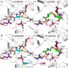
3D modeling of FliY and enantiomer coordination. A–D, FliY was modeled based on the structures of the l-cysteine SBP (A and B, PDB code 2YJP) or the l-cystine SBP (C and D, PDB code 2YLN). The protein backbone is shown as a cartoon representation, and selected ligand-coordinating residues are shown as balls and sticks, colored according to their ConSurf conservation score. The ligands are shown as balls and sticks and are colored cyan (l-cysteine, A; d-cysteine, B) and green (l-cystine, C; d-cystine, D). Also shown at the bottom is the ConSurf color-coded conservation scale (1, variable; 9, conserved).
Figure 7.
Figure 7.
Proposed model for the YecSC-FliY interaction and modulation of ATPase activity. In the absence of ligand, FliY exists in a conformational equilibrium between open and closed unliganded forms, where the majority of the molecules are in the open form (state I). The molecules that are in state I do not interact with the transporter and do not stimulate its ATPase activity. The minority of molecules that are in the closed, unliganded form (state II) interact with the transporter and stimulate its ATPase activity. When ligand is present, its binding induces a population shift toward the closed, liganded form (state III). More molecules are not available for interaction with the transporter, and higher ATPase stimulation is observed. Nevertheless, even in the absence of substrate, when the concentrations of apo FliY are sufficiently high, the concentration of the fraction of the molecules that are in the closed, unliganded form will be higher than the KD for interaction of YecSC with the closed, unliganded FliY and also higher than the concentration of YecSC. Therefore, maximal ATPase rates are achieved (Vmax (apo)Vmax (holo)), and further addition of substrate does not lead to increased activity. KD (apo) and KD (holo) represent the apparent KD for the FliY-YecSC interaction (in the absence or presence of substrate, respectively) as inferred from the apparent Km of FliY-mediated stimulation of ATPase activity.

References

    1. Higgins C. F. (1992) ABC transporters: from microorganisms to man. Annu. Rev. Cell Biol. 8, 67–113 10.1146/annurev.cb.08.110192.000435 - DOI - PubMed
    1. Dassa E., and Bouige P. (2001) The ABC of ABCs: a phylogenetic and functional classification of ABC systems in living organisms. Res. Microbiol. 152, 211–229 10.1016/S0923-2508(01)01194-9 - DOI - PubMed
    1. Holland I. B., Cole S. P. C., Kuchler K., and Higgins C. F. (2003) ABC Proteins: From Bacteria to Man, Academic Press, San Diego, CA
    1. Lewinson O., and Livnat-Levanon N. (2017) Mechanism of action of ABC importers: conservation, divergence, and physiological adaptations. J. Mol. Biol. 429, 606–619 10.1016/j.jmb.2017.01.010 - DOI - PubMed
    1. Locher K. P. (2016) Mechanistic diversity in ATP-binding cassette (ABC) transporters. Nat. Struct. Mol. Biol. 23, 487–493 10.1038/nsmb.3216 - DOI - PubMed

Publication types

MeSH terms

LinkOut - more resources