. 2020 Apr 17;295(16):5278-5291.
doi: 10.1074/jbc.RA119.011916. Epub 2020 Mar 6.

Inter-α-inhibitor heavy chain-1 has an integrin-like 3D structure mediating immune regulatory activities and matrix stabilization during ovulation

Affiliations

Inter-α-inhibitor heavy chain-1 has an integrin-like 3D structure mediating immune regulatory activities and matrix stabilization during ovulation

David C Briggs et al. J Biol Chem. .

Abstract

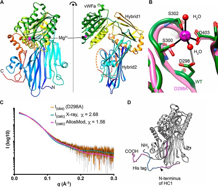
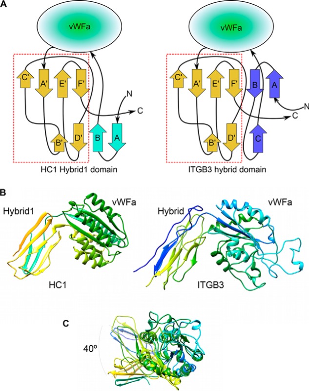
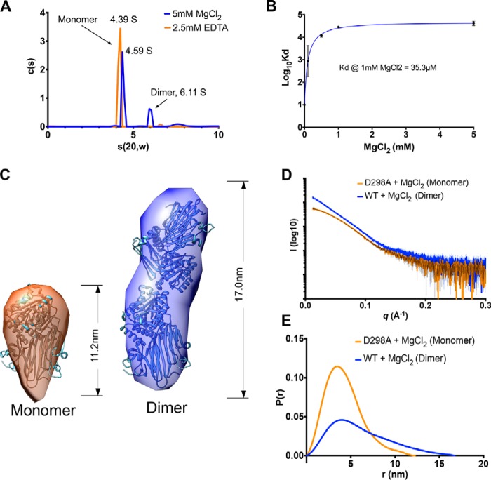
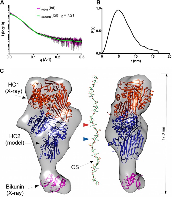
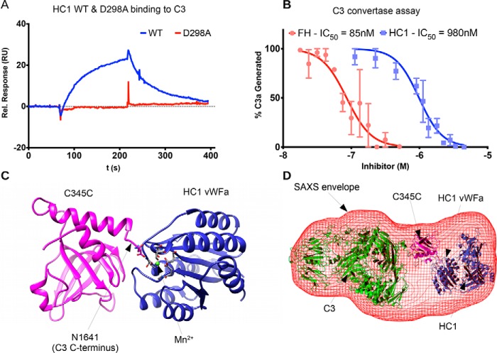
Inter-α-inhibitor is a proteoglycan essential for mammalian reproduction and also plays a less well-characterized role in inflammation. It comprises two homologous "heavy chains" (HC1 and HC2) covalently attached to chondroitin sulfate on the bikunin core protein. Before ovulation, HCs are transferred onto the polysaccharide hyaluronan (HA) to form covalent HC·HA complexes, thereby stabilizing an extracellular matrix around the oocyte required for fertilization. Additionally, such complexes form during inflammatory processes and mediate leukocyte adhesion in the synovial fluids of arthritis patients and protect against sepsis. Here using X-ray crystallography, we show that human HC1 has a structure similar to integrin β-chains, with a von Willebrand factor A domain containing a functional metal ion-dependent adhesion site (MIDAS) and an associated hybrid domain. A comparison of the WT protein and a variant with an impaired MIDAS (but otherwise structurally identical) by small-angle X-ray scattering and analytical ultracentrifugation revealed that HC1 self-associates in a cation-dependent manner, providing a mechanism for HC·HA cross-linking and matrix stabilization. Surprisingly, unlike integrins, HC1 interacted with RGD-containing ligands, such as fibronectin, vitronectin, and the latency-associated peptides of transforming growth factor β, in a MIDAS/cation-independent manner. However, HC1 utilizes its MIDAS motif to bind to and inhibit the cleavage of complement C3, and small-angle X-ray scattering-based modeling indicates that this occurs through the inhibition of the alternative pathway C3 convertase. These findings provide detailed structural and functional insights into HC1 as a regulator of innate immunity and further elucidate the role of HC·HA complexes in inflammation and ovulation.

Keywords: Inter-α-inhibitor Heavy Chain; X-ray crystallography; extracellular matrix; hyaluronan; inflammation; innate immunity; protein stability; proteoglycan; reproduction; small-angle X-ray scattering (SAXS).


Conflict of interest statement

The authors declare that they have no conflicts of interest with the contents of this article

Figures

Figure 1.
Figure 1.
The crystal structure of HC1. A, orthogonal views of the structure of rHC1, colored from the N terminus (blue) to C terminus (red), where domains and the bound Mg2+ ion are labeled; the dotted orange line denotes residues 631–638, which are not visible in the crystal structure. B, close-up of the MIDAS site, showing metal coordination (black) and an important hydrogen bond (gray). The WT structure is shown in green, and the D298A structure (which lacks the Mg2+ ion) is in pink. C, raw SAXS data (orange with black error bars) of a rHC1 monomer (D298A) and back-calculated scattering curves based on the crystal structure of rHC1 alone or the crystal structure with the unstructured/flexible regions modeled in using AllosMod. D, AllosMod model of rHC1 with the N-terminal histidine tag (blue) and residues 35–44, 631–636, and 653–672 (pink) modeled based on SAXS restraints.
Figure 2.
Figure 2.
Integrin-like arrangement of vWFA and Hydrid1 domains in HC1 structure. A, topologies of the HC–Hybrid1 domain from HC1 (left panel) and the hybrid domain from human integrin β3 chain (ITGB3) (right panel); the arrangements of β-strands in the sequences following the vWFA domains are essentially identical (dashed red box). B, side-by-side views of the “hybrid” and vWFA domain pairs of HC1 (left panel) and ITGB3 (right panel). C, the hybrid domains are displaced by ∼40° when the vWFA domains of HC1 and ITGB3 are superimposed.
Figure 3.
Figure 3.
HC1 forms metal ion-dependent dimers. A, a plot of sedimentation coefficient distributions (c(s)) versus s(apparent)) for WT rHC1 derived from velocity AUC analysis. In the presence of 2.5 mm EDTA (orange) 93% of the rHC1 protein is in a monomeric state (s(20,w) = 4.39 S), and there is no detectable dimer present. In 5 mm MgCl2 (blue), 64% of the protein is monomeric (s(20,w) = 4.59 S), and 21% of material is dimeric (s(20,w) = 6.11 S). B, plot of Log10KD versus MgCl2 concentration, derived from equilibrium AUC measurements. At 0 mm MgCl2 (achieved by conducting the experiment in 2.5 mm EDTA), no dimerization was detected. Maximal binding affinity (for self-association of the rHC1 dimer) was reached at ∼1 mm MgCl2, i.e. close to the concentration of free Mg2+ ions in plasma. C, ab initio SAXS models for the HC1 monomer (left panel) and dimer (right panel) where the HC1 structure has been modeled into the SAXS envelopes. D and E, buffer-subtracted SAXS scattering curves for HC1 D298 monomer (orange) and WT dimer (blue) (D) and their derived P(r) versus distance plots (E), consistent with WT HC1 forming an elongated Mg2+-dependent dimer and the MIDAS site mutant (D298A) being monomeric.
Figure 4.
Figure 4.
The quaternary structure of inter-α-inhibitor. A, raw SAXS data for IαI (I(obs)), in the presence of 2 mm MgCl2, fitted to scattering data (pink with black error bars) derived from the pseudo atomic model (I(model)) in C calculated using Allosmod-FoXs. B, P(r) versus distance plot showing that IαI has an elongated and asymmetric shape. C, orthogonal views of the SAXS envelope of IαI (transparent gray surface) determined ab initio from the SAXS scattering curve, with structures of bikunin (PDB code 1BIK; pink) and rHC1 (determined here; orange) and a threading model of HC2 (based upon the structure of HC1; blue), modeled in. The CS chain is shown to indicate its dimensions relative to the SAXS envelope for IαI. Here the CS chain, with a standard tetrasaccharide linker, has been modeled on the sequences determined for bikunin·CS (18) with the GLYCAN-Web GAG Builder modeling tool (82), using the median values established in the Ly et al. study (a = 2, b = 1, c = 4, and d = 1, corresponding to a CS chain of 26 saccharides (18)). The CS chain is attached to S10 (in the mature bikunin sequence), which is not present in the crystal structure (PDB code 1BIK) that corresponds to residues 25–134 (44). The GlcNAc moieties in the CS chain to which HC1 and HC2 are covalently attached, via their unstructured C-terminal peptides (not shown), are indicated by arrowheads (orange and blue, respectively).
Figure 5.
Figure 5.
HC1 inhibits alternative pathway C3 convertase activity through interaction with C3. A, SPR analysis for the interaction of rHC1 (WT and D298A) with C3 in 2 mm MnCl2, where the lack of binding of the D298A mutant indicates an essential role for the MIDAS site. B, rHC1 proteins (WT (blue) and D298A) were compared with FH (red) in an alternative pathway C3 convertase assay, where the proteolytic release of C3a was quantified (by SDS-PAGE) as a surrogate for the conversion of C3 into C3b. The mean values (± S.D.) were derived from independent experiments performed in triplicate. The data were fitted using GraphPad Prism to derive IC50 values for rHC1 and FH control. Only WT rHC1 had inhibitory activity (data for D298A not shown). C, an in silico model of the C3 C-terminal C345C domain (pink) bound to the vWFA domain of HC1 (blue). Here a Mn2+ ion (green) occupies the MIDAS of HC1 (with coordinating residues shown in stick representation) and co-chelates the C-terminal amino acid (Asn) of C3b. D, an ab initio SAXS structure was determined for the rHC1–C3 complex (red mesh), where C3 and HC1 molecules, interacting as in C, could be accommodated.

References

    1. Enghild J. J., Thøgersen I. B., Pizzo S. V., and Salvesen G. (1989) Analysis of inter-α-trypsin inhibitor and a novel trypsin inhibitor, pre-α-trypsin inhibitor, from human plasma: polypeptide chain stoichiometry and assembly by glycan. J. Biol. Chem. 264, 15975–15981 - PubMed
    1. Zhuo L., Yoneda M., Zhao M., Yingsung W., Yoshida N., Kitagawa Y., Kawamura K., Suzuki T., and Kimata K. (2001) Defect in SHAP-hyaluronan complex causes severe female infertility: a study by inactivation of the bikunin gene in mice. J. Biol. Chem. 276, 7693–7696 10.1074/jbc.C000899200 - DOI - PubMed
    1. Sato H., Kajikawa S., Kuroda S., Horisawa Y., Nakamura N., Kaga N., Kakinuma C., Kato K., Morishita H., Niwa H., and Miyazaki J. (2001) Impaired fertility in female mice lacking urinary trypsin inhibitor. Biochem. Biophys. Res. Commun. 281, 1154–1160 10.1006/bbrc.2001.4475 - DOI - PubMed
    1. Chen X., Bonfiglio R., Banerji S., Jackson D. G., Salustri A., and Richter R. P. (2016) Micromechanical analysis of the hyaluronan-rich matrix surrounding the oocyte reveals a uniquely soft and elastic composition. Biophys. J. 110, 2779–2789 10.1016/j.bpj.2016.03.023 - DOI - PMC - PubMed
    1. Russell D. L., and Salustri A. (2006) Extracellular matrix of the cumulus–oocyte complex. Semin. Reprod. Med. 24, 217–227 10.1055/s-2006-948551 - DOI - PubMed

Publication types