. 2020 Apr 10;295(15):5163-5174.
doi: 10.1074/jbc.RA120.012536. Epub 2020 Mar 6.

HIV-1 Nef dimers short-circuit immune receptor signaling by activating Tec-family kinases at the host cell membrane

Affiliations

HIV-1 Nef dimers short-circuit immune receptor signaling by activating Tec-family kinases at the host cell membrane

Wing Fai Li et al. J Biol Chem. .

Abstract

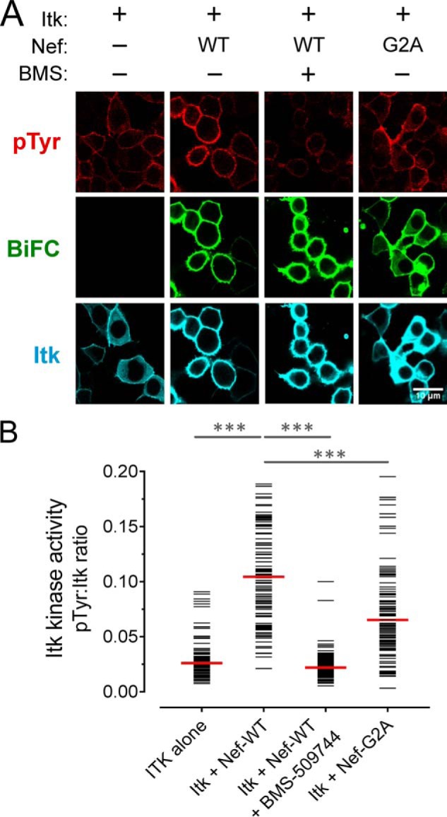
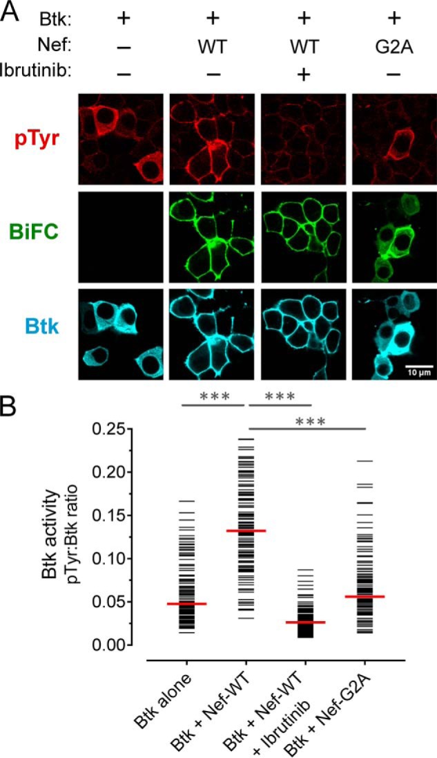
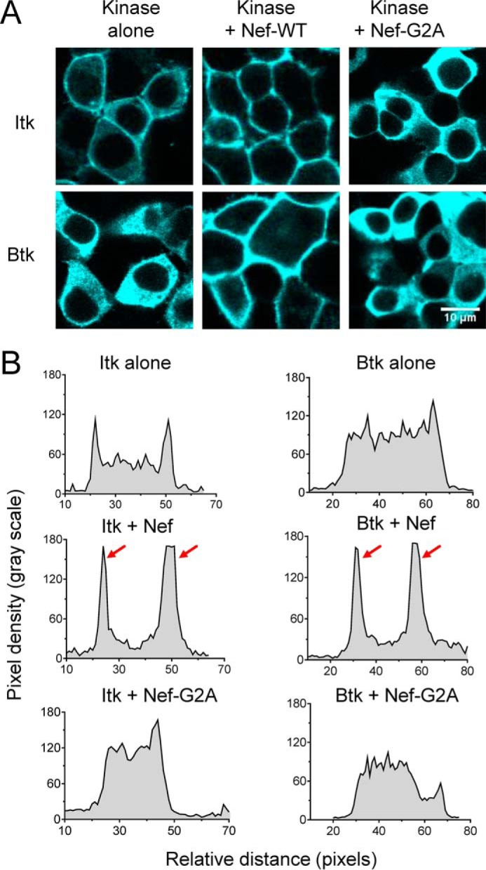
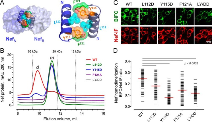
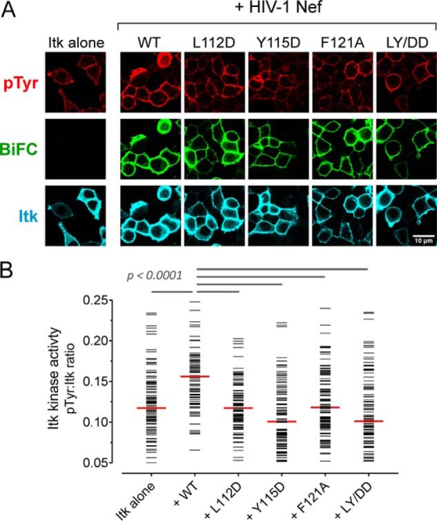
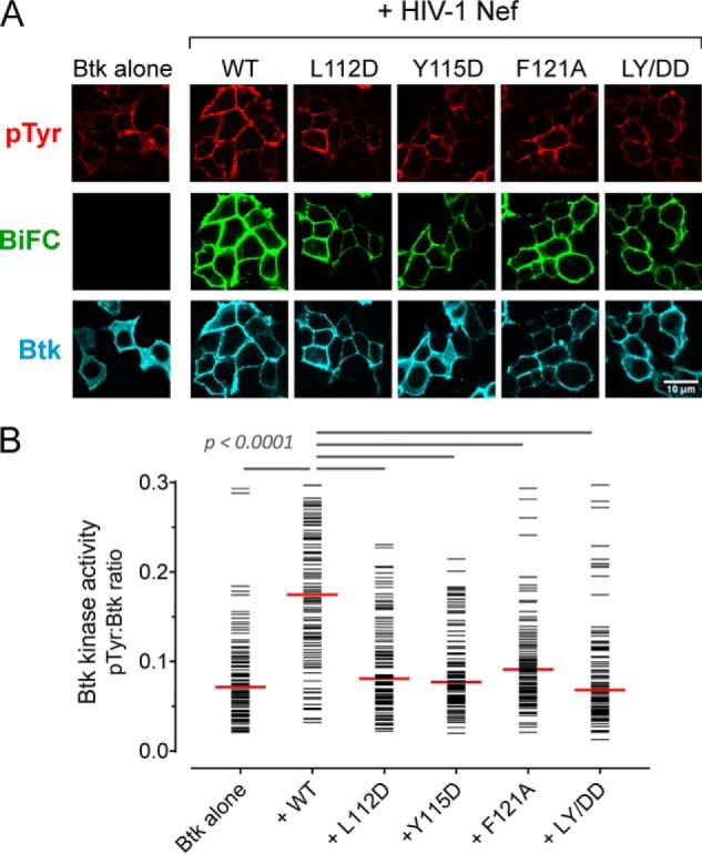
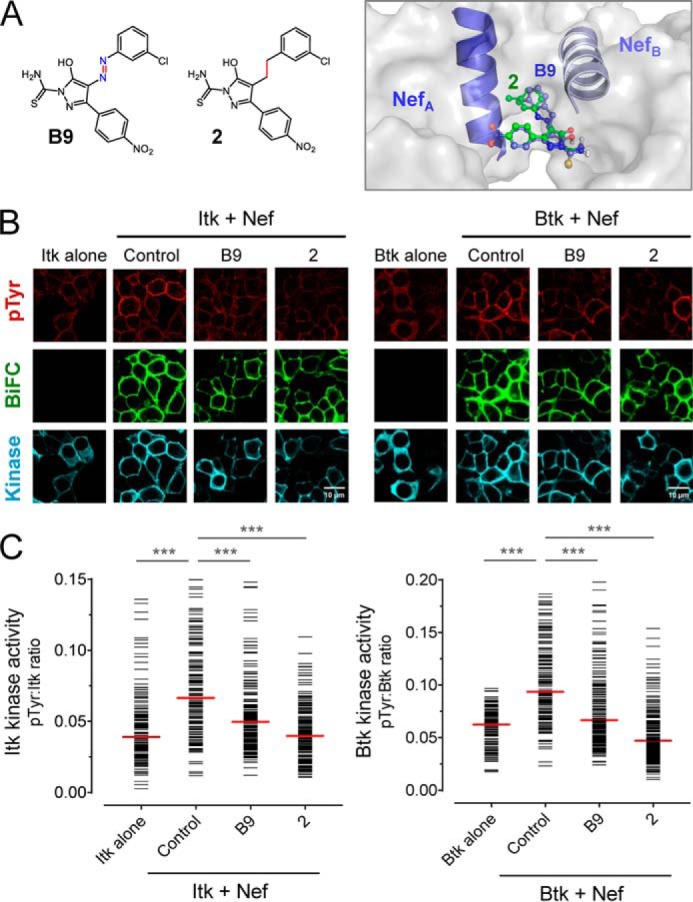
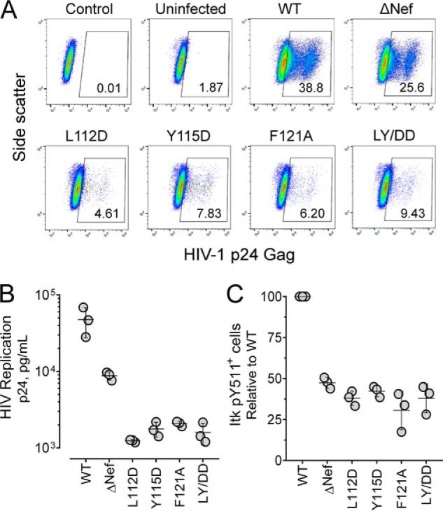
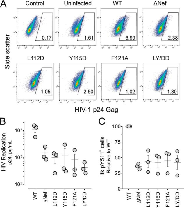
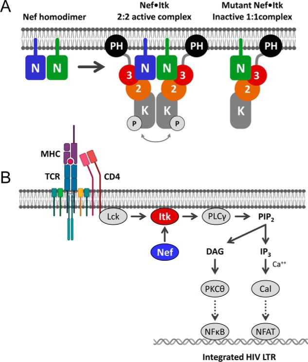
The HIV-1 virulence factor Nef promotes high-titer viral replication, immune escape, and pathogenicity. Nef interacts with interleukin-2-inducible T-cell kinase (Itk) and Bruton's tyrosine kinase (Btk), two Tec-family kinases expressed in HIV-1 target cells (CD4 T cells and macrophages, respectively). Using a cell-based bimolecular fluorescence complementation assay, here we demonstrate that Nef recruits both Itk and Btk to the cell membrane and induces constitutive kinase activation in transfected 293T cells. Nef homodimerization-defective mutants retained their interaction with both kinases but failed to induce activation, supporting a role for Nef homodimer formation in the activation mechanism. HIV-1 infection up-regulates endogenous Itk activity in SupT1 T cells and donor-derived peripheral blood mononuclear cells. However, HIV-1 strains expressing Nef variants with mutations in the dimerization interface replicated poorly and were significantly attenuated in Itk activation. We conclude that direct activation of Itk and Btk by Nef at the membrane in HIV-infected cells may override normal immune receptor control of Tec-family kinase activity to enhance the viral life cycle.

Keywords: Bruton's tyrosine kinase (BTK); HIV-1 Nef; T-cell receptor (TCR); Tec-family kinase; bimolecular fluorescence complementation (BiFC); dimerization; human immunodeficiency virus (HIV); infectious disease; interleukin-2-inducible kinase (ITK); protein kinase; protein–protein interaction; signal transduction.


Conflict of interest statement

The authors declare that they have no conflicts of interest with the contents of this article

Figures

Figure 1.
Figure 1.
Nef induces constitutive activation of Itk at the cell membrane. A, Itk was expressed either alone or together with WT or myristoylation-defective Nef (G2A) as BiFC pairs in the absence or presence of the Itk inhibitor BMS-509744 (1 μm) in 293T cells. The cells were fixed and stained for confocal microscopy with anti-pTyr antibodies as a measure of kinase activity (red) and anti-V5 antibodies to verify Itk protein expression (blue). Nef interaction with Itk is observed as fluorescence complementation of the YFP variant, Venus (BiFC; green). B, single-cell image analysis. Mean fluorescence intensities for the pTyr and Itk signals were determined for ≥100 cells from each condition using ImageJ. The fluorescence intensity ratio (pTyr:Itk expression) for each cell is shown as a horizontal bar, with the median value indicated by the red bar. Student's t tests were performed on the groups indicated by horizontal lines above the plot; p < 0.0001 in each case (***).
Figure 2.
Figure 2.
Nef induces constitutive activation of Btk at the cell membrane. A, Btk was expressed either alone or together with WT or myristoylation-defective Nef (G2A) as BiFC pairs in the absence or presence of the Btk inhibitor, ibrutinib (1 μm) in 293T cells. The cells were fixed and stained for confocal microscopy with anti-pTyr antibodies as a measure of kinase activity (red) and anti-V5 antibodies to verify Btk protein expression (blue). Nef interaction with Btk is observed as fluorescence complementation of the YFP variant, Venus (BiFC; green). B, single-cell image analysis was performed as per the legend to Fig. 1. The fluorescence intensity ratio (pTyr:Btk expression) for each cell is presented as a horizontal bar, with each median value indicated by the red bar. Student's t tests were performed on the groups indicated by horizontal lines above the plot; p < 0.0001 in each case (***).
Figure 3.
Figure 3.
Membrane recruitment of Btk and Itk requires myristoylation of HIV-1 Nef. A, Itk or Btk were expressed either alone or in combination with WT or myristoylation-defective Nef (G2A) in 293T cells. The cells were stained with antibodies to the V5 tag present in each kinase protein, and imaged via confocal microscopy. Representative images are shown. B, using the Plot Profile tool from ImageJ, a line was drawn across the cytoplasm of representative cells from each condition, excluding the nucleus. The change in fluorescence intensity along this line was then plotted as relative pixel density as a function of distance. Very similar results were observed with at least 10 cells from each condition, and a representative example is shown. Strong membrane localization of both Itk and Btk was observed in the presence of WT Nef and is reflected by the prominent peaks indicated by the red arrows.
Figure 4.
Figure 4.
Conserved hydrophobic residues in the folded Nef core are required for homodimer formation. A, Nef homodimers present in the X-ray crystal structure of the HIV-1 Nef core in complex with a Src-family kinase SH3 domain (PDB code 1EFN). An overview of the Nef dimer structure is shown on the left, with the αB helices that form the dimer interface highlighted; SH3 domains are not shown for clarity. The αB helices are enlarged on the right. Side chains of Leu112, Tyr115, and Phe121 from each Nef monomer form the hydrophobic core of the interface. B, analytical size-exclusion chromatography of WT Nef and dimerization interface mutants. WT Nef and the L112D, Y115D, F121A, and L112D/Y115D (LY/DD) mutants were expressed in E. coli and purified (see “Experimental procedures”). Each Nef protein was then characterized by analytical size-exclusion chromatography. Elution peaks of standard proteins are indicated by the vertical dotted lines and molecular weight. m, monomer; d, dimer. C, hydrophobic residues in the Nef core are required for homodimer formation in cells. WT Nef and the dimer interface mutants were expressed as BiFC pairs in 293T cells. The cells were stained for Nef expression with an anti-Nef antibody and imaged by confocal microscopy to detect Nef homodimer formation (BiFC, green) and Nef expression as immunofluorescence (Nef-IF, red). D, single-cell image analysis. Mean fluorescence intensities for the BiFC (interaction) and Nef-IF signals were determined with ImageJ for ≥100 cells. The Nef-BiFC:Nef-IF fluorescence intensity ratio for each cell is presented as a horizontal bar, with the median value indicated by the red bar. Student's t tests were performed on the groups indicated by horizontal lines above the plot; p < 0.0001 in each case.
Figure 5.
Figure 5.
Dimerization-defective Nef mutants interact with Itk but fail to induce kinase activation. A, Itk was expressed either alone or together with WT Nef or dimerization-defective mutants as BiFC pairs in 293T cells. The cells were fixed and stained for confocal microscopy with anti-pTyr antibodies as a measure of kinase activity (red) and anti-V5 antibodies to verify Itk protein expression (blue). Nef interaction with Itk is observed as Venus fluorescence (BiFC; green). B, single-cell image analysis was performed as per the legend to Fig. 1. The fluorescence intensity ratio (pTyr:Itk expression) for each cell is shown as a horizontal bar, with the median value indicated by the red bar. Student's t tests were performed on the groups indicated by horizontal lines above the plot; p < 0.0001 in each case.
Figure 6.
Figure 6.
Dimerization-defective Nef mutants interact with Btk but fail to induce kinase activation. A, Btk was expressed either alone or together with WT Nef or Nef dimerization-defective mutants as BiFC pairs in 293T cells. The cells were fixed and stained for confocal microscopy with anti-pTyr antibodies as a measure of kinase activity (red) and anti-V5 antibodies to verify Btk protein expression (blue). Nef interaction with Btk is observed as Venus fluorescence (BiFC; green). B, single-cell image analysis was performed as per the legend to Fig. 2. The fluorescence intensity ratio (pTyr:Btk expression) for each cell is shown as a horizontal bar, with the median value indicated by the red bar. Student's t tests were performed on the groups indicated by horizontal lines above the plot; p < 0.0001 in each case.
Figure 7.
Figure 7.
Small molecule Nef inhibitors block Nef-mediated Itk and Btk activation. A, structures of small molecule Nef inhibitors, B9 and compound 2, are shown on the left. Computational docking predicts interaction of both inhibitors with the Nef dimerization interface (model at right; Nef αB helices that form this interface are also shown). B and C, Itk and Btk were expressed in 293T cells either alone or together with WT Nef as BiFC pairs in the absence (Control) or presence of B9 or compound 2 (final concentration of 1.0 μm). Following inhibitor treatment, the cells were fixed and stained for confocal microscopy with anti-pTyr antibodies as a measure of kinase activity (red) and anti-V5 antibodies to verify kinase protein expression (blue). Nef interaction with each kinase was visualized as fluorescence complementation of the YFP variant, Venus (BiFC; green). B, representative images from each experiment. C, single-cell image analysis. Mean fluorescence intensities for the pTyr and Itk or Btk signals were determined for ≥100 cells from each condition using ImageJ. The fluorescence intensity ratio (pTyr:Itk expression) for each cell is shown as a horizontal bar, with the median value indicated by the red bar. Student's t tests were performed on the groups indicated by horizontal lines above the plot; p < 0.0001 in each case (***).
Figure 8.
Figure 8.
HIV-1 expressing dimerization-defective Nef mutants show attenuated replication and reduced endogenous Itk activation in SupT1 cells. SupT1 cells were infected with HIV-1 expressing WT Nef, dimerization-defective mutants of Nef or virus that does not express Nef (ΔNef; viral input in each case 5000 pg/ml p24). Four days later, infected cells were primed with anti-CD3/anti-CD28 antibodies, fixed, and stained with antibodies to HIV-1 p24 Gag and active Itk (anti-pTyr511 antibody). A, representative flow cytometry plots for unstained, uninfected cells (Control), uninfected stained cells (Uninfected), and cells infected with WT, ΔNef, and dimerization-defective Nef mutant viruses. The number in the gating box indicates the percentage of p24+ infected cells present in each culture. B, viral replication was quantified as p24 Gag release into the culture supernatant. The results from three independent determinations are shown with the horizontal bar representing the mean values ± S.E. Student's t tests show that p24 values for ΔNef and each Nef mutant virus are significantly different from WT (p < 0.05 in each case). In addition, comparison of the p24 values for ΔNef with each of the Nef mutants is also significant (p < 0.0005 in each case). C, percentage of Itk pTyr511+ cells present in each infected (p24+) cell population. The results of three independent experiments are plotted as percentages of pTyr511+ cells relative to control cells infected with WT HIV-1. Mean values are shown as the horizontal bar ± S.E. The values for ΔNef and each Nef mutant virus are significantly different from WT (p < 0.0005 in each case).
Figure 9.
Figure 9.
HIV-1–expressing dimerization-defective Nef mutants show attenuated replication and endogenous Itk activation in donor PBMCs. PBMCs were isolated from normal donors and stimulated with PHA and IL-2 for 3 days. The cells were then infected with HIV-1–expressing WT Nef, dimerization-defective mutants of Nef or virus that does not express Nef (ΔNef; viral input in each case 40 pg/ml p24). Infected PBMCs were primed with anti-CD3/anti-CD28 antibodies on day 5, and on day 7 they were fixed and stained for HIV-1 p24 Gag and active Itk (anti-pTyr511 antibody) followed by flow cytometry. A, representative flow cytometry plots for unstained, uninfected cells (Control), uninfected stained cells (Uninfected), and cells infected with WT, ΔNef, and dimerization-defective Nef mutant viruses. The number in the gating box indicates the percentage of p24+ infected cells present in each culture. B, viral replication was quantified as p24 Gag release into the culture supernatant. The results from three independent determinations are shown with the horizontal bar representing the mean value ± S.E. Student's t tests show that p24 values for ΔNef and each Nef mutant virus are significantly different from WT (p < 0.05 in each case). This experiment was repeated three times with comparable results; a representative example is shown. C, percentage of Itk pTyr511+ cells present in each infected (p24+) cell population. This experiment was performed three times, and the results are plotted as percentages of pTyr511+ cells relative to control cells infected with WT HIV-1 ± S.E. The values for ΔNef and each Nef mutant virus are significantly different from WT (p < 0.01 in each case).
Figure 10.
Figure 10.
Models of Nef-mediated Itk activation and consequences for HIV-1 transcription. A, Nef forms homodimers at the cytoplasmic face of the plasma membrane, which stabilize Itk homodimers as a 2:2 complex. Regulatory domain displacement and kinase domain juxtaposition contribute to sustained kinase activation via autophosphorylation in trans. Dimerization-defective Nef mutants retain interaction with Itk at the membrane but form inactive 1:1 complexes. N, Nef; 3, Src homology 3 domain; 2, Src homology 2 domain; K, kinase domain; P, activation loop phosphorylation. B, proposed consequences of constitutive Itk activation by Nef for HIV-1 transcription in CD4 T cells. The T-cell receptor (TCR) complex is normally activated by antigen-loaded MHC molecules. The TCR activates the T cell–specific Src-family kinase Lck, which is associated with the cytosolic tail of the co-receptor, CD4. Active Lck directly phosphorylates Itk on its activation loop, which in turn phosphorylates and activates phospholipase Cγ (PLCγ). Active PLCγ generates the second messenger diacylglycerol (DAG) and inositol triphosphate (IP3) via hydrolysis of membrane phosphatidylinositol 4,5-bisphosphate (PIP2), ultimately leading to activation of protein kinase Cθ (PKCθ) and the calcium-dependent protein serine/threonine phosphatase, calcineurin (Cal), respectively. Protein kinase Cθ promotes activation of NF-κB via the CARMA1/BCL10/MALT1 complex (not shown), whereas calcineurin dephosphorylates NFAT to drive nuclear localization. Both NF-κB and NFAT participate in transcription of the integrated HIV-1 provirus. The data presented here support direct activation of Itk by Nef at the membrane downstream of the TCR. Additional details of TCR signaling are omitted for clarity. The cartoon was adapted from Gaud et al. (46). LTR, long terminal repeat.

References

    1. Basmaciogullari S., and Pizzato M. (2014) The activity of Nef on HIV-1 infectivity. Front. Microbiol. 5, 232 - PMC - PubMed
    1. Pawlak E. N., and Dikeakos J. D. (2015) HIV-1 Nef: a master manipulator of the membrane trafficking machinery mediating immune evasion. Biochim. Biophys. Acta 1850, 733–741 10.1016/j.bbagen.2015.01.003 - DOI - PubMed
    1. Kestler H. 3rd, Ringler D. J., Mori K., Panicali D. L., Sehgal P. K., Daniel M. D., and Desrosiers R. C. (1991) Importance of the nef gene for maintenance of high viral loads and for development of AIDS. Cell 65, 651–662 10.1016/0092-8674(91)90097-I - DOI - PubMed
    1. Kirchhoff F., Greenough T. C., Brettler D. B., Sullivan J. L., and Desrosiers R. C. (1995) Absence of intact nef sequences in a long-term survivor with nonprogressive HIV-1 infection. N. Engl. J. Med. 332, 228–232 10.1056/NEJM199501263320405 - DOI - PubMed
    1. Deacon N. J., Tsykin A., Solomon A., Smith K., Ludford-Menting M., Hooker D. J., McPhee D. A., Greenway A. L., Ellett A., Chatfield C., Lawson V. A., Crowe S., Maerz A., Sonza S., Learmont J., et al. (1995) Genomic structure of an attenuated quasi species of HIV-1 from a blood transfusion donor and recipients. Science 270, 988–991 10.1126/science.270.5238.988 - DOI - PubMed

Publication types

MeSH terms

Substances