Review
. 2020 Apr 17;295(16):5377-5389.
doi: 10.1074/jbc.REV119.007741. Epub 2020 Mar 6.

Evolution, expression, and substrate specificities of aldehyde oxidase enzymes in eukaryotes

Affiliations
Review

Evolution, expression, and substrate specificities of aldehyde oxidase enzymes in eukaryotes

Mineko Terao et al. J Biol Chem. .

Abstract

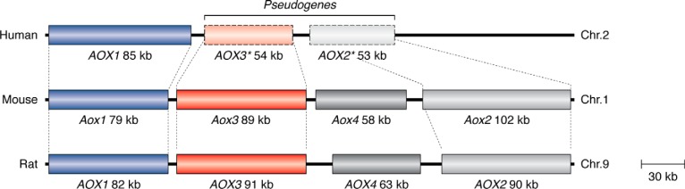
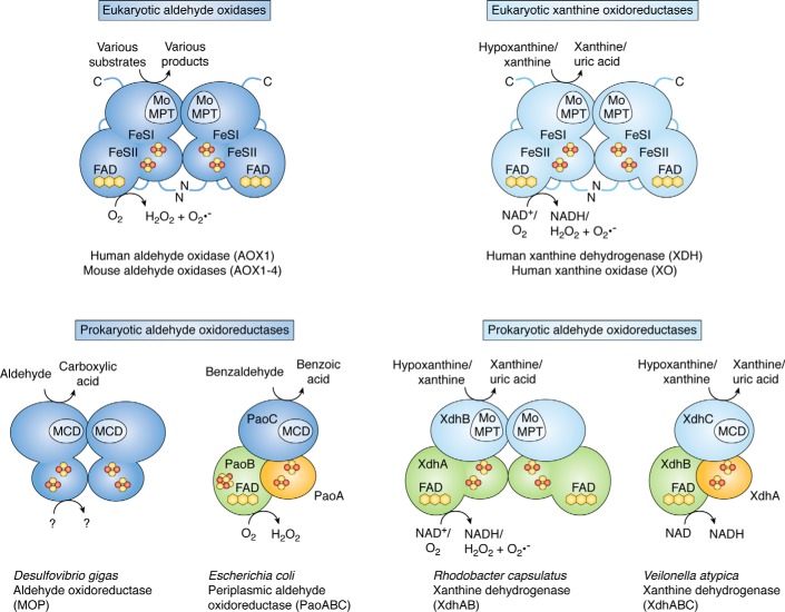
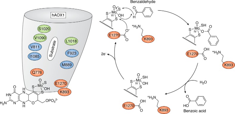
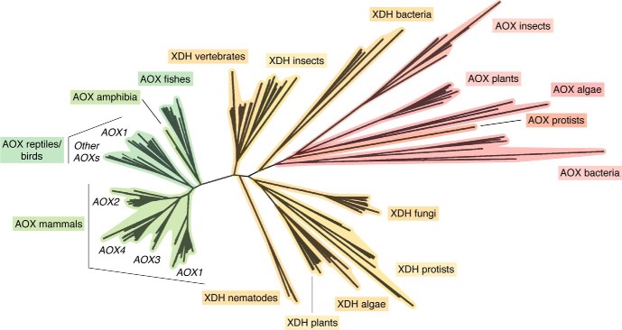
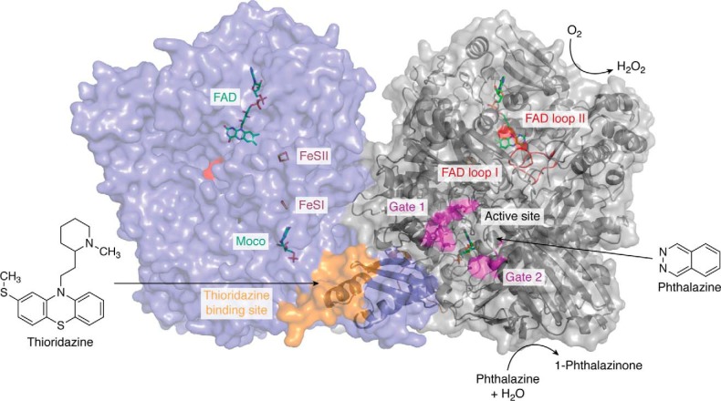
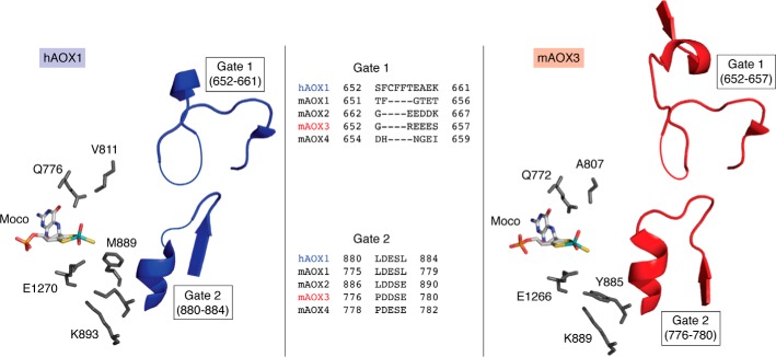
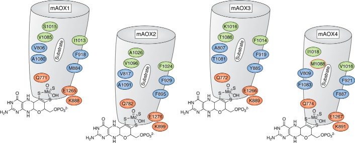
Aldehyde oxidases (AOXs) are a small group of enzymes belonging to the larger family of molybdo-flavoenzymes, along with the well-characterized xanthine oxidoreductase. The two major types of reactions that are catalyzed by AOXs are the hydroxylation of heterocycles and the oxidation of aldehydes to their corresponding carboxylic acids. Different animal species have different complements of AOX genes. The two extremes are represented in humans and rodents; whereas the human genome contains a single active gene (AOX1), those of rodents, such as mice, are endowed with four genes (Aox1-4), clustering on the same chromosome, each encoding a functionally distinct AOX enzyme. It still remains enigmatic why some species have numerous AOX enzymes, whereas others harbor only one functional enzyme. At present, little is known about the physiological relevance of AOX enzymes in humans and their additional forms in other mammals. These enzymes are expressed in the liver and play an important role in the metabolisms of drugs and other xenobiotics. In this review, we discuss the expression, tissue-specific roles, and substrate specificities of the different mammalian AOX enzymes and highlight insights into their physiological roles.

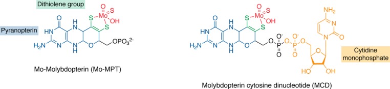
Keywords: 2Fe-2S cluster; aldehyde oxidase (AOX); drug metabolism; enzyme evolution; flavin adenine dinucleotide (FAD); flavoprotein; iron-sulfur protein; metal-containing enzyme; metalloenzyme; molybdenum; molybdenum cofactor (Moco); molybdo-flavoenzyme; mouse; oxidase; oxygen radicals; xenobiotic.


Conflict of interest statement

The authors declare that they have no conflicts of interest with the contents of this article

Figures

Figure 1.
Figure 1.
Organization of the human, mouse, and rat AOX genes. Shown is a schematic representation of the Homo sapiens, Mus musculus, and Rattus norvegicus AOX genes and pseudogenes. Pseudogenes are marked by an asterisk.
Figure 2.
Figure 2.
Overview of the subunit composition and cofactor organization of aldehyde oxidases and xanthine oxidoreductases. In eukaryotes, the catalytically active forms of AOXs and XORs consist of α2 homodimers. Each subunit of the dimer consists of separate domains containing the Moco catalytic site, two distinct [2Fe-2S] redox centers, and the FAD-binding site. In the prokaryotic enzymes, the [2Fe-2S] cluster-binding subunits are shown in orange, the subunits binding the FAD cofactor are colored in green, and the subunits containing the Moco are colored in blue. The FAD domain is absent in the D. gigas aldehyde oxidoreductase. In the R. capsulatus xanthine dehydrogenase, the iron-sulfur and flavin-binding domains of the protein constitute one subunit (XdhA), and the Mo-MPT–binding domain constitutes a second (XdhB) subunit. In the V. atypica xanthine dehydrogenase, the iron-sulfur centers are located in one subunit (XdhA), the flavin in a second (XdhB), and the Moco as MCD in a third (XdhC) subunit. In the PaoABC from E. coli, a [4Fe-4S] cluster is present in proximity to the FAD cofactor on the PaoB subunit. Potential substrates and electron acceptors are indicated.
Figure 3.
Figure 3.
Forms of the molybdenum cofactor present in enzymes of the xanthine oxidase family. Characteristic to enzymes of the xanthine oxidase family is a sulfido ligand at the equatorial position of the molybdenum atom. In eukaryotic enzymes of this family, the basic form of Moco is a pyranopterin, named Mo-MPT, which coordinates the molybdenum atom (colored in red) by the characteristic dithiolene group (colored in green) at the C1′ and C2′ positions of the pyranopterin ring (colored in blue). In bacteria, the MPT core can be modified by an additional CMP nucleotide (colored in orange) at the phosphate group (colored in black), forming MCD.
Figure 4.
Figure 4.
The catalytic mechanism of human AOX1. Shown is a representation of the proposed reaction mechanism for human AOX1, as exemplified for the substrate benzaldehyde. For details, see “Domain structure and catalytic mechanism of the AOX enzymes.”
Figure 5.
Figure 5.
Phylogenetic tree of AOX and XDH proteins in prokaryotes and eukaryotes. The unrooted phylogenetic tree was generated from the available prokaryotic as well as eukaryotic AOX and XDH protein sequences. The phylogenetic tree consists of all of the AOX and XDH proteins whose structure could be predicted from the cloning of the corresponding cDNAs or deduced from genome-sequencing data.
Figure 6.
Figure 6.
The crystal structure of the human AOX1 homodimer in complex with phthalazine (substrate) and thioridazine (inhibitor). The different protein cofactors (FAD, FeSII, FeSI, and Moco) are indicated and are shown in a color-coded stick representation on the left monomer. The phthalazine substrate as well as the substrate-binding site, including the flexible Gates 1 and 2, are marked in pink on the right monomer. The thioridazine-binding site is marked in orange. The flexible loops at the FAD site (loop I and loop II) are marked in red (surface representation on the left monomer, cartoon representation on the right monomer). The figure was generated using PDB entry 1UHW published by Coelho et al. (3).
Figure 7.
Figure 7.
The active site of hAOX1 and mAOX3. Shown are the residues of hAOX1 (left) and mAOX3 (right) surrounding the Moco and the location of Gate 1 and Gate 2 at the entrance of the substrate funnel. Missing residues in the electron density (and therefore not present in the coordinate files) are indicated by thin lines. An amino acid sequence alignment of Gate 1 and Gate 2 of the human and mouse enzymes is given. The figure was created using PyMOL version 2.1.1.
Figure 8.
Figure 8.
Active site and substrate binding funnel in mouse AOX enzymes. Representation of the substrate binding sites in mouse AOX1, AOX2, AOX3, and AOX4 in addition to residues in the conserved and nonconserved substrate-binding funnel, which selects the substrate specificity. The representation of mAOX3 is taken from the crystal structure (PDB: 3ZYV) (38), whereas those of mAOX1, mAOX2, and mAOX4 are taken from the modeled structures (54). Residues in red indicate the amino acids whose nature is conserved in all mouse AOX enzymes; residues in blue are hydrophobic residues, partially conserved and involved in substrate orientation; and residues in green are those specific for AOX4, with the AOX4-Met1088 residue highlighted in red. The funnel for mAOX4 is predicted to be smaller compared with the other enzymes.

References

    1. Hille R., Hall J., and Basu P. (2014) The mononuclear molybdenum enzymes. Chem. Rev. 114, 3963–4038 10.1021/cr400443z - DOI - PMC - PubMed
    1. Terao M., Romão M. J., Leimkühler S., Bolis M., Fratelli M., Coelho C., Santos-Silva T., and Garattini E. (2016) Structure and function of mammalian aldehyde oxidases. Arch. Toxicol. 90, 753–780 10.1007/s00204-016-1683-1 - DOI - PubMed
    1. Coelho C., Foti A., Hartmann T., Santos-Silva T., Leimkühler S., and Romão M. J. (2015) Structural insights into xenobiotic and inhibitor binding to human aldehyde oxidase. Nat. Chem. Biol. 11, 779–783 10.1038/nchembio.1895 - DOI - PubMed
    1. Enroth C., Eger B. T., Okamoto K., Nishino T., Nishino T., and Pai E. F. (2000) Crystal structures of bovine milk xanthine dehydrogenase and xanthine oxidase: structure-based mechanism of conversion. Proc. Natl. Acad. Sci. U.S.A. 97, 10723–10728 10.1073/pnas.97.20.10723 - DOI - PMC - PubMed
    1. Kisker C., Schindelin H., Pacheco A., Wehbi W. A., Garrett R. M., Rajagopalan K. V., Enemark J. H., and Rees D. C. (1997) Molecular basis of sulfite oxidase deficiency from the structure of sulfite oxidase. Cell 91, 973–983 10.1016/S0092-8674(00)80488-2 - DOI - PubMed

Publication types

Substances

LinkOut - more resources