Review
. 2020 May;21(5):268-283.
doi: 10.1038/s41580-020-0227-y. Epub 2020 Mar 6.

Cellular adaptation to hypoxia through hypoxia inducible factors and beyond

Affiliations
Review

Cellular adaptation to hypoxia through hypoxia inducible factors and beyond

Pearl Lee et al. Nat Rev Mol Cell Biol. 2020 May.

Abstract

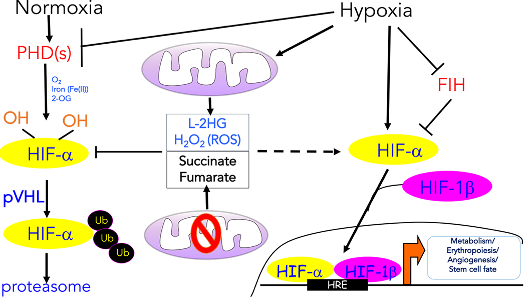
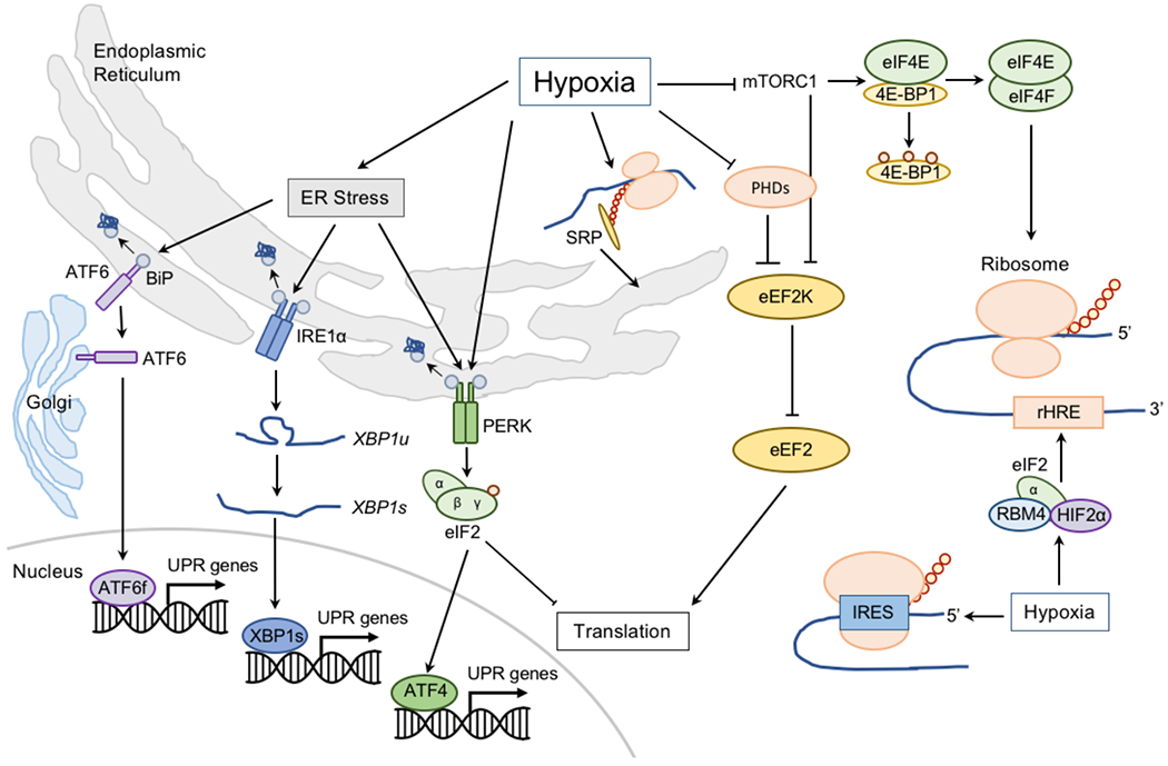
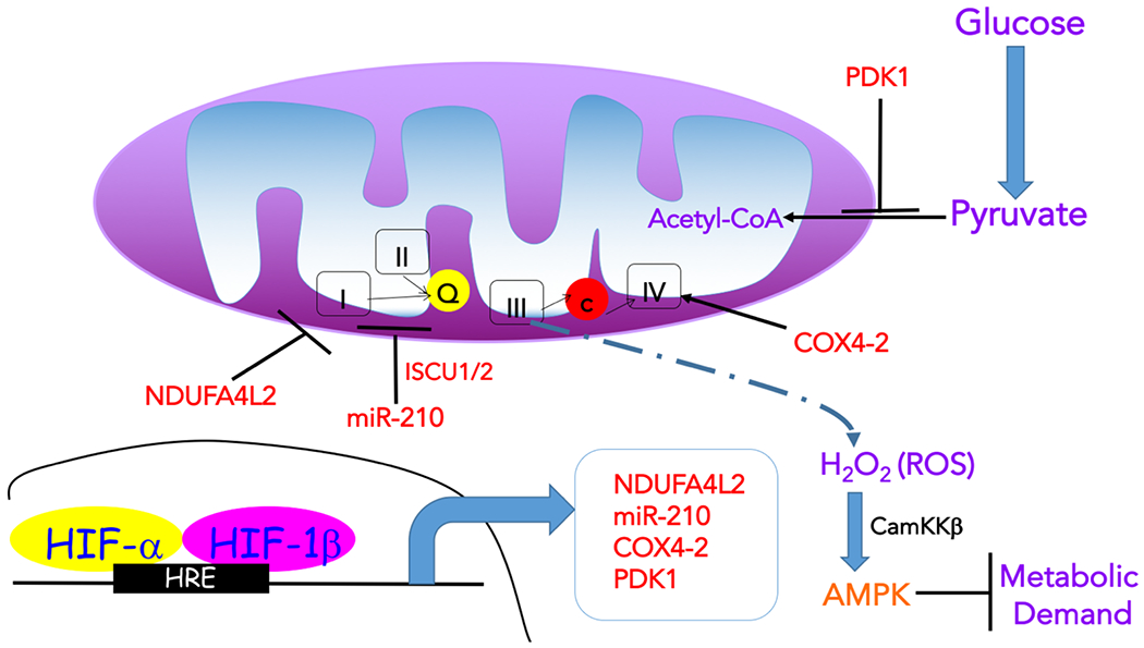
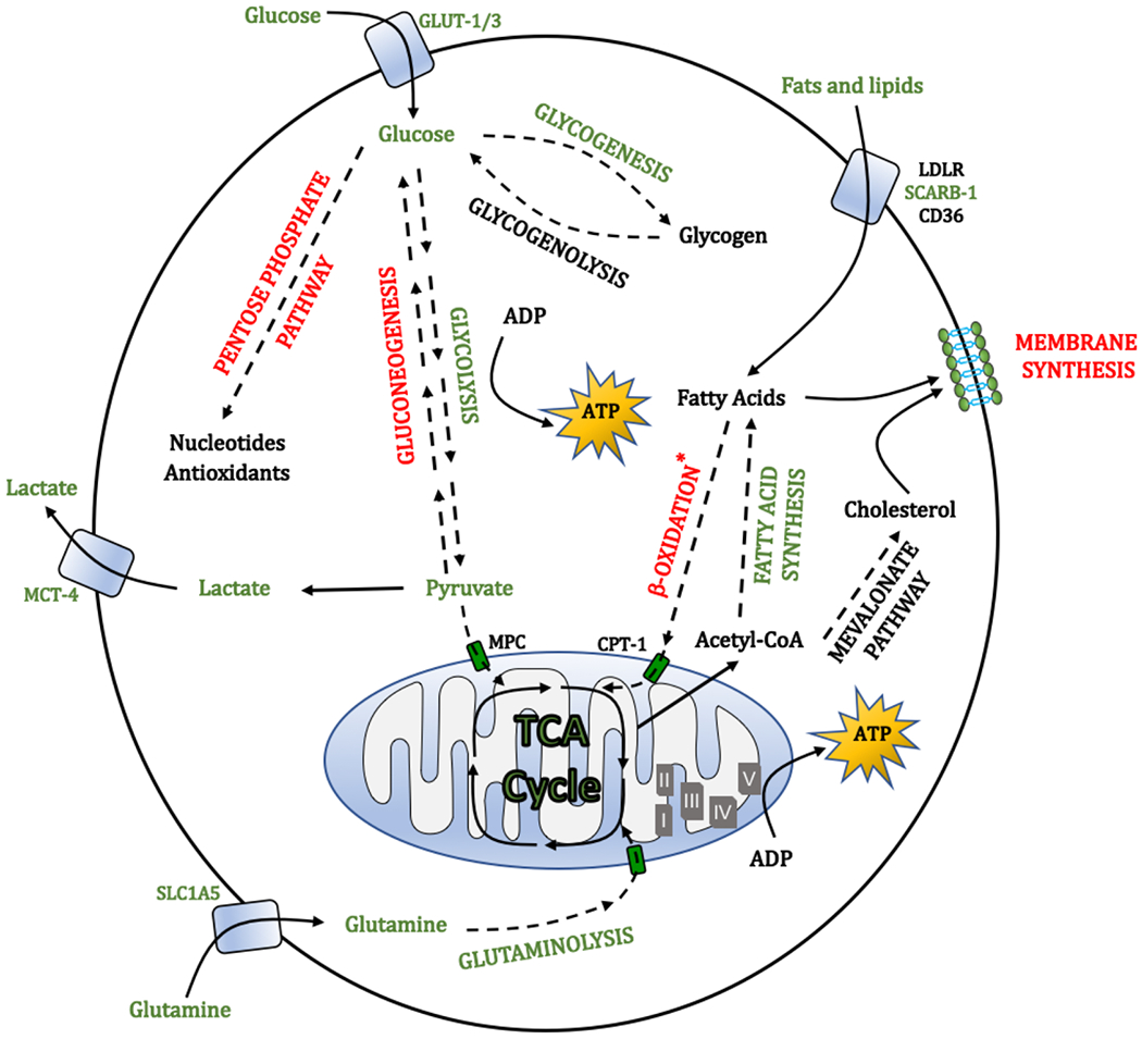
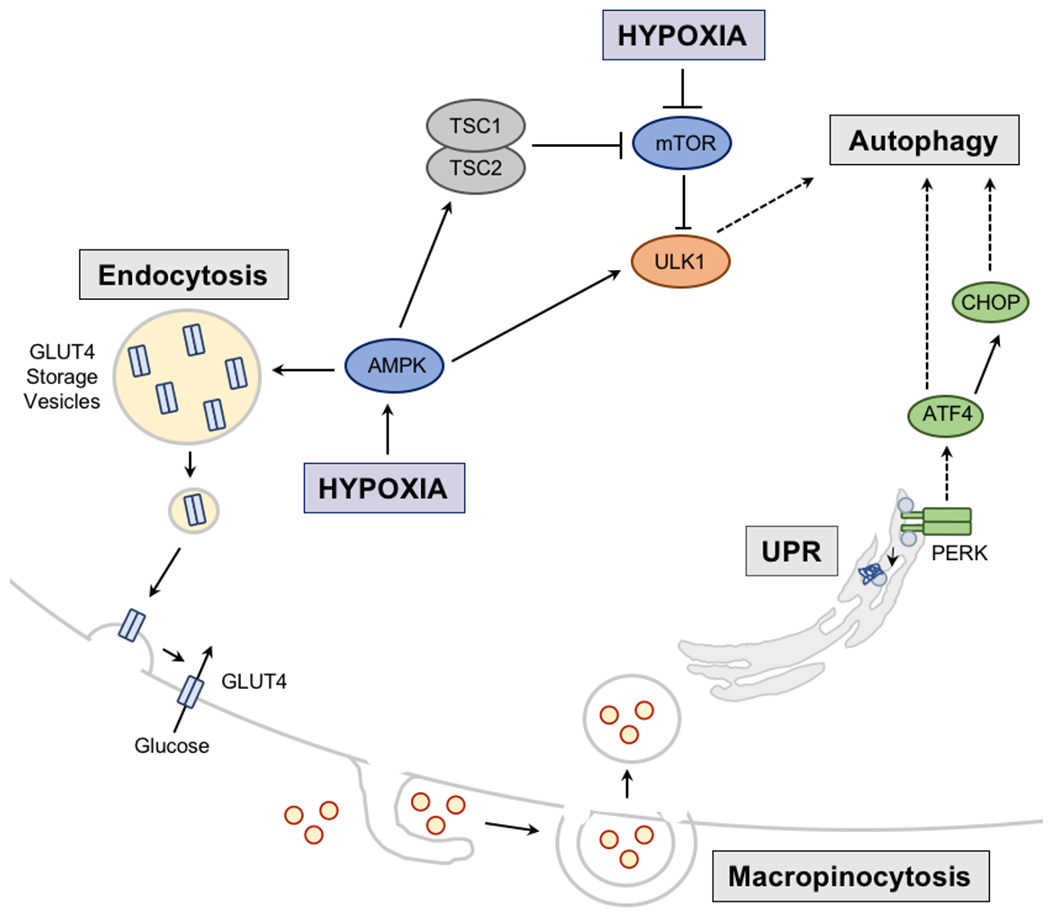
Molecular oxygen (O2) sustains intracellular bioenergetics and is consumed by numerous biochemical reactions, making it essential for most species on Earth. Accordingly, decreased oxygen concentration (hypoxia) is a major stressor that generally subverts life of aerobic species and is a prominent feature of pathological states encountered in bacterial infection, inflammation, wounds, cardiovascular defects and cancer. Therefore, key adaptive mechanisms to cope with hypoxia have evolved in mammals. Systemically, these adaptations include increased ventilation, cardiac output, blood vessel growth and circulating red blood cell numbers. On a cellular level, ATP-consuming reactions are suppressed, and metabolism is altered until oxygen homeostasis is restored. A critical question is how mammalian cells sense oxygen levels to coordinate diverse biological outputs during hypoxia. The best-studied mechanism of response to hypoxia involves hypoxia inducible factors (HIFs), which are stabilized by low oxygen availability and control the expression of a multitude of genes, including those involved in cell survival, angiogenesis, glycolysis and invasion/metastasis. Importantly, changes in oxygen can also be sensed via other stress pathways as well as changes in metabolite levels and the generation of reactive oxygen species by mitochondria. Collectively, this leads to cellular adaptations of protein synthesis, energy metabolism, mitochondrial respiration, lipid and carbon metabolism as well as nutrient acquisition. These mechanisms are integral inputs into fine-tuning the responses to hypoxic stress.


Conflict of interest statement

Competing interests

The authors declare no competing interests.

Figures

Figure 1:
Figure 1:. Transcription regulation induced by hypoxia
The oxygen-dependent interaction between the hypoxia inducible transcription factor α (HIF-α) subunits and the von Hippel–Lindau (VHL) tumour suppressor protein (pVHL) complex requires hydroxylation of two HIF-α proline residues by a family of α-ketoglutarate -dependent dioxygenases termed prolyl hydroxylases (PHDs), which requires oxygen (O2), iron (Fe2+), and α-ketoglutarate to function. Following hydroxylation, HIF-α subunits are polyubiquitylated by pVHL and targeted for proteasomal degradation. Hypoxia prevents the hydroxylation of the HIF-α protein subunits and their ubiquitin-mediated proteasomal degradation. As a result, the HIF-α protein subunits are allowed to dimerize with the HIF-1β protein subunits to form transcriptionally active complexes that bind to hypoxia response elements (HREs) to coordinate the induction of a large network of genes involved in metabolism, erythropoiesis, angiogenesis, and cell fate. Factor inhibiting HIF-1 (FIH-1) hydroxylates HIF-α subunits under normoxia to prevent recruitment of the transcription coactivators. Various mitochondrial products can also influence the hypoxic response. The production of reactive oxygen species (ROS) by mitochondrial complex III and L-2-hydroxyglutarate (L-2HG) under hypoxia can promote the stabilization of HIF-α protein levels. ROS likely inhibit PHDs by Cys oxidation, while L-2HG competes with α-ketoglutarate. Mutations in tricarboxylic acid (TCA) cycle components result in the accumulation of succinate and fumarate, which also inhibit PHD activity by competing with α-ketoglutarate, thereby causing an accumulation of the HIF-α protein even under normoxia.
Figure 2:
Figure 2:. Hypoxic adaptations in proteostasis.
Low oxygen (O2) can induce endoplasmic reticulum (ER) stress and the unfolded protein response (UPR; Supplementary Box 1). Misfolded peptides bind to binding immunoglobulin protein (BiP) and causes it to activate the stress sensors: activating transcription factor 6 (ATF6), inositol-requiring protein 1α (IRE1α), and protein kinase RNA-like ER kinase (PERK) to initiate responses to restore ER homeostasis. ATF6 is transported to the Golgi apparatus, where it is processed to release its active transcriptional form (ATF6f). IRE1α activation and dimerization, triggers its RNase activity, which processes unspliced X box-binding protein 1 (XBP1u) to produce an active transcription factor, spliced XBP1 (XBP1s). Upon activation, PERK phosphorylates the initiation factor eukaryotic translation initiator factor 2α (eIF2α) to attenuate general peptide translation and promote the expression of transcription factor ATF4. Activation of the UPR alleviates the burden of misfolded and/or unfolded proteins, whereas translation inhibition reduces energy expenditure. Hypoxia also negatively regulates translation initiation by controlling the formation of the mRNA cap-binding eIF4F complex, comprising eIF4E, eIF4A and eIF4G. Formation of this complex is promoted by the release of eIF4E from its inhibitors, eIF4E-binding proteins (4E-BPs). This release is regulated by phosphorylation of 4E-BPs by mechanistic target of rapamycin (mTOR). Hypoxia inhibits mTOR activity, thereby interfering with eIF4E release. Hypoxia also promotes nuclear sequestration of eIF4E by its transporter, 4-ET. Elongation of mRNA translation is also regulated during hypoxia by modulating the activity of eukaryotic elongation factor 2 (eEF2) kinase (eEF2K) — an inhibitor of eEF2. eEF2K is negatively regulated by prolyl hydroxylases (PHDs) and mTOR, and inhibition of both during hypoxia increases eEF2K activity, which by phosphorylating eEF2, prevents mRNA elongation. Finally, translation termination is negatively affected by hypoxia due to decreased hydroxylation of eukaryotic release factor 1 (eRF1) by Jumonji domain-containing 4 (JMJD4) — α-ketoglutarate and Fe2+-dependent oxygenase that like PHDs is inhibited in hypoxia. Importantly, some mRNAs need to overcome the translation repression induced by hypoxia, prominently including those encoding mediators of hypoxia, such as hypoxia inducible transcription factor (HIF)-responsive genes. HIF-responsive mRNAs contain hypoxia response element (rHREs). Low O2 stimulates the formation of a complex including HIF-2α, RBM4, and eIF4E2 (eIF4E homolog) that assembles at these rHREs to promote translation initiation. Hypoxia also promotes a formation of hypoxia-specific eIF4F complex (eIF4FH) that binds rHREs. Selective hypoxia-responsive mRNA translation can also occur by direct binding of ribosomes to internal ribosome entry sites (IRES) encoded within the 5′-UTR of certain mRNAs (such as VEGF, eIF4G, and C-MYC). Adaptive protein synthesis during hypoxia is further regulated by the partitioning and recruitment of mRNAs to the ER by signal recognition particles (SRPs) which deliver mRNAs, such as those encoding VEGF, HIF1, and P4HA1 to SRP-binding proteins on the ER membrane.
Figure 3:
Figure 3:. Impact of hypoxia on mitochondrial function.
Hypoxia is characterized by decreased flux through the tricarboxylic acid (TCA) cycle in mitochondria, leading to the reduction of metabolites, such as acetyl-CoA and aspartate, required for anabolic processes. Specifically, hypoxia inducible transcription factor 1 (HIF-1) induces the expression of lactate dehydrogenase A (LDHA) and pyruvate dehydrogenase kinase 1 (PDK1), which then negatively regulates the entry of pyruvate to the TCA cycle (by promoting lactate generation and inhibiting its conversion to acetyl-CoA, respectively). Hypoxia also impacts on the activity of the electron transport chain (ETC). Under acute hypoxia, ETC activity is maintained by hypoxia-induced expression of the complex IV (COX) subunit COX4I2, which substitutes the COX4I1 subunit allowing for a more efficient transfer of electrons to oxygen (O2) during hypoxia. Another HIF-1-dependent protein that enhances COX activity through unknown mechanisms is the hypoxia-inducible gene domain family member 1A (HIGD1A). This allows cells to maintain energy homeostasis in the event of short-term stresses. However, under prolonged hypoxia, ETC activity is diminished by inducing NDUFA4L2 (NADH dehydrogenase [ubiquinone] 1 alpha subcomplex, 4-like 2) in a HIF-1-dependent manner to decrease complex I activity and respiration, as well as several microRNAs (miRNAs), including mir-210, which represses ETC complex assembly. This repression of ETC activity has the potential benefit of decreasing the production of reactive oxygen species (ROS), which are generated by complex III under hypoxia (although mechanisms of hypoxic induction of ROS remain poorly understood) and can be potentially toxic by mediating damage to macromolecules. Nevertheless, hypoxia-induced ROS, and in particular hydrogen peroxide (H2O2), can also have various signalling roles. One of the consequences of mitochondrial ROS increase in hypoxia is the elevation in calcium (Ca2+), which activates Ca2+/calmodulin-dependent protein kinase kinase (CaMKK). CAMKK then activates AMP-activated protein kinase (AMPK) to suppress ATP consuming processes (metabolic demand). Ca2+ is also a signal for firing of carotid body nerves to stimulate ventilation and for vasoconstriction of pulmonary arteries. Moderate amounts of mitochondrial ROS were also shown to have anti-ageing functions. In addition to metabolism, hypoxia modulates mitochondrial dynamics (fission), likely to enhance quality control of damaged mitochondria via mitophagy, which can aid in limiting ROS generation. HRE, hypoxia response element; ISCU1/2, Iron-sulphur cluster assembly scaffold protein 1/2.
Figure 4.
Figure 4.. Overview of sugar and lipid metabolic pathways affected by oxygen availability.
The figure highlights the upregulated (green) and downregulated (red) metabolic pathways under hypoxia. Hypoxic cells increase the uptake and utilization of glucose via glycolysis to obtain energy, while reducing their metabolism in mitochondria (Fig. 3). This leads to the generation of large amounts of lactate, which is secreted and can support metabolism of neighbouring cells. As a protective mechanism, glucose is also diverted towards the serine synthesis pathway to overcome the loss of cellular antioxidant capacity when pentose phosphate pathway activity is decreased. Hypoxia also promotes glycogenesis, which could provide a mechanism of energy storage to survive prolonged stress. Under hypoxic inhibition of the tricarboxylic acid (TCA) cycle, generation of anabolic metabolites, such as acetyl-CoA, which is key for the synthesis of fatty acids, is largely supported by the increased uptake and metabolism of glutamine. Concomitantly, catabolism of fatty acids via β-oxidation is suppressed, while the uptake of lipids from the exterior increased. Lipid desaturation, allowing generation of unsaturated fatty acids is inhibited in hypoxia (owing to the requirement for oxygen by stearoyl-CoA desaturase). To counteract potential lipotoxicity associated with the accumulation of saturated lipids and disruption of membrane structure and integrity, cells increase the uptake of unsaturated lipids from the environment and increase the formation of lipid droplets, which can act as buffers for saturated lipid species. ADP, adenosine diphosphate; ATP, adenosine triphosphate; CD36, cluster of differentiation 36; CPT-1, carnitine palmitoyltransferase 1; ETC, electron transport chain; GLUT-1, glucose transporter 1; LDLR, low density lipoprotein receptor; MCT4, monocarboxylate transporter 4; MPC, mitochondrial pyruvate carrier; SCARB-1, scavenger receptor B1; SLC1A5, solute carrier family 1 (neutral amino acid transporter) member 5.
Figure 5:
Figure 5:. Regulation of nutrient acquisition and use under hypoxia.
Hypoxia induces nutrient recycling via autophagy. In this case, the unfolded protein response (UPR) resulting from hypoxia, activates protein kinase RNA-like ER kinase (PERK), which induces transcription factor ATF4 and subsequently C/EBP homologous protein (CHOP) to activate autophagic machinery. Hypoxia, via activation of hypoxia inducible factors (HIFs) further regulates autophagy by HIF-mediated regulation of expression of two mitochondrial proteins BNIP3 and BNIP3L that have been linked to autophagy of mitochondria (mitophagy). Autophagy can also be induced during hypoxia via activation of AMP kinase (AMPK) which subsequently activates autophagy directly or indirectly through inhibition of mechanistic target of rapamycin (mTOR), a negative regulator of the Unc-51 like autophagy activating kinase 1 (ULK1). AMPK can also promote the movement of the glucose transporter GLUT4 from its storage vesicles to the cell membrane to promote glucose uptake. Nutrient acquisition is also achieved via macropinocytosis, which is promoted by hypoxia and supports the ingestion of extracellular macromolecules, such as amino acids and fatty acids. TSC1/2, Tuberous sclerosis 1/2.

References

    1. Pouyssegur J & López-Barneo J Hypoxia in health and disease. Molecular Aspects of Medicine 47–48, 1–2 (2016). - PubMed
    1. Taylor CT, Doherty G, Fallon PG & Cummins EP Hypoxia-dependent regulation of inflammatory pathways in immune cells. J. Clin. Invest. 126, 3716–3724 (2016). - PMC - PubMed
    1. Semenza GL Hypoxia-inducible factors in physiology and medicine. Cell 148, 399–408 (2012). - PMC - PubMed
    1. Keith B, Johnson RS & Simon MC HIF1α and HIF2α: sibling rivalry in hypoxic tumour growth and progression. Nat. Rev. Cancer 12, 9–22 (2011). - PMC - PubMed
    1. Kaelin WG et al. Oxygen sensing by metazoans: the central role of the HIF hydroxylase pathway. Mol Cell 30, 393–402 (2008). - PubMed
    2. Reviews by Semenza (2012) and Kaelin et al, (2008) provide an excellent outline of the regulation and importance of HIFs in physiology and diseases.

Publication types

MeSH terms