Review
. 2021 Apr 19;60(17):9215-9246.
doi: 10.1002/anie.202000451. Epub 2020 Nov 19.

Superoxide Dismutase 1 in Health and Disease: How a Frontline Antioxidant Becomes Neurotoxic

Affiliations
Review

Superoxide Dismutase 1 in Health and Disease: How a Frontline Antioxidant Becomes Neurotoxic

Benjamin G Trist et al. Angew Chem Int Ed Engl. .

Abstract

Cu/Zn superoxide dismutase (SOD1) is a frontline antioxidant enzyme catalysing superoxide breakdown and is important for most forms of eukaryotic life. The evolution of aerobic respiration by mitochondria increased cellular production of superoxide, resulting in an increased reliance upon SOD1. Consistent with the importance of SOD1 for cellular health, many human diseases of the central nervous system involve perturbations in SOD1 biology. But far from providing a simple demonstration of how disease arises from SOD1 loss-of-function, attempts to elucidate pathways by which atypical SOD1 biology leads to neurodegeneration have revealed unexpectedly complex molecular characteristics delineating healthy, functional SOD1 protein from that which likely contributes to central nervous system disease. This review summarises current understanding of SOD1 biology from SOD1 genetics through to protein function and stability.

Keywords: Cu/Zn superoxide dismutase; antioxidants; copper; neurodegeneration; protein misfolding.


Conflict of interest statement

The authors declare no conflict of interest.

Figures

Figure 1
Figure 1
Structure of the human SOD1 gene and its main regulatory elements. Included: untranslated regions (red), exons (orange), introns (grey), transcription start site (red arrow), transcription factors (blue), polyadenylation (PolyA) sites (yellow) and AU‐rich elements (ARE; green). Exact binding sites for transcription factors are listed in Table 1. Figure adapted from ref. [17]. Copyright Milani et al (2011); Creative Commons Attribution 3.0 licence.
Figure 2
Figure 2
Structural elements within SOD1 protein. A,B) Mature SOD1 is dimeric, and comprises an eight‐stranded β‐barrel (grey) with one Cu (orange) and Zn (cyan) ion bound in each monomer. The electrostatic loop (blue; A, B, C) contains charged (dark blue) and polar (light blue) residues important for guiding anionic O2 .− towards the active site. The metal‐binding loop (green; B) mediates Zn coordination through three histidine residues and one aspartic acid residue (cyan; A, D). The disulfide loop (yellow; D) is a substructure within the metal‐binding loop, and contains one of the two cysteine residues forming a stabilizing intramonomeric disulfide bond (yellow; A, B). Cu coordination is facilitated by four histidine residues (orange; A, E), one of which is shared with Zn (His63). Multiple residues within β‐strand 8 and the disulfide loop participate in reciprocal hydrogen bonding between SOD1 monomers (red; F, G), creating a tight interface that underlies the exceptional thermodynamic stability of the holo‐SOD1 dimer. The Greek key loop (pink; A, B) forms a plug at one pole of the β‐barrel and contributes to dimer interface stability. Exact residues pertaining to each structure detailed in Supplementary Table 1.
Figure 3
Figure 3
Transitional free‐energy landscapes of SOD1 metalloforms and CCS‐dependent SOD1 maturation. Free energy landscapes of SOD1 metalloforms demonstrate the existence of multiple conformers, which interchange in complex equilibria. Zn incorporation into the native monomeric SOD1 and CCS apo proteins occurs via unknown mechanisms, and elicits conformational change in each protein to prepare them for coupling and Cu+ insertion. Zn insertion into apo‐CCS prompts homodimerisation and migration to the plasma membrane, where it forms a stable complex with the copper transporter Ctr1 via its C‐terminal domain (CCS‐Cu+‐Ctr1) and acquires Cu+ in D1. Zn incorporation into apo‐SOD1 is promoted by CCS coupling, mediated by D2 and D3 of CCS protein. Apo‐ or Zn‐bound‐SOD1 may couple with CCS within a preformed CCS‐Cu+‐Ctr1 complex at the plasma membrane, forming a ternary SOD1‐CCS‐Cu+‐Ctr1 complex, or alternatively may couple with CCS within the cytosol and subsequently migrate to the plasma membrane to complex with Ctr1. Zn first binds transiently to the Cu‐binding site of SOD1, triggering the formation of the Zn‐binding site and the subsequent migration of Zn to its physiological Zn centre. Zn binding reduces structural flexibility in SOD1 protein and prevents transitions to non‐native excited SOD1 conformers. SOD1‐CCS coupling and SOD1 Zn binding trigger conformational change within CCS, whereby D3 arches over the disulfide loop of SOD1 to stabilise SOD1‐CCS coupling, uncover an “entry” binding site for Cu+ in SOD1, and expose the SOD1 Cys146 residue to solvent. Oxidation of the exposed thiol side chain of SOD1 Cys146 triggers disulfide exchange reactions between the CXC motif in CCS D3, and adjacent Cys146/Cys57 residues of SOD1 protein, culminating in the formation of a disulfide bond between SOD1 Cys57 and Cys146. SOD1 disulfide bond formation promotes Cu+ migration from the entry site to the active site. Together these events trigger SOD1 and CCS dissociation from Ctr1, producing fully metalated holo‐SOD1 monomers that can now homodimerize.
Figure 4
Figure 4
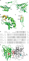
Structural elements within CCS protein and its structural homology to SOD1 protein. A) CCS protein consists of three distinct domains (D1–D3). D1 (B) and D3 (C) bind Cu+ (orange spheres) by virtue of MXCXXC and CXC binding motifs, respectively. D) D2 exhibits a high sequence homology to SOD1 protein (homologous residues indicated by black bolding), facilitating SOD1–CCS heterodimerisation prior to Cu transfer and disulfide bond formation. E) Conserved arginine residues at positions 104 and 232 of CCS D2, together with a GDNT motif within SOD1 protein, mediate strong stabilizing interactions across the heterodimer interface (residues highlighted in red). Zn2+ represented by cyan spheres in (A) and (E).
Figure 5
Figure 5
Superoxide dismutation catalyzed by SOD1. A) Redox cycling of Cu within the active site of SOD1 enables sequential superoxide (O2 .−) oxidation to molecular oxygen (O2; reaction 1) and O2 .− reduction to hydrogen peroxide (H2O2; reaction 2). B) Charged and polar residues (red), especially within the electrostatic loop, in the active site channel provide electrostatic guidance for O2 .− towards the catalytic Cu co‐factor (orange). Lys136 (K136) and Glu133 (E133) direct the long‐range approach of O2 .−, whereas Arg143 (R143) is important in hydrogen bonding with incoming O2 .− and limits incoming anion size.
Figure 6
Figure 6
SOD1 and CCS transport and retention within the mitochondrial intermembrane space (IMS). Apo‐CCS and apo‐SOD1 protein import into the mitochondrial IMS from the cytoplasm (cyto) is facilitated by translocator of the outer membrane (TOM). The Mia40/Erv1 disulfide relay system is thought to trigger apo‐CCS maturation through disulfide bond oxidation (2SH→SS) and potentially metalation of apo‐CCS within the IMS, trapping it within this compartment. Holo‐CCS and oxidative stress work together to trigger SOD1 maturation within the IMS, mitigating the build‐up of reactive oxygen species (ROS) in this compartment produced primarily by the electron transport chain. IMM: inner mitochondrial membrane; Cyt C: cytochrome C.
Figure 7
Figure 7
SOD1 antioxidant function and redox signaling. Aside from the enzymatic breakdown of superoxide (O2 .−) radicals, SOD1 combats the accumulation of reactive oxygen species (ROS) by acting as a transcription factor for multiple genes involved in adaptive stress responses, including a large number of antioxidant genes. This occurs following phosphorylation of SOD1 by Cds1 kinase, which is itself phosphorylated and activated by ATM kinase upon increases in cellular ROS levels. By directly and indirectly influencing the levels of cellular ROS, especially O2 .− and hydrogen peroxide (H2O2), SOD1 is also an important component of redox signaling pathways governing processes such as cell proliferation and adaptive stress responses.
Figure 8
Figure 8
Distribution of 46 well‐studied wild‐type‐like (purple; A4V–L144F) and metal‐binding region (black; H46R–C146R) SOD1 mutations (A) and, where known, their effect on Cu and Zn binding (B). Corresponding biochemical data for all mutations detailed in Table 2. Mutant metal binding values were averaged for mutants where metal binding data was available from more than one expression system.
Figure 9
Figure 9
Distribution of known sites of post‐translational modification for human SOD1 protein. Included: oxidation (A, B; yellow), phosphorylation (C; blue), ubiquitylation (D; purple), acetylation (E; light green), deamidation (E; light pink) succinylation (F; dark pink), methylgloxalation (F; violet), glycation (G; brown), palmitoylation (H; dark green). Residues labeled in all panels were identified from analyses of human SOD1 protein using mass spectrometry (PhosphoSitePlus®, Cell Signalling Technology),[ 102a , 120 ] with the exception of panel B which was performed in silico. [121] Specific residue numbers listed in Supplementary Table 1.
Figure 10
Figure 10
Heterogeneity in mechanisms of mutant and wild‐type SOD1 aggregation derives from differences in the biochemistry of soluble forms of each misfolded species, and may partially underlie variation in disease duration (onset to death) between individual SOD1‐linked familial ALS and sporadic ALS patients. Data was obtained from Table 2 in this review, grey shaded boxes represent no data available. Oxidised wild‐type SOD1 (Ox‐WT) onset to death (years) represented by average sporadic ALS disease duration. Coloured regions of grey bars represent residues of SOD1 protein implicated in self‐assembly for that misfolded variant, confirmed by mass spectrometry as reported by Furukawa and colleagues [1] (specific residue numbers listed Supplementary Table 2).
Figure 11
Figure 11
Mitochondrial SOD1 biology under physiological and pathological conditions. In a healthy cell apo‐SOD1 is transferred from the cytosol (Cyto) into the mitochondrial intermembrane space (IMS) by translocase of the outer membrane (TOM). Cu chaperone for SOD1 (CCS) facilitates metal insertion and maturation of apo‐SOD1 to holo‐SOD1 in the IMS, which detoxifies reactive oxygen species (ROS) produced by the electron transport chain (ETC) in the inner mitochondrial membrane (IMM). ATP produced by the ETC is essential for maintaining many cellular metabolic processes, and is shuttled between the mitochondrial matrix and the cytosol by adenine nucleotide translocator (ANT) and voltage‐dependent anion‐selective channel 1 (VDAC1), located in the IMM and outer mitochondrial membrane (OMM), respectively. VDAC1 is also crucial for ADP and Ca2+ import, which together drive mitochondrial energy production. Calcium may be stored in the mitochondrial matrix, with fluxes across the IMM controlled by mitochondrial calcium uniporter (MCU) and the mitochondrial sodium calcium exchanger NCLX. SOD1 gene mutations or atypical post‐translational modifications to SOD1 protein result in misfolded SOD1 (mis‐SOD1) protein, which either remains in the cytosol or enters the mitochondrial IMS, where it is shown to aggregate. Aggregated SOD1 is unable to detoxify ROS in the IMS, and is proposed to impair ETC function to promote the accumulation of ROS and pro‐apoptotic cytochrome c (Cyt C) in this compartment. Cytosolic mis‐SOD1 binds to Bcl‐2, an anti‐apoptotic protein which normally inhibits the function of pro‐apoptotic proteins Bax and Bak. SOD1 binding causes conformational change in Bcl‐2, exposing its toxic BH3 domain. SOD1‐Bcl‐2‐BH3 complexes localise to the OMM where they inhibit VDAC1 and promote the formation of Bax and Bak pores. Together, these actions disrupt mitochondrial calcium homeostasis and ATP production, and facilitate the release of Cyt C into the cytosol, triggering cell death.

References

    1. Furukawa Y., Kaneko K., Yamanaka K., Nukina N., J. Biol. Chem. 2010, 285, 22221–22231. - PMC - PubMed
    1. Karnati S., Luers G., Pfreimer S., Baumgart-Vogt E., Histochem. Cell Biol. 2013, 140, 105–117. - PubMed
    1. Oury T. D., Day B. J., Crapo J. D., Free Radical Biol. Med. 1996, 20, 957–965. - PubMed
    1. Mann T., Kleinin D., Proc. R. Soc. London Ser. B 1938, 126, 303–315.
    1. McCord J. M., Fridovic I., J. Biol. Chem. 1969, 244, 6049. - PubMed

Publication types

MeSH terms