. 2020 May 6;106(3):526-536.e4.
doi: 10.1016/j.neuron.2020.02.011. Epub 2020 Mar 6.

Propagating Motor Cortical Dynamics Facilitate Movement Initiation

Affiliations

Propagating Motor Cortical Dynamics Facilitate Movement Initiation

Karthikeyan Balasubramanian et al. Neuron. .

Erratum in

  • Propagating Motor Cortical Dynamics Facilitate Movement Initiation.
    Balasubramanian K, Papadourakis V, Liang W, Takahashi K, Best MD, Suminski AJ, Hatsopoulos NG. Balasubramanian K, et al. Neuron. 2025 Aug 6;113(15):2546. doi: 10.1016/j.neuron.2025.07.007. Epub 2025 Jul 12. Neuron. 2025. PMID: 40652940 Free PMC article. No abstract available.

Abstract

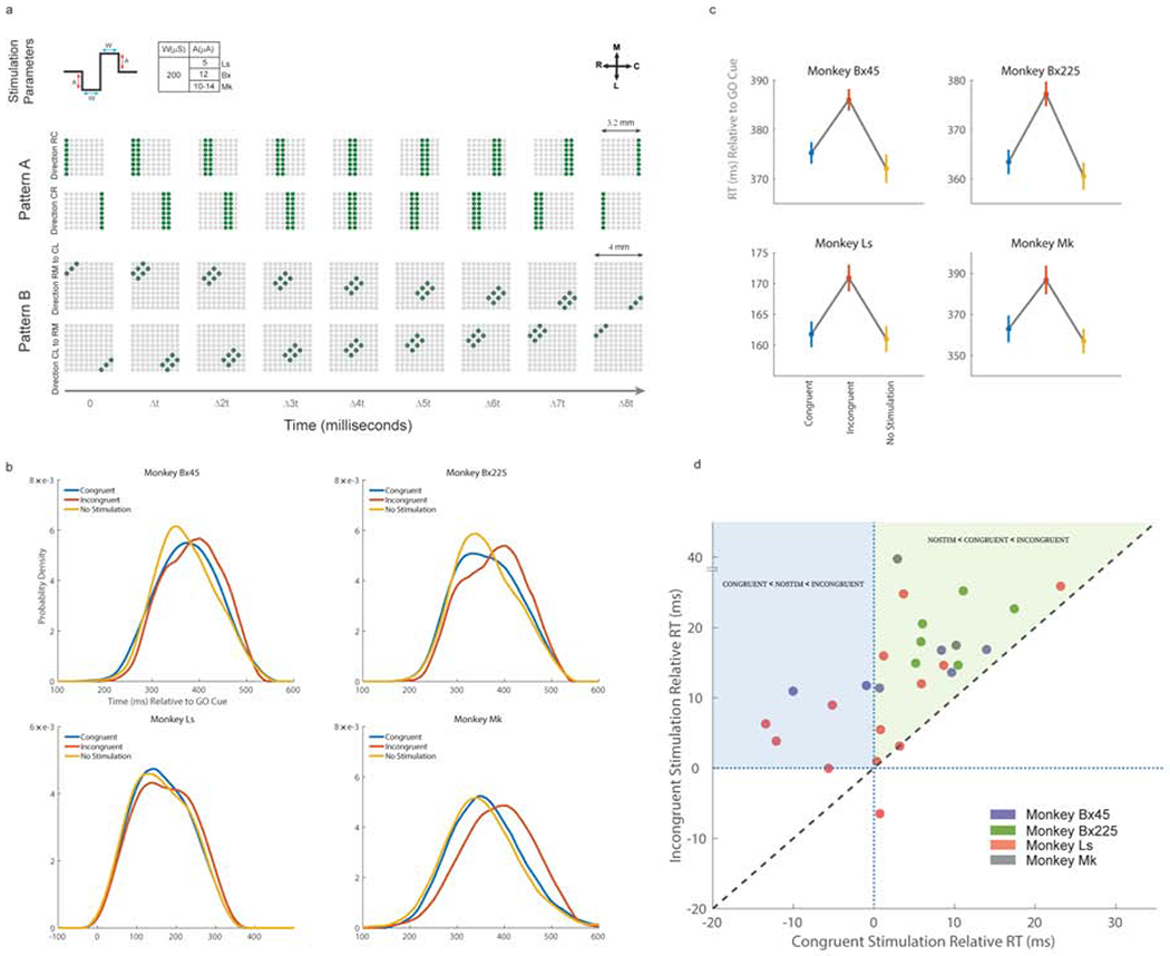
Voluntary movement initiation involves the modulations of large groups of neurons in the primary motor cortex (M1). Yet similar modulations occur during movement planning when no movement occurs. Here, we show that a sequential spatiotemporal pattern of excitability propagates across M1 prior to the movement initiation in one of two oppositely oriented directions along the rostro-caudal axis. Using spatiotemporal patterns of intracortical microstimulation, we find that reaction time increases significantly when stimulation is delivered against, but not with, the natural propagation direction. Functional connections among M1 units emerge at movement that are oriented along the same rostro-caudal axis but not during movement planning. Finally, we show that beta amplitude profiles can more accurately decode muscle activity when they conform to the natural propagating patterns. These findings provide the first causal evidence that large-scale, propagating patterns of cortical excitability are behaviorally relevant and may be a necessary component of movement initiation.

Keywords: beta attenuation; beta oscillations; functional connectivity; intracortical microstimulation; motor cortex; movement initiation; propagating patterns; spatiotemporal patterns.


Conflict of interest statement

Declaration of Interests N.G.H. serves as a consultant for BlackRock Microsystems, Inc., the company that sells the multi-electrode arrays and acquisition system used in this study.

Figures

Figure 1.
Figure 1.. Beta band power and synchrony during rest and movement.
a. Beta frequency oscillations (gray) and amplitude envelope (red) on one trial from one electrode. When the beta envelope drops below a threshold amplitude (blue dotted line shows a hypothetical threshold value), the corresponding timestamp denotes beta attenuation timing (BATs; see Methods on how BATs were determined). MO-movement onset. b. Spike-triggered beta phase histograms (see Methods) from multi-unit activity on a single electrode during movement preparation (early; −500 to −400 millisecond window w.r.t. MO) and during movement initiation (late; −100 to 0 millisecond window w.r.t. MO). c. Spike-triggered beta phase histograms pooled over electrodes showing a beta phase preference angle of 120 to 180 degrees in the early window (blue) exhibited desynchronization during the late window prior to movement initiation (red). Monkeys Bx and Ls showed biased distributions (Rayleigh’s test; p=1.5e-309 for Bx and p=3.2e-15 for Ls; resultant direction (blue arrow) of 1460 for Bx and 1560 for Ls) during the early window, and, as neurons desynchronized, both resulted in uniform distributions (Rayleigh’s test; p=0.504 for Bx and p=0.73 for Ls) during the late window. d. Normalized beta envelopes averaged over different trial groups (trials sorted by reaction time, 50 trials per group) aligned on the GO cue (top left) or on movement onset (bottom left) from one session in Ls. (Right) Channel-averaged beta attenuation time (BAT) regressed on mean reaction times (big colored dots) in each trial group with respect to the GO cue (black line, r2= 0.996, p=0.00018) or movement onset (red line, r2= 0.126, p=0.558). Black dots represent single channel values (offset by 10ms and jittered for clarity).
Figure 2.
Figure 2.. Single trial beta attenuation propagates along one of the two directions along a rostrocaudal axis.
a. Beta envelopes of two trials (left; inset shows spatial positions of three electrodes with early (blue), intermediate (green), and late (red) attenuation times) and their corresponding spatial maps of beta attenuation times (right) are shown for monkey Ls. Regression fits to the pattern show the beta attenuation orientation (BAO) directed rostrally or caudally (red arrows). b. Polar histograms show the distributions of BAOs across multiple single trials and recording sessions for three monkeys (a total of 1369 and 1417 trials in 11 sessions in Bx45 and Bx225, respectively; 1626 trials in 9 sessions in Ls; and 278 trials in 4 sessions for Mk). A mixture of two von Mises functions was fit to the BAO distributions (red curves) to estimate the two direction modes of the BAO (blue arrows). Insets represent the initial hold (filled red) and target positions (filled gray) of the hand.
Figure 3.
Figure 3.. Spatio-temporal stimulation affects reaction time.
a. The stimulus was a biphasic pulse with sub-threshold currents (top), delivered concurrently over sets of electrodes with a propagation latency (Δt) of 10 milliseconds (4 milliseconds for Monkey Mk). Stimulation patterns were delivered to a group of electrodes propagating from rostral to causal sites or vice versa (Pattern A, middle) or diagonally (Pattern B, bottom). The stimulus patterns were delivered either along the BAO direction (CONGRUENT) or against the BAO direction (INCONGRUENT). b. The distributions of reaction times across trials for the two stimulation conditions along with those trials that received no stimulation (NO STIMULATION) are shown for Bx45 (731, 727, and 676 trials for CONGRUENT, INCONGRUENT, and NO STIMULATION), Bx225 (674, 650, and 595 trials), Ls (1028, 1048, and 1038 trials), and Mk (106, 91, and 168 trials). c. Mean (standard error) reaction time in the three conditions show that the INCONGRUENT stimulation resulted in delayed reaction compared to the other two conditions. d. Scatter plot comparing the mean reaction times between the two stimulation conditions from 27 individual experimental sessions over all three monkeys. The mean reaction time of the NO STIMULATION from each session was subtracted from the mean reaction times of the other two conditions. The diagonal dashed line represents the identity line, and the dotted lines show the zero mark. The shaded areas highlight regions where reaction times in the INCONGRUENT condition are longer than in the CONGRUENT condition but also reaction times in the NO STIMULATION condition are longer than in the CONGRUENT condition (blue shaded area) or, alternatively, reaction times in the CONGRUENT condition are longer than in the NO STIMULATION condition (green shaded area).
Figure 4.
Figure 4.. Functional connections among neurons emerge along the BAO axis during movement onset.
a. Multi-unit activity (MUA) from a population of M1 neurons was used to estimate functional connectivity during movement preparation and movement onset epochs. MUA for each electrode was normalized to its peak activity and sorted by time of peak activity. MUA from 60 and 64 electrodes are shown for Bx and Ls, respectively. b. Polar histograms of the orientation of functional connections during the movement preparation epoch (top, Bx for 45 degree movements; middle, Bx for 225 degree movements; bottom, Ls for 45 degree movements). The von Mises fits (red curves and dashed blue arrows) show functional connectivity distributions that were different from the BAO modes (blue arrows). A total of 95, 128, and 54 significant connections were found for Bx45, Bx225, and Ls, respectively. c. During the movement onset epoch, the neuronal functional connection distributions were oriented similar to the BAO distributions. A total of 52, 102, and 36 significant connections were found for Bx45, Bx225, and Ls, respectively.
Figure 5.
Figure 5.. Propagating sequence directions do not follow the somatotopic organization.
The somatotopic maps across the arrays implanted in the three monkeys show a proximal-to-distal somatotopy (colored dots) oriented along the medio-lateral axes. The somatotopic maps for Bx and Ls are based on two 8×8 arrays implanted medio-laterally in M1. The BAO axes (blue arrows), computed for the medial arrays in Ls and Bx and the one array in Mk are nearly orthogonal to the proximal-to-distal gradient (brown arrows). (see Figure S5 for statistical robustness of the somatotopic gradient estimates)
Figure 6.
Figure 6.. EMG prediction is more sensitive to perturbations along the BAO axis.
a. A feedforward back propagation neural network architecture that maps instantaneous beta amplitudes to electromyographic (EMG) from 5 muscles that were active during the particular movement condition. b. Trial-averaged predicted EMG values (red) are overlaid on the actual trial-averaged EMG signals (blue) for Bx. c. Perturbation of the spatiotemporal pattern was performed along the BAO axis (parallel swap) or orthogonal to it (orthogonal swap), and FVaF of the model’s performance was computed. d. Distributions of FVaFs over 5000 swaps are shown for the parallel swap condition (yellow; mean FVaF of 6.5%) and the orthogonal swap condition (blue; mean FVaF of 12%) along with the FVaF of the original unperturbed model (red line; mean FVaF of 40.5%). Inset represent the initial hold (filled red) and target positions (filled gray) of the hand.

References

    1. Baker SN, Olivier E, Lemon RN, 1997. Coherent oscillations in monkey motor cortex and hand muscle EMG show task-dependent modulation. J. Physiol 501, 225–241. 10.1111/j.1469-7793.1997.225bo.x - DOI - PMC - PubMed
    1. Best MD, Suminski AJ, Takahashi K, Brown KA, Hatsopoulos NG, 2017. Spatio-Temporal Patterning in Primary Motor Cortex at Movement Onset. Cereb. Cortex 27 10.1093/cercor/bhv327 - DOI - PMC - PubMed
    1. Bullock D, Grossberg S, 1988. Neural dynamics of planned arm movements: Emergent invariants and speed-accuracy properties during trajectory formation. Psychol. Rev 95, 49–90. 10.1037/0033-295X.95.1.49 - DOI - PubMed
    1. Chase MH, 1983. Synaptic mechanisms and circuitry involved in motoneuron control during sleep. Int Rev Neurobiol 24, 213–258. - PubMed
    1. Chen R, Yaseen Z, Cohen LG, Hallett M, 1998. Time course of corticospinal excitability in reaction time and self-paced movements. Ann. Neurol 44, 317–325. 10.1002/ana.410440306 - DOI - PubMed

Publication types

LinkOut - more resources