. 2020 May 6;28(5):1251-1262.
doi: 10.1016/j.ymthe.2020.02.016. Epub 2020 Feb 24.

Oncolytic Adenovirus Armed with BiTE, Cytokine, and Checkpoint Inhibitor Enables CAR T Cells to Control the Growth of Heterogeneous Tumors

Affiliations

Oncolytic Adenovirus Armed with BiTE, Cytokine, and Checkpoint Inhibitor Enables CAR T Cells to Control the Growth of Heterogeneous Tumors

Caroline E Porter et al. Mol Ther. .

Abstract

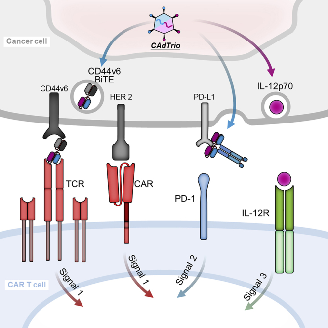
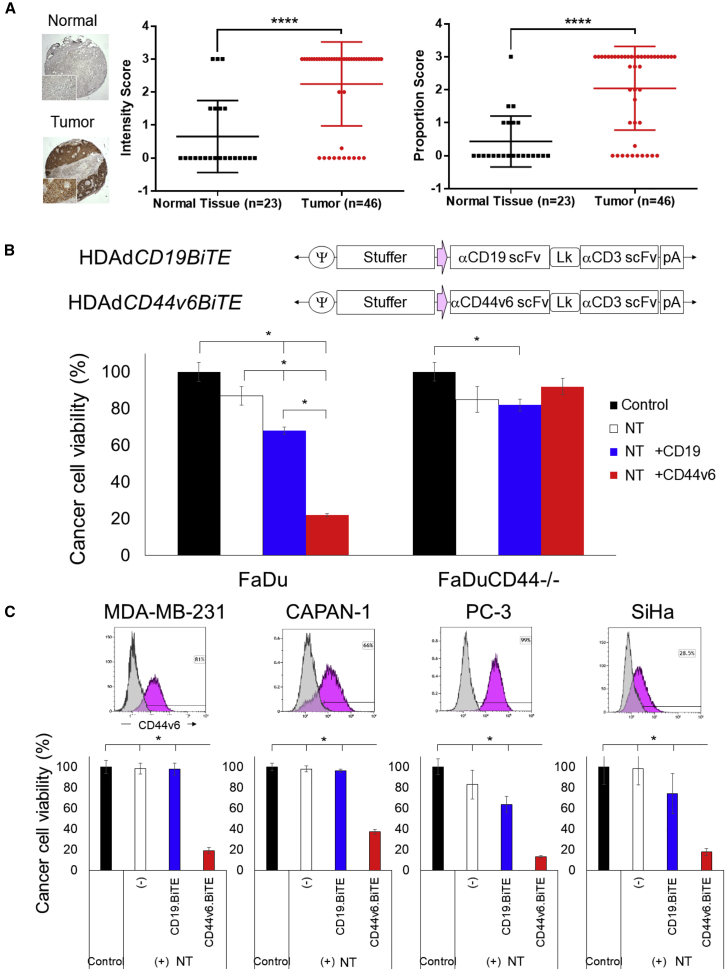
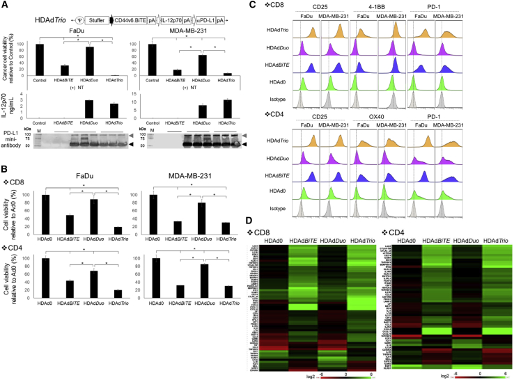
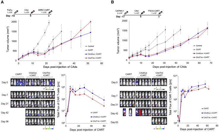
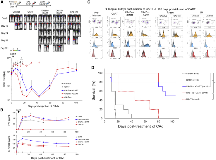
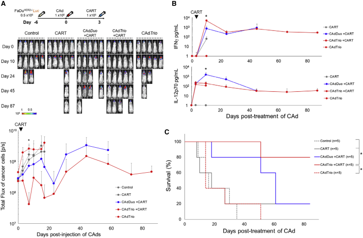
No single cancer immunotherapy will likely defeat all evasion mechanisms of solid tumors, including plasticity of tumor antigen expression and active immune suppression by the tumor environment. In this study, we increase the breadth, potency, and duration of anti-tumor activity of chimeric antigen receptor (CAR) T cells using an oncolytic virus (OV) that produces cytokine, checkpoint blockade, and a bispecific tumor-targeted T cell engager (BiTE) molecule. First, we constructed a BiTE molecule specific for CD44 variant 6 (CD44v6), since CD44v6 is widely expressed on tumor but not normal tissue, and a CD44v6 antibody has been safely administered to cancer patients. We then incorporated this BiTE sequence into an oncolytic-helper binary adenovirus (CAdDuo) encoding an immunostimulatory cytokine (interleukin [IL]-12) and an immune checkpoint blocker (PD-L1Ab) to form CAdTrio. CD44v6 BiTE from CAdTrio enabled HER2-specific CAR T cells to kill multiple CD44v6+ cancer cell lines and to produce more rapid and sustained disease control of orthotopic HER2+ and HER2-/- CD44v6+ tumors than any component alone. Thus, the combination of CAdTrio with HER2.CAR T cells ensures dual targeting of two tumor antigens by engagement of distinct classes of receptor (CAR and native T cell receptor [TCR]), and significantly improves tumor control and survival.

Keywords: oncolytic viro-immunotherapy, CAR T cell, BiTE molecule, cytokine, checkpoint inhibitor, CD44 variant 6.


Figures

None
Graphical abstract
Figure 1
Figure 1
CD44v6 Is Highly Expressed on HNSCC Tumors, and CD44v6.BiTE Enables T Cell Killing of Multiple Cancer Cell Lines (A) CD44v6 immunohistochemistry of larynx and salivary glands TMA. Data are presented as means ± SD. ∗∗∗∗p < 0.001. (B) Schematic structure of HDAd encoding a BiTE expression cassette. FaDu- and FaDuCD44−/−-expressing ffLuc cells were infected with 100 vp/cell of HDAds (n = 4 per group). Non-transduced T cells (NTs) were added with an effector-to-target ratio of 1:10 (E:T = 1:10) at 24 h post-infection. Cells were harvested 72 h post-co-culture with T cells, and viable cancer cells were analyzed by a luciferase assay. Data are presented as means ± SD. ∗p < 0.001. (C) CD44v6 expression was analyzed by flow cytometry on MDA-MB-231, CAPAN-1, SiHa, and PC-3 cells. These cells expressing ffLuc were infected with 100 vp/cell HDAds (n = 4 per group). NTs were added at 24 h post-infection (E:T = 1:10). Cells were harvested 72 h post-co-culture, and viable cancer cells were analyzed by a luciferase assay. Data are presented as means ± SD. ∗p < 0.001.
Figure 2
Figure 2
HDAd-Derived CD44v6.BiTE, IL-12p70, and PD-L1 Blocking Antibody Increase the Anti-tumor Effects of Non-transduced T Cell In Vitro (A) Schematic structure of HDAd encoding CD44v6.BiTE, human IL-12p70, and anti-PD-L1 mini-antibody expression cassettes (HDAdTrio). FaDu- and MDA-MB-231-expressing ffLuc cells were infected with 100 vp/cell HDAdCD44v6.BiTE, HDAdDuo, or HDAdTrio (n = 4 per group). NTs were added at 24 h post-infection (E:T = 1:10). Cells were harvested 72 h post-co-culture with T cells, and viable cancer cells were analyzed by a luciferase assay. Data are presented as means ± SD. ∗p < 0.001. FaDu and MDA-MB-231 cells were infected with 200 vp/cell HDAdCD44v6.BiTE, HDAd12_PDL1, or HDAdTrio (n = 4 per group), and media samples collected 48 h post-infection were subjected to IL-12p70 ELISA and western blotting for PD-L1 mini-antibody, which was detected by anti-HA antibody. IL-12p70 data are presented as means ± SD. (B) FaDu- and MDA-MB-231-expressing ffLuc cells were infected with 100 vp/cell HDAd0 (no transgene), HDAdCD44v6.BiTE, HDAdDuo, or HDAdTrio (n = 5 per group). Either CD8 or CD4 NTs were added at 24 h post-infection (E:T = 1:10). Cells were harvested 72 h post-co-culture with T cells, and viable cancer cells were analyzed by a luciferase assay. Data are presented as means ± SD. ∗p < 0.001. (C) T cells were harvested 72 h post-co-culture, and CD25, PD-1, 4-1BB, and OX40 expression were analyzed by flow cytometry. (D) RNA was extracted from T cells 72 h post-co-culture, and gene expression was profiled with NanoString. Genes showing more than 75% coefficient of variation (CV) compared to pre-treatment T cells are shown.
Figure 3
Figure 3
Addition of BiTE to CAdDuo Construct Does Not Increase Anti-tumor Activity in Subcutaneous Solid Tumor Models (A) FaDu cells were transplanted into the right flank of NSG mice (n = 5 per group). A total of 1 × 108 vp of CAdDuo or CAdTrio (Onc:HD = 1:20) were injected into the tumor. A total of 1 × 106 HER2.CARTs expressing ffLuc were systemically administered 3 days post-injection of CAd. Tumor volumes and bioluminescence of HER2.CARTs were monitored at different time points. Data are presented as means ± SD, (B) CAPAN-1 cells were transplanted into the right flank of NSG mice (n = 5 per group). A total of 1 × 108 vp of CAdDuo or CAdTrio (Onc:HD = 1:20) were injected into the tumor. A total of 1 × 106 PSCA.CARTs expressing ffLuc were systemically administered 3 days post-injection of CAd. Tumor volumes and bioluminescence of PSCA.CARTs were monitored at different time points. Data are presented as means ± SD.
Figure 4
Figure 4
CAd-Derived CD44v6.BiTE Induces Early Activation of HER2.CAR T Cells and Enhances Their Anti-tumor Effects in an Orthotopic HNSCC Model (A) FaDu cells expressing ffLuc were transplanted into the tongues of NSG mice. A total of 1 × 108 vp of CAdDuo or CAdTrio (Onc:HD = 1:20) were injected into the tongue. A total of 0.2 × 106 HER2.CARTs were systemically administered 3 days post-injection of CAds. Bioluminescence of FaDu cells was monitored at different time points (n = 5 per group). Data are presented as means ± SD. ∗p = 0.002. (B) To monitor tumor growth (n = 5) and HER2.CART persistence (n = 5), serum samples were collected from mice at 0, 3, 10, 24, 45, 66, and 87 days post-injection of CAd, and IFNγ and IL-12p70 levels in serum were measured by ELISA. Data are presented as means ± SD. ∗p < 0.001. (C) T cells were isolated from tongue and lymph node sites at 8 and 105 days post-infusion of HER2.CARTs, and HER2.CAR, CD25, and PD-1 expression were analyzed by flow cytometry. (D) Kaplan-Meier survival curve after CAdDuo or CAdTrio administration in mice (n = 5–10). Data are presented as means ± SD. ∗p < 0.01.
Figure 5
Figure 5
CAd-Derived CD44v6.BiTE Improves the Anti-tumor Effects of HER2.CARTs in an Orthotopic HER2−/− HNSCC model (A) FaDuHER2−/− cells expressing ffLuc were transplanted into the tongues of NSG mice. A total of 1 × 108 vp of CAdDuo or CAdTrio (Onc:HD = 1:20) were injected into the tongue (n = 5 per group). A total of 1 × 106 HER2.CARTs were systemically administered 3 days post-injection of CAds. Bioluminescence of FaDuHER2−/− cells was monitored at different time points. Data are presented as means ± SD. ∗p < 0.01. (B) Serum samples were collected at 0, 3, 10, 24, 45, 66, and 87 days post-injection of CAds (n = 5 per group), and IFNγ and IL-12p70 levels in serum were measured by ELISA. Data are presented as means ± SD. ∗p < 0.001. (C) Kaplan-Meier survival curve after CAdDuo or CAdTrio administration. Data are presented as means ± SD. ∗p = 0.012.

Comment in

References

    1. Farzad L., Cerullo V., Yagyu S., Bertin T., Hemminki A., Rooney C., Lee B., Suzuki M. Combinatorial treatment with oncolytic adenovirus and helper-dependent adenovirus augments adenoviral cancer gene therapy. Mol. Ther. Oncolytics. 2014;1:14008. - PMC - PubMed
    1. Velasquez M.P., Torres D., Iwahori K., Kakarla S., Arber C., Rodriguez-Cruz T., Szoor A., Bonifant C.L., Gerken C., Cooper L.J. T cells expressing CD19-specific engager molecules for the immunotherapy of CD19-positive malignancies. Sci. Rep. 2016;6:27130. - PMC - PubMed
    1. Zhu M., Wu B., Brandl C., Johnson J., Wolf A., Chow A., Doshi S. Blinatumomab, a bispecific T-cell Engager (BiTE®) for CD-19 targeted cancer immunotherapy: clinical pharmacology and its implications. Clin. Pharmacokinet. 2016;55:1271–1288. - PubMed
    1. Slaney C.Y., Wang P., Darcy P.K., Kershaw M.H. CARs versus BiTEs: a comparison between T cell-redirection strategies for cancer treatment. Cancer Discov. 2018;8:924–934. - PubMed
    1. Harrington K., Freeman D.J., Kelly B., Harper J., Soria J.C. Optimizing oncolytic virotherapy in cancer treatment. Nat. Rev. Drug Discov. 2019;18:689–706. - PubMed

Publication types

MeSH terms