Review
. 2020 Mar 7;17(1):80.
doi: 10.1186/s12974-020-01752-1.

Autonomic nervous system and inflammation interaction in endometriosis-associated pain

Affiliations
Review

Autonomic nervous system and inflammation interaction in endometriosis-associated pain

Yajing Wei et al. J Neuroinflammation. .

Abstract

Endometriosis is a chronic inflammatory disease. Pain is the most common symptom in endometriosis. Endometriosis-associated pain is caused by inflammation, and is related to aberrant innervation. Although the specific mechanism between endometriosis-associated pain and the interaction of aberrant innervation and inflammation remains unclear, many studies have confirmed certain correlations between them. In addition, we found that some chronic inflammatory autoimmune diseases (AIDs) such as inflammatory bowel disease (IBD) and rheumatoid arthritis (RA) share similar characteristics: the changes in dysregulation of inflammatory factors as well as the function and innervation of the autonomic nervous system (ANS). The mechanisms underlying the interaction between the ANS and inflammation have provided new advances among these disorders. Therefore, the purpose of this review is to compare the changes in inflammation and ANS in endometriosis, IBD, and RA; and to explore the role and possible mechanism of sympathetic and parasympathetic nerves in endometriosis-associated inflammation by referring to IBD and RA studies to provide some reference for further endometriosis research and treatment.

Keywords: Autonomic nervous system; Endometriosis; Inflammation; Pain.


Conflict of interest statement

The authors declare that they have no competing interests.

Figures

Fig. 1
Fig. 1
Endometriosis and inflammation menstrual materials retrograde flow to the peritoneal cavity and implant into tissues. Inflammatory cells are recruited to lesions and stimulate multiple inflammatory mediators. These substances form an inflammatory microenvironment and promote the development of lesion that retroact on inflammatory cells and mediators. This vicious cycle contributes to the aggregation of endometriosis-associated inflammation
Fig. 2
Fig. 2
Inflammatory cells in endometriosis macrophages, MCs, and neutrophils are recruited into endometriotic lesions. After macrophage polarization, M2 macrophages secrete multiple cytokines and reduce the expression of CHUN and NFKBIA mRNA and the ratio of P-NFKBIA/NFKBIA. The latter can also promote the secretion of cytokines. Recruited MCs secrete and induce the production of cytokines, chemokines, and other mediators, resulting in angiogenesis, neurogenesis, and hypersensitivity reactions. Under the influence of a high level of E2 (estradiol), MCs degranulate and secrete a large amount of NGF. NGF upregulates NAV1.8, leading to neurogenesis and hyperalgesia. Neutrophils mainly promote the production of VEGF and IL-17α. IL-17α promotes proliferation of endometrial stromal cells (ESCs) and stimulates Gro-α, IL-8, and COX-2 secretion. In turn, the latter can promote the recruitment of neutrophils. These mediators are involved in the formation of the inflammatory microenvironment in endometriotic lesions, causing neurogenesis, angiogenesis and pain
Fig. 3
Fig. 3
Inflammation-associated mediators in endometriosis abnormal secretion of inflammation-associated mediators can be found in endometriotic lesions. Increased TGF-β1 promotes the activation and expression of RHOGTPases and induces Aα2-6-sialylation through TGF-βRI-SMAD2/3 signaling. TGF-β1 can also activate the P38MAPK molecular target to pro-inflammatory cytokines. IL-1β stimulates the production of BDNF through JNK, NF-κB, and mechanistic target of rapamycin signal transduction pathways. IL-27 promotes IL-10 production in Th17 cells by c-Maf/RORyt/Blimp-1 signaling. IL-25 and IL-17α enhance the secretion of Gro-α to recruit more neutrophils. PGs can also activate NF-κB to promote the expression of inflammation-related genes, and then, more inflammatory cells are recruited (such as neutrophils and macrophages) and stimulate further cytokine release. In addition, NGF, TNF-α, and other inflammation-associated mediators are increased in endometriotic lesions, resulting in the formation of inflammation and angiogenesis and leading to pain feelings in women with endometriosis
Fig. 4
Fig. 4
Relevant mechanisms of IBD Sympathetic nerve fibers (SNFs) are increased in UC, leading to an enhanced concentration of CA. Parasympathetic nerve fibers (PNFs) are decreased in UC and CD, resulting in inhibition of the vagus pathway through autonomic-related projection neurons of the PVH to the dorsal motor nucleus of the vagus nerve. This inhibition of the vagus pathway suppresses the secretion of ACh, which promotes increased cytokines and contributes to persistent intestinal inflammation as well as inappropriate responses. Decreased PNFs also occur in CD. Reduction in SNFs in CD lead to increased SP-positive nerve fibers, elevated TRPV1, and reduced CA concentrations. The former promotes the secretion of TNF, IL-1β, IL-6, and IL-8. Elevated TRPV1 contributes to the enhancement of TRPV1-immunorective fibers, which can reduce the threshold of pain via regulation of action potentials production. These processes can enhance visceral hypersensitivity. These factors can work on IBD lesions, form a persistent intestinal inflammatory environment and make patients feel pain
Fig. 5
Fig. 5
Relevant mechanisms of RA In RA, reduced PNS activity can inhibit the vagus pathway, contributing to the formation of inflammatory lesions. The overactivated SNS, as well as sema3C, sema3F, and TNF secreted by macrophages play important roles in the reduction in sympathetic nerve fibers. Then, the concentration of NE is decreased. Decreased NE promotes the secretion of IL-22 and IL-17 by Th17 cells. In addition, NE can activate α-AR and reduce the expression of β-AR. The latter inhibits the β2-AR-cAMP-PKA pathway and suppresses NE and β-AR binding to Gαs-coupled proteins by transforming Gαs to Gαi. All these processes are beneficial to the formation and persistence of inflammation in RA
Fig. 6
Fig. 6
Possible relationship between the ANS and endometriosis-associated inflammation endometriotic lesions are formed after retrograde menstruation. Inflammatory cells (neutrophils, macrophages, MCs, and other cells) are recruited to endometriotic lesions, and total nerve fiber density (NFD) is increased. Sympathetic and parasympathetic nerve fiber densities are reduced in endometriotic lesions. Decreased SNF can induce an enhancement of SP-positive nerve fibers and a decreased in CA concentrations. Sema3C, Sema3F, and TNF secreted by macrophages also play vital roles in the reduction in SNF. The binding between ACh and the α-7-nicotinic ACh receptor of macrophages is restrained when the vagal reflex pathway is suppressed, causing an increase in TNF. The three main parts of endometriotic lesions cause the formation and persistence of inflammation, angiogenesis and neurogenesis. Ultimately, most women with endometriosis have various pain symptoms

References

    1. Symons LK, Miller JE, Kay VR, Marks RM, Liblik K, Koti M, Tayade C. The Immunopathophysiology of endometriosis. Trends Mol Med. 2018;24:748–762. doi: 10.1016/j.molmed.2018.07.004. - DOI - PubMed
    1. Morotti M, Vincent K, Brawn J, Zondervan KT, Becker CM. Peripheral changes in endometriosis-associated pain. Hum Reprod Update. 2014;20:717–736. doi: 10.1093/humupd/dmu021. - DOI - PMC - PubMed
    1. McKinnon BD, Bertschi D, Bersinger NA, Mueller MD. Inflammation and nerve fiber interaction in endometriotic pain. Trends Endocrinol Metab. 2015;26:1–10. doi: 10.1016/j.tem.2014.10.003. - DOI - PubMed
    1. Zhang T, De Carolis C, Man G, Wang CC. The link between immunity, autoimmunity and endometriosis: a literature update. Autoimmun Rev. 2018;17:945–955. doi: 10.1016/j.autrev.2018.03.017. - DOI - PubMed
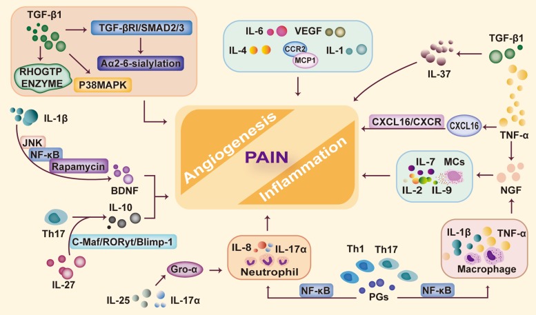
    1. Yu J, Francisco A, Patel BG, Cline JM, Zou E, Berga SL, Taylor RN. IL-1beta stimulates brain-derived neurotrophic factor production in eutopic endometriosis stromal cell cultures: a model for cytokine regulation of neuroangiogenesis. Am J Pathol. 2018;188:2281–2292. doi: 10.1016/j.ajpath.2018.06.011. - DOI - PMC - PubMed

MeSH terms