

Long non-coding RNA SNHG6 promotes the growth and invasion of non-small cell lung cancer by downregulating miR-101-3p
- PMID: 32147945
- PMCID: PMC7180593
- DOI: 10.1111/1759-7714.13371
Long non-coding RNA SNHG6 promotes the growth and invasion of non-small cell lung cancer by downregulating miR-101-3p
Abstract
Background: The aim of this study was to determine the function of long non-coding RNA small nucleolar RNA host gene 6 (SNHG6) in non-small cell lung cancer (NSCLC) and its underlying mechanisms.
Methods: The association of SNHG6 or miR-101-3p with clinicopathological characteristics and prognosis in patents with NSCLC was assessed by TCGA dataset. Cell proliferation and invasion were evaluated by MTT and Transwell assays and SNHG6-specific binding with miR-101-3p was verified by bioinformatic analysis, luciferase gene report and RNA immunoprecipitation assays. qRT-PCR and Western blot was used to assess the effects of SNHG6 on the expression of miR-101-3p and chromodomain Y like (CDYL) in NSCLC cells. A xenograft tumor model in vivo was established to observe the effects of SNHG6 knockdown on tumor growth.
Results: We found that increased expression of SNHG6 was associated with pathological stage and lymph node infiltration, and acted as an independent prognostic factor of tumor recurrence in patients with NSCLC. Silencing SNHG6 expression repressed cell growth and invasion in vitro and in vivo, but overexpression of SNHG6 reversed these effects. Furthermore, SNHG6 was identified to act as a sponge of miR-101-3p, which could reduce cell proliferation and attenuate SNHG6-induced CDYL expression. Low expression of miR-101-3p or high expression of CDYL was related to poor survival in patients with NSCLC.
Conclusions: Our findings demonstrated that lncRNA SNHG6 contributed to the proliferation and invasion of NSCLC by downregulating miR-101-3p.
Keywords: Growth; SNHG6; invasion; miR-101-3p; non-small cell lung cancer.
© 2020 The Authors. Thoracic Cancer published by China Lung Oncology Group and John Wiley & Sons Australia, Ltd.
Figures

 low SNHG6 expression,
  high SNHG6 expression), (
 low SNHG6 expression,
  high SNHG6 expression).
 BEAS‐2B,
  A549,
A549,  NCI‐H23,
  NCI‐H1993,
  NCI‐H522,
  NCI‐H460). (b) qRT‐PCR analysis of the overexpression efficiencies of SNHG6 plasmids in A549 cell line or knockdown efficiency of sh‐SNHG6 in NCI‐H460 cell line (
 pcDNA3.1,
  SNHG6,
  sh‐NC,
  sh‐SNHG6). (c), (d), MTT and Transwell analysis of the effects of SNHG6 overexpression or knockdown on cell proliferation and invasion in A549 and NCI‐H460 cells (
 pcDNA3.1,
  SNHG6,
  sh‐NC,
  sh‐SNHG6). (e) Western blot analysis of the effects of SNHG6 overexpression or knockdown on PCNA and MMP2 expression in A549 or NCI‐H460 cell line (
 pcDNA3.1,
  SNHG6,
  sh‐NC,
  sh‐SNHG6), (
 pcDNA3.1,
  SNHG6,
  sh‐NC,
  sh‐SNHG6). *P < 0.05; **P < 0.01.
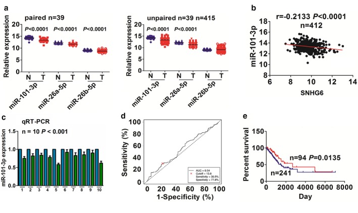
 ) Adjacent normal and (
 ) LAC. (d) The cutoff value of miR‐101‐3p was acquired by ROC curve in LAC according to the miR‐101‐3p expression, and the patients' survival time and survival status by Cutoff Finder. (e) Kaplan‐Meier analysis demonstrated that the patients with low miR‐101‐3p expression had a poorer survival as compared with those with high miR‐101‐3p expression. (
 ) miR‐101‐3p high expression and (
 ) miR‐101‐3p low expression.
 ) miR‐NC, (
 ) miR‐101‐3p‐mimic, (
 ) NC and (
 ) miR‐101‐3p‐inhibitor. (c) qRT‐PCR analysis of the overexpression efficiency of miR‐101‐3p mimic in A549 cell line or knockdown efficiency of miR‐101‐3p inhibitor in NCI‐H460 cell line. (
 ) miR‐NC, (
 ) miR‐101‐3p‐mimic, (
 ) NC and (
 ) miR‐101‐3p‐inhibitor. (d) qRT‐PCR analysis of the effects of SNHG6 overexpression or knockdown on miR‐101‐3p expression in A549 or NCI‐H460 cell line. (
 ) pcDNA3.1, (
 ) SNHG5, (
 ) sh‐NC and (
 ) sh‐SNHG6. (e) RIP assay and qRT‐PCR analysis of the increased enrichment levels of SNHG6 and miR‐101‐3p pulled down from Ago2 or IgG protein in A549 and NCI‐H460 cells. (f) MTT analysis of the cell viability after cotransfection with SNHG6 plasmids and miR‐101‐3p mimic in A549 cell line or sh‐SNHG6 and miR‐101‐3p inhibitor in NCI‐H460 cell line.(
 ) pcDNA3.1+miR‐NC, (
 ) SNHG6+miR‐NC, (
 ) pcDNA3.1+miR‐101‐3p mimic and (
 ) SNHG6+miR‐101‐3P mimic. (
 ) sh‐NC+NC, (
 ) sh‐SNHG6+NC, (
 ) sh‐NC+miR‐101‐3p inhibitor and (
 ) sh‐SNHG6+miR‐101‐3p inhibitor. *P < 0.05, **P < 0.01.
 ) CDYL high expression and (
 ) CDYL low expression.
 ) miR‐NC, (
 ) miR‐101‐3p‐mimic, (
 ) NC and (
 ) miR‐101‐3p‐inhibitor. (c),(d) qRT‐PCR and western blot analysis of the CDYL expression levels after cotransfection with SNHG6 plasmids and miR‐101‐3p mimic in A549 cell line or sh‐SNHG6 and miR‐101‐3p inhibitor in NCI‐H460 cell line. (
 ) pcDNA3.1+miR‐NC, (
 ) SNHG6+miR‐NC, (
 ) pcDNA3.1+miR‐101‐3p mimic and (
 ) SNHG6+miR‐101‐3P mimic. (
 ) sh‐NC+NC, (
 ) sh‐SNHG6+NC, (
 ) sh‐NC+miR‐101‐3p inhibitor and (
 ) sh‐SNHG6+miR‐101‐3p inhibitor. *P < 0.05; **P < 0.01.
 sh‐NC and
  sh‐SNHG6). (c),(d), Tumor volume and weight were lower in the sh‐SNHG6 group as compared with the sh‐NC group. (e), IHC analysis demonstrated that Ki‐67 proliferation index was decreased in the sh‐SNHG6 group as compared with the sh‐NC group. *P < 0.05.
References
-
- Chen W, Zheng R, Zhang S et al Cancer incidence and mortality in China, 2013. Cancer Lett 2017; 401: 63–71. - PubMed
-
- Siegel RL, Miller KD, Jemal A. Cancer statistics, 2018. CA Cancer J Clin 2018; 68 (1): 7–30. - PubMed
-
- Wu DW, Hsu NY, Wang YC et al C‐Myc suppresses microRNA‐29b to promote tumor aggressiveness and poor outcomes in non‐small cell lung cancer by targeting FHIT. Oncogene 2015; 34 (16): 2072–82. - PubMed
-
- Yu W, Ding J, He M et al Estrogen receptor β promotes the vasculogenic mimicry (VM) and cell invasion via altering the lncRNA‐MALAT1/miR‐145‐5p/NEDD9 signals in lung cancer. Oncogene 2019; 38 (8): 1225–38. - PubMed
-
- Shi SL, Zhang ZH. Long non‐coding RNA SNHG1 contributes to cisplatin resistance in non‐small cell lung cancer by regulating miR‐140‐5p/Wnt/β‐catenin pathway. Neoplasma 2019; 66 (5): 756–65. - PubMed
Publication types
MeSH terms
Substances
LinkOut - more resources
Full Text Sources
Medical
