. 2020 Feb 19:2020:6401969.
doi: 10.1155/2020/6401969. eCollection 2020.

Phenotypic and Functional Changes in Peripheral Blood Natural Killer Cells in Crohn Disease Patients

Affiliations

Phenotypic and Functional Changes in Peripheral Blood Natural Killer Cells in Crohn Disease Patients

Suzanne Samarani et al. Mediators Inflamm. .

Abstract

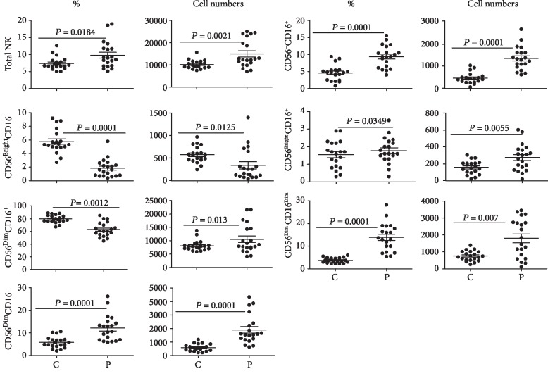
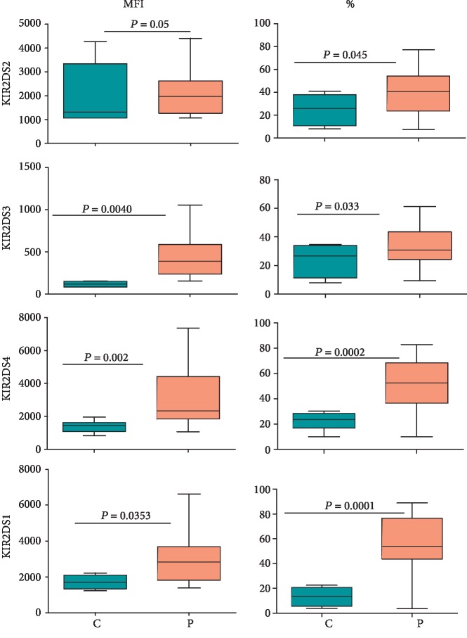
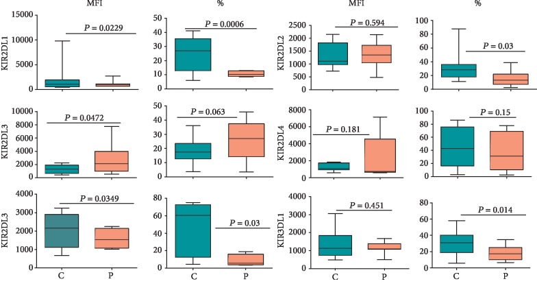
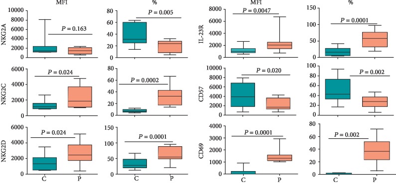
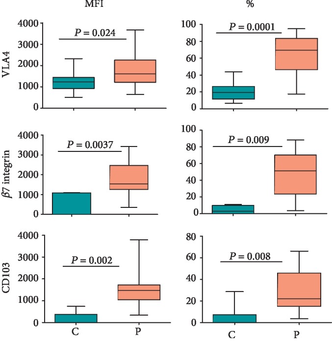
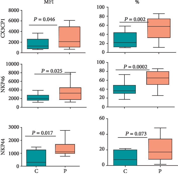
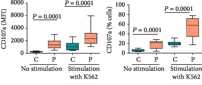
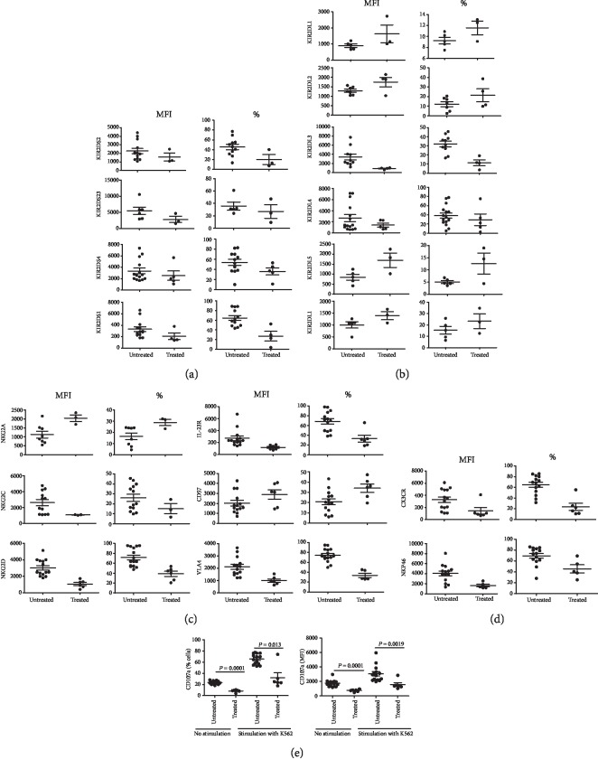
We investigated activation status, cytotoxic potential, and gut homing ability of the peripheral blood Natural Killer (NK) cells in Crohn disease (CD) patients. For this purpose, we compared the expression of different activating and inhibitory receptors (KIR and non-KIR) and integrins on NK cells as well as their recent degranulation history between the patients and age-matched healthy controls. The study was conducted using freshly obtained peripheral blood samples from the study participants. Multiple color flow cytometry was used for these determinations. Our results show that NK cells from treatment-naïve CD patients expressed higher levels of activating KIR as well as other non-KIR activating receptors vis-à-vis healthy controls. They also showed increased frequencies of the cells expressing these receptors. The expression of several KIR and non-KIR inhibitory receptors tended to decrease compared with the cells from healthy donors. NK cells from the patients also expressed increased levels of different gut-homing integrin molecules and showed a history of increased recent degranulation events both constitutively and in response to their in vitro stimulation. Furthermore, treatment of the patients tended to reverse these NK cell changes. Our results demonstrate unequivocally, for the first time, that peripheral blood NK cells in treatment-naïve CD patients are more activated and are more poised to migrate to the gut compared to their counterpart cells from healthy individuals. Moreover, they show that treatment of the patients tends to normalize their NK cells. The results suggest that NK cells are very likely to play a role in the immunopathogenesis of Crohn disease.


Conflict of interest statement

The authors declare that they have no conflicts of interest.

Figures

Figure 1
Figure 1
Percentages and absolute numbers of NK cells and their subsets. The figure shows percentages and absolute numbers (per ml) of the peripheral blood NK cells and their subsets (with respect to the expression of CD16 and CD56) in 20 CD patients (P) vis-à-vis 20 healthy control subjects (C). The figure shows means ± SE, individual data points, and p values.
Figure 2
Figure 2
Expression of activating KIR on NK cells in CD patients and healthy controls. Fifty microliters of blood samples was used and stained for CD3, CD14, and CD19 and activating KIR (PE or FITC-conjugated) as described in Materials and Methods. The expression was determined on CD3-CD14-CD19-CD56bright/dim (NK) cells. The figure shows mean fluorescence intensities (MFIs) and percentages of NK cells expressing the receptor from 15 treatment-naïve patients and 20 healthy controls. Each box and whisker plot indicates median, upper and lower values, interquartile range, and p values.
Figure 3
Figure 3
Expression of inhibitory KIR on NK cells in 15 treatment-naïve CD patients and 15 healthy controls. The cells were stained and gated as described in the legend of Figure 2 and stained for antibodies specific for inhibitory KIR as described in Materials and Methods. The figure shows median, upper and lower values, interquartile range, and p values.
Figure 4
Figure 4
Expression of non-KIR markers on CD56bright/dim NK cells in CD patients and healthy controls. The figure shows MFI and percentages of NK cells expressing the indicated marker in 15 treatment-naïve CD patients and 20 healthy controls. Each box and whisker plot indicates median, interquartile ranges, and p values.
Figure 5
Figure 5
Expression of different integrins on CD56bright/dim NK cells in CD patients and healthy controls. The figure shows MFI and percentages of NK cells expressing indicated integrins. Each box and whisker plot indicates median, interquartile ranges, and p values for 15 treatment-naïve CD patients and 20 healthy control donors.
Figure 6
Figure 6
Expression of NKp46, NKp44, and CX3CR1 on CD56bright/dim NK cells in CD patients and healthy controls. The figure shows MFI and percentages of NK cells expressing the markers. The box and whisker plots indicate median, interquartile ranges, and p values for 15 treatment-naïve CD patients and 15 healthy controls.
Figure 7
Figure 7
The surface expression of CD107a on CD56bright/dim NK cells in CD patients vis-à-vis healthy controls with and without prior stimulation with K562. The panels show MFI of CD107a expression and percentages of CD107a expressed in NK cells. The box and whisker plots indicate median, interquartile ranges, and p values from ten treatment-naïve CD patients and the same number of healthy control donors.
Figure 8
Figure 8
Comparison of NK cell expression of different parameters between treatment-naïve (untreated) and under-treatment (treated) CD patients. The figure shows NK cell expression of activating KIR (a), inhibitory KIR (b), non-KIR receptors and VLA-4 (c), NKP46 and CX3CR1 (d), and CD107a with and without stimulation with K562 cells (e). This figure shows means ± SE and p values.

References

    1. Campbell K. S., Hasegawa J. Natural killer cell biology: an update and future directions. Journal of Allergy and Clinical Immunology. 2013;132(3):536–544. doi: 10.1016/j.jaci.2013.07.006. - DOI - PMC - PubMed
    1. Zitti B., Bryceson Y. T. Natural killer cells in inflammation and autoimmunity. Cytokine & Growth Factor Reviews. 2018;42:37–46. doi: 10.1016/j.cytogfr.2018.08.001. - DOI - PubMed
    1. Choucair K., Duff J. R., Cassidy C. S., et al. Natural killer cells: a review of biology, therapeutic potential and challenges in treatment of solid tumors. Future Oncology. 2019;15(26):3053–3069. doi: 10.2217/fon-2019-0116. - DOI - PubMed
    1. Wilk E., Kalippke K., Buyny S., Schmidt R. E., Jacobs R. New aspects of NK cell subset identification and inference of NK cells' regulatory capacity by assessing functional and genomic profiles. Immunobiology. 2008;213(3-4):271–283. doi: 10.1016/j.imbio.2007.10.012. - DOI - PubMed
    1. Ochoa M. C., Minute L., Rodriguez I., et al. Antibody-dependent cell cytotoxicity: immunotherapy strategies enhancing effector NK cells. Immunology and Cell Biology. 2017;95(4):347–355. doi: 10.1038/icb.2017.6. - DOI - PubMed