

Bone Formation in Grafts with Bio-Oss and Autogenous Bone at Different Proportions in Rabbit Calvaria
- PMID: 32148500
- PMCID: PMC7049819
- DOI: 10.1155/2020/2494128
Bone Formation in Grafts with Bio-Oss and Autogenous Bone at Different Proportions in Rabbit Calvaria
Abstract
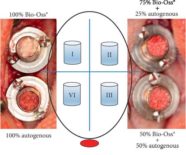
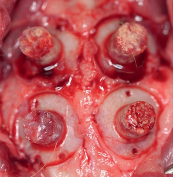
Background: The aim of this study was to assess the volumetric stability and bone formation in grafts with Bio-Oss and autogenous bone at different proportions in rabbit calvaria. Material and Methods. Ten rabbits received four titanium cylinders in their calvaria and randomly divided into the following groups: Group I: Bio-Oss (100%), Group II: Bio-Oss (75%) + autogenous bone (25%), Group III: Bio-Oss (50%) + autogenous bone (50%), and Group IV: autogenous bone (100%). After twelve weeks, the animals were euthanized, and samples were collected for clinical and histological analysis.
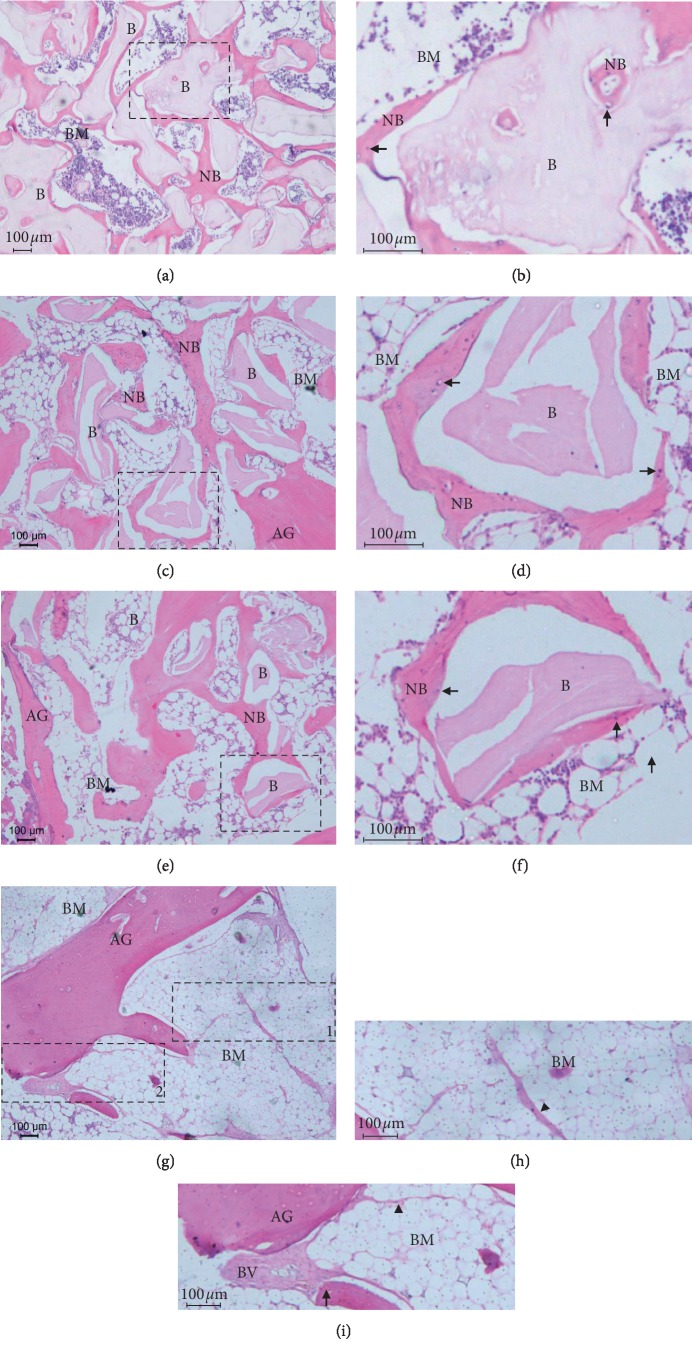
Results: Clinical analysis showed that Groups I (90.43 ± 8.99) and II (90.87 ± 7.43) had greater dimensional stability compared to Group IV (P=0.0005). Histologically, Groups I, II, and III showed areas of bone formation with particles of biomaterial remaining in close contact with the newly formed bone. However, there were no significant differences between the groups regarding the newly formed bone area.
Conclusion: It was concluded that the use of Bio-Oss either alone or associated with the autogenous bone at a proportion of 25% showed superior dimensional stability compared to the use of autogenous bone in the proposed experimental model.
Copyright © 2020 Yeon Jung Kim et al.
Conflict of interest statement
The authors declare no conflicts of interest.
Figures
References
-
- Carmagnola D., Abati S., Celestino S., Chiapasco M., Bosshardt D., Lang N. P. Oral implants placed in bone defects treated with Bio-Oss, ostim-paste or perioglas: an experimental study in the rabbit tibiae. Clinical Oral Implants Research. 2008;19(12):1246–1253. doi: 10.1111/j.1600-0501.2008.01584.x. - DOI - PubMed
-
- Schlegel A., Hamel J., Wichmann M., Eitner S. Comparative clinical results after implant placement in the posterior maxilla with and without sinus augmentation. The International Journal of Oral & Maxillofacial Implants. 2008;23(2):289–298. - PubMed
LinkOut - more resources
Full Text Sources
