. 2020 Feb 27:8:e8326.
doi: 10.7717/peerj.8326. eCollection 2020.

Gene expression patterns of novel visual and non-visual opsin families in immature and mature Japanese eel males

Affiliations

Gene expression patterns of novel visual and non-visual opsin families in immature and mature Japanese eel males

Jun-Hwan Byun et al. PeerJ. .

Abstract

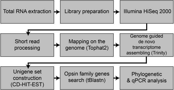
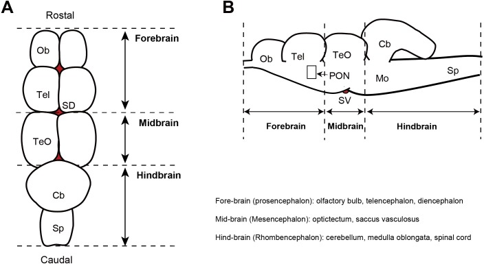
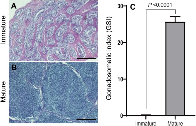
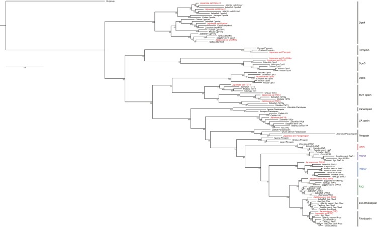
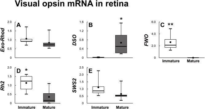
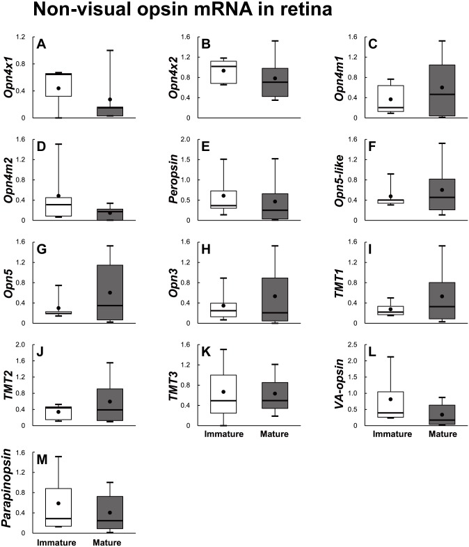
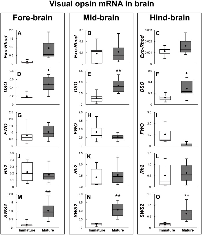
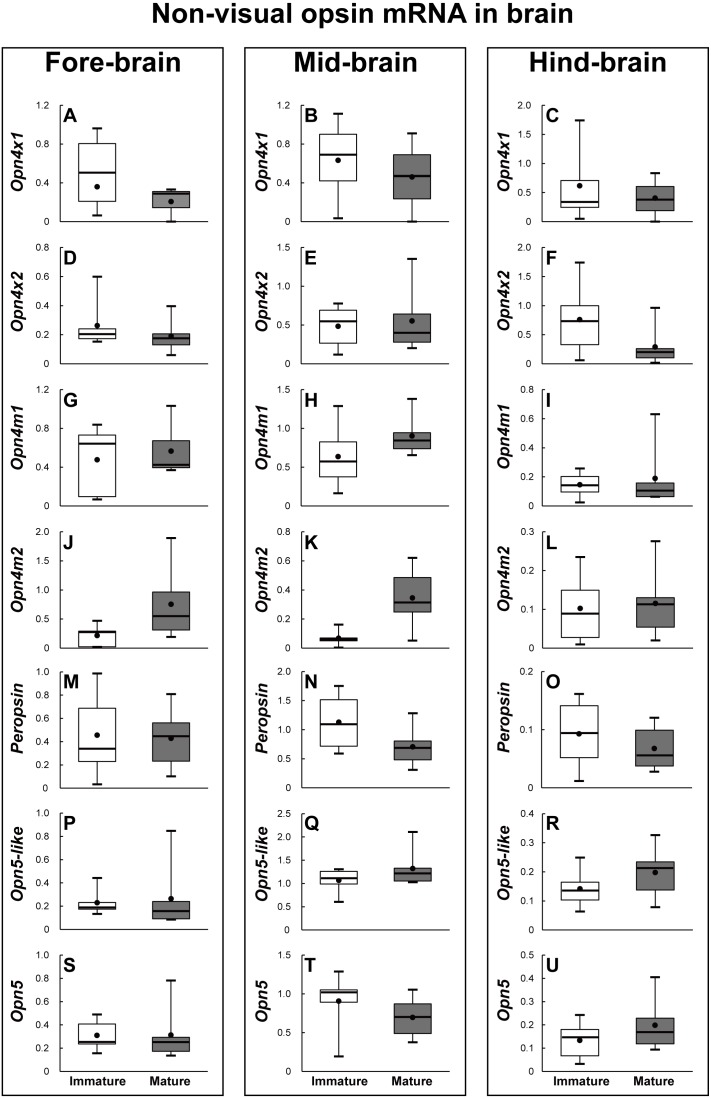
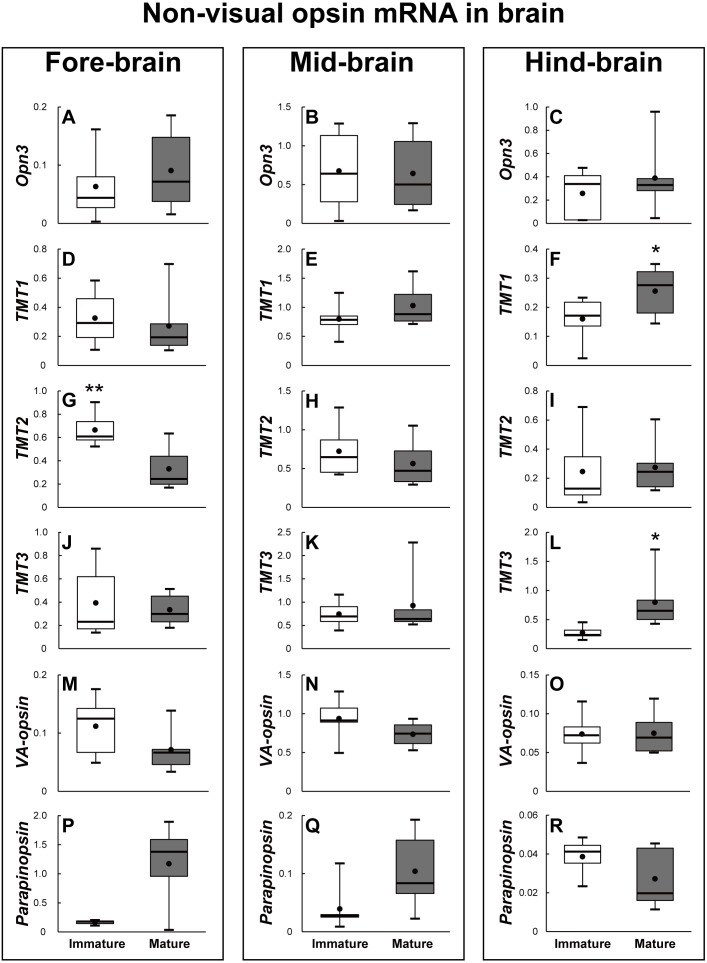
This study was carried out to identify and estimate physiological function of a new type of opsin subfamily present in the retina and whole brain tissues of Japanese eel using RNA-Seq transcriptome method. A total of 18 opsin subfamilies were identified through RNA-seq. The visual opsin family included Rh2, SWS2, FWO, DSO, and Exo-Rhod. The non-visual opsin family included four types of melanopsin subfamily (Opn4x1, Opn4x2, Opn4m1, and Opn4m2), peropsin, two types of neuropsin subfamily (Opn5-like, Opn5), Opn3, three types of TMT opsin subfamily (TMT1, 2, 3), VA-opsin, and parapinopsin. In terms of changes in photoreceptor gene expression in the retina of sexually mature and immature male eels, DSO mRNA increased in the maturation group. Analysis of expression of opsin family gene in male eel brain before and after maturation revealed that DSO and SWS2 expression in terms of visual opsin mRNA increased in the sexually mature group. In terms of non-visual opsin mRNA, parapinopsin mRNA increased whereas that of TMT2 decreased in the fore-brain of the sexually mature group. The mRNA for parapinopsin increased in the mid-brain of the sexually mature group, whereas those of TMT1 and TMT3 increased in the hind-brain of the sexually mature group. DSO mRNA also increased in the retina after sexual maturation, and DSO and SWS2 mRNA increased in whole brain part, suggesting that DSO and SWS2 are closely related to sexual maturation.

Keywords: Anguilla japonica; Japanese eel; Opsin; Photoreceptor; Sex maturation.


Conflict of interest statement

The authors declare there are no competing interests

Figures

Figure 1
Figure 1. Flowchart of the present study.
Figure 2
Figure 2. Diagram showing the dorsal view (A) and sagittal plane (B) of the eel brain.
Ob, olfactory bulb; Tel, telencephalon; TeO, optic tectum; Cb, cerebellum; Mo, medulla oblongata; P, pineal gland; SD, saccus dorsalis; PON, preoptic nucleus; SV, saccus vasculosus.
Figure 3
Figure 3. Microphotographs of histological sections of different stages of eel testis and changes of gonadosomatic index (GSI) after hormonally induced sexual maturation.
(A) Immature testis, (B) mature testis, (C) GSI. Scale bar = 200 µm.
Figure 4
Figure 4. Phylogeny of vertebrate visual and non-visual opsins. One thousand bootstrap repetitions were performed and values are shown at the inner nodes.
The zebrafish beta 1 adrenergic receptor was used as an outgroup to root the tree. Analysis was performed with multiple alignments from the amino acid sequence by using ClustalW program. Bold is indicated the visual and non-visual opsin families of Japanese eel, A. japonica.
Figure 5
Figure 5. Visual opsin mRNA level in the retina of sexually immature and mature male Japanese eel.
For the artificially induced sexual maturation, hCG was intraperitoneally injected to the experimental fish group (n = 6) at a concentration of 1 IU/g body weight. Immature fish was sampled before hCG injection (n = 6). Eight weeks after injection, retina was sampled and used for total RNA extraction and cDNA synthesis. The mRNA expression of visual opsin (A–E) in each sample was measured real-time qPCR. Boxplots show min and max values (whiskers), first and third quartiles (box limits), and median (box inner line) of mRNA levels. The asterisk above each bar indicates significant differences according to the unpaired t test (*P < 0.05, **P < 0.01).
Figure 6
Figure 6. Non-visual opsin mRNA level in the retina of sexually immature and mature male Japanese eel.
For the artificially induced sexual maturation, hCG was intraperitoneally injected to the experimental fish group (n = 6) at a concentration of 1 IU/g body weight. Immature fish was sampled before hCG injection (n = 6). Eight weeks after injection, retina was sampled and used for total RNA extraction and cDNA synthesis. The mRNA expression of non-visual opsin (A–M) in each sample was measured real-time qPCR. Boxplots show min and max values (whiskers), first and third quartiles (box limits), and median (box inner line) of mRNA levels. The asterisk above each bar indicates significant differences according to the unpaired t test (*P < 0.05, **P < 0.01).
Figure 7
Figure 7. Visual opsin mRNA level in the brain of sexually immature and mature male Japanese eel.
For the artificially induced sexual maturation, hCG was intraperitoneally injected to the experimental fish group (n = 6) at a concentration of 1 IU/g body weight. Immature fish was sampled before hCG injection (n = 6). Eight weeks after injection, brain was sampled and used for total RNA extraction and cDNA synthesis. The mRNA expression of visual opsin (A–O) in each sample was measured real-time qPCR. Boxplots show min and max values (whiskers), first and third quartiles (box limits), and median (box inner line) of mRNA levels. The asterisk above each bar indicates significant differences according to the unpaited t test (*P < 0.05, **P < 0.01).
Figure 8
Figure 8. Non-visual opsin mRNA level in the brain of sexually immature and mature male Japanese eel male.
Boxplots show min and max values (whiskers), first and third quartiles (box limits), and median (box inner line) of mRNA levels. For the artificially induced sexual maturation, hCG was intraperitoneally injected to the experimental fish group (n = 6) at a concentration of 1 IU/g body weight. Immature fish was sampled before hCG injection (n = 6). Eight weeks after injection, brain was sampled and used for total RNA extraction and cDNA synthesis. The mRNA expression of non-visual opsin (A–U) in each sample was measured real-time qPCR. Boxplots show min and max values (whiskers), first and third quartiles (box limits), and median (box inner line) of mRNA levels. The asterisk above each bar indicate significant differences according to the unpaired t test (*P < 0.05, **P < 0.01).
Figure 9
Figure 9. Non-visual opsin mRNA level in the brain of sexually immature and mature male Japanese eel male.
Boxplots show min and max values (whiskers), first and third quartiles (box limits), and median (box inner line) of mRNA levels. For the artificially induced sexual maturation, hCG was intraperitoneally injected to the experimental fish group (n = 6) at a concentration of 1 IU/g body weight. Immature fish was sampled before hCG injection (n = 6). Eight weeks after injection, brain was sampled and used for total RNA extraction and cDNA synthesis. The mRNA expression of non-visual opsin (A–R) in each sample was measured real-time qPCR. Boxplots show min and max values (whiskers), first and third quartiles (box limits), and median (box inner line) of mRNA levels. The asterisk above each bar indicate significant differences according to the unpaired t test (*P < 0.05, **P < 0.01).

Similar articles

Cited by

References

    1. Beaudry FEG, Iwanicki TW, Mariluz BRZ, Darnet S, Brinkmann H, Schneider P, Taylor JS. The non-visual opsins: eighteen in the ancestor of vertebrates, astonishing increase in ray-finned fish, and loss in amniotes. Journal of Experimental Zoology Part B: Molecular and Developmental Evolution. 2017;328(7):685–696. doi: 10.1002/jez.b.22773. - DOI - PubMed
    1. Bellingham J, Chaurasia SS, Melyan Z, Liu C, Cameron MA, Tarttelin EE, Iuvone PM, Hankins MW, Tosini G, Lucas RJ. Evolution of melanopsin photoreceptors: discovery and characterization of a new melanopsin in nonmammalian vertebrates. PLOS Biology. 2006;4(8):e254. doi: 10.1371/journal.pbio.0040254. - DOI - PMC - PubMed
    1. Bellingham J, Tarttelin EE, Foster RG, Wells DJ. Structure and evolution of the Teleost extraretinal rod-like opsin (errlo) and ocular rod opsin (rho) genes: is teleost rho a retrogene? Journal of Experimental Zoology Part B: Molecular and Developmental Evolution. 2003;297(1):1–10. - PubMed
    1. Benoit JM. Chronobiologic study in the domestic duck. II. Physiological mechanism of the chronobiologic action of visible light on the gonads of the male duck. Chronobiologia. 1978;5:158–168. - PubMed
    1. Bolger AM, Lohse M, Usadel B. Trimmomatic: a flexible trimmer for Illumina sequence data. Bioinformatics. 2014;30:2114–2120. doi: 10.1093/bioinformatics/btu170. - DOI - PMC - PubMed

LinkOut - more resources