Review
. 2019 Feb 23;1(1):30-43.
doi: 10.1016/j.jhepr.2019.02.004. eCollection 2019 May.

Macrophages in obesity and non-alcoholic fatty liver disease: Crosstalk with metabolism

Affiliations
Review

Macrophages in obesity and non-alcoholic fatty liver disease: Crosstalk with metabolism

Sander Lefere et al. JHEP Rep. .

Abstract

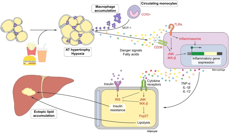
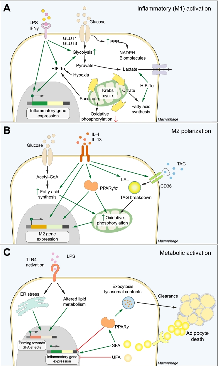
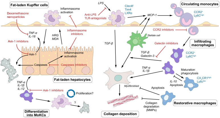
Non-alcoholic fatty liver disease (NAFLD) is the most prevalent liver disease worldwide, and a major cause of liver cirrhosis and hepatocellular carcinoma. NAFLD is intimately linked with other metabolic disorders characterized by insulin resistance. Metabolic diseases are driven by chronic inflammatory processes, in which macrophages perform essential roles. The polarization status of macrophages is itself influenced by metabolic stimuli such as fatty acids, which in turn affect the progression of metabolic dysfunction at multiple disease stages and in various tissues. For instance, adipose tissue macrophages respond to obesity, adipocyte stress and dietary factors by a specific metabolic and inflammatory programme that stimulates disease progression locally and in the liver. Kupffer cells and monocyte-derived macrophages represent ontologically distinct hepatic macrophage populations that perform a range of metabolic functions. These macrophages integrate signals from the gut-liver axis (related to dysbiosis, reduced intestinal barrier integrity, endotoxemia), from overnutrition, from systemic low-grade inflammation and from the local environment of a steatotic liver. This makes them central players in the progression of NAFLD to steatohepatitis (non-alcoholic steatohepatitis or NASH) and fibrosis. Moreover, the particular involvement of Kupffer cells in lipid metabolism, as well as the inflammatory activation of hepatic macrophages, may pathogenically link NAFLD/NASH and cardiovascular disease. In this review, we highlight the polarization, classification and function of macrophage subsets and their interaction with metabolic cues in the pathophysiology of obesity and NAFLD. Evidence from animal and clinical studies suggests that macrophage targeting may improve the course of NAFLD and related metabolic disorders.

Keywords: NASH; immunometabolism; innate immunity; liver immunology; steatohepatitis, Kupffer cells.


Figures

Fig. 1
Fig. 1
Macrophages in obese adipose tissue. In obesity, chemokines stimulate the infiltration of macrophages into the AT. Various danger molecules and fatty acids promote a pro-inflammatory phenotype in ATMs, which contributes to the development of a persistent, low-grade inflammation. Pro-inflammatory mediators secreted by ATMs induce insulin resistance by signalling through the JNK and IKK-β pathways. Inhibition of lipid droplet-associated peptides, such as fsp27, enhance lipolysis. Downstream, insulin resistance and lipolysis lead to ectopic lipid accumulation in various organs, including the liver. AT, adipose tissue; ATMs, adipose tissue macrophages; IRS, insulin receptor substrates; TLRs, Toll-like receptors.
Fig. 2
Fig. 2
Metabolic coordination of macrophage activation. (A) Inflammatory (M1) activation. LPS and/or IFNγ stimulation increases glucose consumption, which is converted to lactate instead of entering the Krebs cycle as pyruvate. The pentose phosphate pathway and fatty acid synthesis are activated to generate biomolecules. These metabolic alterations depend in part on HIF-1α, which stimulates glucose entry and conversion to lactate and directly promotes M1 gene expression. (B) M2 polarization depends on increased β-oxidation and oxidative phosphorylation of fatty acids. This is fuelled by the uptake and lysosomal breakdown of TAGs and the de novo synthesis from glucose. PPARγ and PPARδ bind these intracellular lipids and are key effectors of the metabolic and phenotype switch upon IL-4 stimulation. (C) Macrophages display a metabolically activated phenotype upon stimulation with SFAs. In the adipose tissue, one mechanism of SFA uptake is through the digestion of dead adipocytes at crown-like structures. Binding of LPS primes macrophages towards the pro-inflammatory effects of SFAs, leading to a distinct macrophage phenotype that secretes inflammatory mediators such as IL-1β. PPARγ has a dual role in stimulating the clearance and breakdown of dead adipocytes, and inhibiting the production of inflammatory cytokines. ER, endoplasmic reticulum; LPS, lipopolysaccharide; PPP, pentose phosphate pathway; SFA, saturated fatty acid; TAGs, triacylglycerides; UFA, unsaturated fatty acid.
Fig. 3
Fig. 3
Macrophage-mediated inflammation as a therapeutic target in NASH progression. In NASH, KCs are activated by bacterial LPS and by danger signals from fat-laden hepatocytes, leading to the production of pro-inflammatory mediators that promote disease progression. Chemokines induce the infiltration of monocytes, which differentiate into monocyte-derived macrophages. These cells contribute to the progression of NASH by aggravating hepatic steatosis, activating stellate cells and promoting fibrosis. Ly6Chigh macrophages can mature into Ly6Clow restorative macrophages that endorse disease resolution through secretion of anti-inflammatory cytokines and collagen degrading factors. Monocyte-derived macrophages can differentiate into KCs (monocyte-derived KCs, MoKC), although it is unclear at this point if this population is capable of long-term self-renewal or if they are short-lived. Selected drugs that target inflammatory pathways and which are currently under investigation are indicated. HRG, histidine-rich glycoprotein; KC, Kupffer cell; LPS, lipopolysaccharide; MDA, malonyldialdehyde; MMPs, matrix metalloproteinases; MoKC, monocyte-derived Kupffer cell; NASH, non-alcoholic steatohepatitis; TLR, Toll-like receptor.

Similar articles

Cited by

References

    1. Yki-Jarvinen H. Non-alcoholic fatty liver disease as a cause and a consequence of metabolic syndrome. Lancet Diabetes Endocrinol. 2014;2:901–910. - PubMed
    1. Younossi Z, Anstee QM, Marietti M, Hardy T, Henry L, Eslam M. Global burden of NAFLD and NASH: trends, predictions, risk factors and prevention. Nat Rev Gastroenterol Hepatol. 2018;15:11–20. - PubMed
    1. Estes C, Razavi H, Loomba R, Younossi Z, Sanyal AJ. Modeling the epidemic of nonalcoholic fatty liver disease demonstrates an exponential increase in burden of disease. Hepatology. 2018;67:123–133. - PMC - PubMed
    1. Angulo P, Kleiner DE, Dam-Larsen S, Adams LA, Bjornsson ES, Charatcharoenwitthaya P. Liver Fibrosis, but No Other Histologic Features, Is Associated With Long-term Outcomes of Patients With Nonalcoholic Fatty Liver Disease. Gastroenterology. 2015;149:389–397. e10. - PMC - PubMed
    1. Ekstedt M, Hagstrom H, Nasr P, Fredrikson M, Stal P, Kechagias S. Fibrosis stage is the strongest predictor for disease-specific mortality in NAFLD after up to 33 years of follow-up. Hepatology. 2015;61:1547–1554. - PubMed

LinkOut - more resources